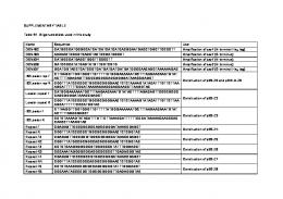
Table S1. AR and pattern of cell arrangement in different nematode species genera/ .... For statistical analysis, normality was tested with a ShapiroâWilk test,.
Development 144: doi:10.1242/dev.154609: Supplementary information
Supplementary Tables
genera/species
aspect ratio
pattern
references
Enoplus brevis
1.17
Pyramid
(Schulze and Schierenberg, 2011)
Halicephalobus
1.37
Diamond
(Goldstein, 2001)
PS1010
1.39
Diamond
(Goldstein, 2001)
Goodeyus
1.39
Diamond
(Goldstein, 2001)
Caenorhabditis
1.40
Diamond
(Goldstein, 2001)
Bunonema
1.45
Diamond
(Goldstein, 2001)
Rhabditis
1.45
Diamond
(Goldstein, 2001)
Teratorhabditis
1.51
Diamond
(Goldstein, 2001)
Plectonchus
1.62
Diamond
(Goldstein, 2001)
Cruznema
1.62
Diamond
(Goldstein, 2001)
Panagrellus
1.67
Diamond
(Goldstein, 2001)
Mesorhabditis
1.76
Diamond
(Goldstein, 2001)
Panagrobelus
1.77
Diamond
(Goldstein, 2001)
Pristionchus
1.77
Diamond
(Goldstein, 2001)
Aduncospiculum
1.78
Diamond
(Goldstein, 2001)
Nothacrobeles
1.84
Diamond
(Goldstein, 2001)
Diploscapter
1.88
Linear
(Goldstein, 2001)
Diploscapter coronata
1.90
Linear
This study
Acrobeloides
1.91
T-shaped
(Goldstein, 2001)
Protorhabditis
1.91
Linear
(Goldstein, 2001)
Cephalobus
1.97
T-shaped
(Goldstein, 2001)
Rhabditella
2.02
Diamond
(Goldstein, 2001)
Eucephalobus
2.03
T-shaped
(Goldstein, 2001)
Pseudoacrobeles
2.06
T-shaped
(Goldstein, 2001)
Panagrolaimus
2.09
Diamond
(Goldstein, 2001)
Cervidellus
2.18
T-shaped
(Goldstein, 2001)
Acrobeles
2.21
T-shaped
(Goldstein, 2001)
Diplenteron
2.31
Diamond
(Goldstein, 2001)
1
Development • Supplementary information
Table S1. AR and pattern of cell arrangement in different nematode species
Development 144: doi:10.1242/dev.154609: Supplementary information
Aphelenchoides
2.44
T-shaped
(Goldstein, 2001)
Meloidogyne
2.47
Linear
(Goldstein, 2001)
Zeldia
2.47
Linear
(Goldstein, 2001)
Chiloplacus
2.48
T-shaped
(Goldstein, 2001)
Nacobbus
2.52
Linear
(Goldstein, 2001)
Pratylenchus
2.56
Linear
(Goldstein, 2001)
Teratocephalus
2.69
Diamond
(Goldstein, 2001)
Aphelenchus
2.79
Diamond
(Goldstein, 2001)
Belonolaimus
4.11
Linear
(Goldstein, 2001)
Aphelenchoides besseyi
4.40
Linear
This study
References Goldstein, B. (2001). On the evolution of early development in the Nematoda. Philos. Trans. R. Soc. Lond. B Biol. Sci. 356, 1521–1531. Schulze, J. and Schierenberg, E. (2011). Evolution of embryonic development in nematodes.
Development • Supplementary information
Evodevo 2, 18.
2
Development 144: doi:10.1242/dev.154609: Supplementary information
Table S2. Caenorhabditis elegans strains used in this study name
genotype
sources
N2
wild type
CGC*
CB185
lon-1(e185)III
CGC
CB207
dpy-11(e207)V
CGC
EG4601
oxIs279 [pie-1p::GFP::H2B + unc-119(+)]II; unc-119(ed3)III
CGC
FT741
xnSi6 [mex-5P::hmr-1::GFP::hmr-1 3´-UTR + unc-119(+)]II;
CGC
unc-119(ed3)III GR1034
ceh-18(mg57)X
CGC
LP172
hmr-1(cp21[hmr-1::GFP + LoxP]) I.
CGC
LP316
hmp-2(cp78[GFP::hmp-2a + LoxP]) III.
CGC
RB1353
spv-1(ok1498)II
CGC
CAL1661
dpy-11(e207)V; oxIs279 [pie-1p::GFP::H2B + unc-119(+)]II;
This study
unc-119(ed3)III CAL1671
lon-1(e185)III; oxIs279 [pie-1p::GFP::H2B + unc-119(+)]II;
This study
unc-119(ed3)III
Development • Supplementary information
* CGC = Caenorhabditis Genetics Center
3
Development 144: doi:10.1242/dev.154609: Supplementary information
Table S3. Parameters implemented in the computer simulation parameters
values
references
Force Force by the eggshell
K0/γ
0.2 (µm/sec)
(Fickentscher et al., 2013)
Force by cells
F0/γ
0.1 (µm/sec)
(Fickentscher et al., 2013)
Random fluctuation
ξ
0 ± 0.027(µm)
(Fickentscher et al., 2013)
Stable repulsion ratio
α
0.7–1.0
This study
Δt
5 (sec)
(Fickentscher et al., 2013)
at 2-cell stage
186 (step)
(Fickentscher et al., 2013)
at 3-cell stage
72 (step)
(Fickentscher et al., 2013)
at 4-cell stage
172 (step)
(Fickentscher et al., 2013)
0 (step)
This study
156 (step)
This study
Time Time interval per step Duration time
at 3-cell stage (par-2, par-3 RNAi*) at 4-cell (par-2, par-3 RNAi) Size and Geometry Aspect ratio of the eggshell
AR
1.0–4.0
This study
Total volume of cells
Volume
23,550 (µm3)
(Fickentscher et al., 2013)
of AB cell
R_AB
15.00 (µm)
(Fickentscher et al., 2013)
of P1 cell
R_P1
13.10 (µm)
(Fickentscher et al., 2013)
of ABa cell
R_ABa
11.90 (µm)
(Fickentscher et al., 2013)
of ABp cell
R_ABp
11.90 (µm)
(Fickentscher et al., 2013)
of EMS cell
R_EMS
11.05 (µm)
(Fickentscher et al., 2013)
of P2 cell
R_P2
9.65 (µm)
(Fickentscher et al., 2013)
R_2cell
14.12 (µm)
This study
R_4cell
11.20 (µm)
This study
at 2-cell stage (par-2, par-3 RNAi**) at 4-cell (par-2, par-3 RNAi**)
4
Development • Supplementary information
Cell radius
Development 144: doi:10.1242/dev.154609: Supplementary information
* Under the par-2 and par-3 (RNAi) conditions, the two cells at the 2-cell stage divide simultaneously, and there is no three-cell stage. ** Under the par-2 and par-3 (RNAi) conditions, there are no asymmetry in volume at the 2-cell or 4-cell stage. Reference Fickentscher, R., Struntz, P. and Weiss, M. (2013). Mechanical Cues in the Early Embryogenesis
Development • Supplementary information
of Caenorhabditis elegans. Biophys. J. 105, 1805–1811.
5
Development 144: doi:10.1242/dev.154609: Supplementary information
Development • Supplementary information
Supplemental Figures
6
Development 144: doi:10.1242/dev.154609: Supplementary information
Figure S1. Eggshell shapes in mutant and RNAi-treated strains of C. elegans and the model that explains the variation in eggshell shapes (A) The diamond-type cell arrangement is formed in C. elegans embryo as follows: At the one-cell stage, the P0 cell divides parallel to the anterior-posterior axis. The anterior daughter cell of P0 is named AB, and the posterior daughter cell is P1. AB divides vertically to the anterior-posterior axis, and during cell elongation, the orientation of cell division axis is skewed. The anterior daughter cell of AB is named ABa, and the posterior daughter cell ABp. Then, P1 divides parallel with the anterior-posterior axis. The daughter cell of P1 in contact with ABa and ABp is named EMS. The posterior daughter cell of P1 is named P2. At the 4-cell stage, all cells, except ABa and P2, are in contact with each other. Theoretically, the diamond-type-arrangement with ABa-P2 contact (and loss of ABp-EMS contact) is possible. However, such an arrangement has never been observed experimentally throughout this study. (B) Histograms showing ARs in C. elegans mutants and RNAi-treated strains. In a previous study, an spv-1 mutant was reported to produce embryos of various shapes (Tan and Zaidel-Bar, 2015). We did not use the spv-1 mutant for the other part of the present study as this strain produces embryos with non-ellipsoidal shape and/or with very small or very large volumes. The means ± SDs of the eggshell shapes in each strain are as follows: N2 (1.6 ± 0.1, n = 281), spv-1(ok1498) (1.9 ± 0.4, n = 283), ceh-18(mg57) (1.6 ± 0.2, n = 183), C27D9.1 (RNAi) (1.9 ± 0.2, n = 90). For statistical analysis, normality was tested with a Shapiro–Wilk test, and homoscedasticity was confirmed using an F test; ***P < 0.001 vs. N2 (wild type). Student’s t-test was used forC27D9.1 (RNAi). Wilcoxon’s rank-sum test was used for spv-1(ok1498), and ceh-18(mg57). (C) A model that explains how eggshell shape is defined: The following two factors volume; dpy-11 mutants are considered to have thick gonads and lon-1 mutants slender gonads. Increased volume of oocyte induced by C27D9.1 (RNAi) might be squeezed in the gonad. (D) Quantification of the aspect ratio of the cross-sectional area (cAR) of the eggshell perpendicular to the long axis; upper left panel shows the definition of axes. The x-y plane is parallel to the focal planes of the microscope. Upper middle and right panels are images of the eggshell visualized by Texas Red-Dextran staining (bar = 10 µm). The white lines with arrows represent long and short axis of the eggshells in x-y (upper middle) or y-z (upper right) plane. Lower left panel is the plot of cAR against aspect ratio of x-y plane, and lower right panels are histograms of cAR. The measurements were performed in four kinds of C. elegans strains. N2 (black circle, n = 15), dpy-11(e207) (red square, n = 22), lon-1(e185) (blue triangle, n = 22), lon-1(e185);
7
Development • Supplementary information
are considering determine eggshell shape: (1) the cross-sectional area of the gonad and (2) oocyte
Development 144: doi:10.1242/dev.154609: Supplementary information
C27D9.1(RNAi) (green cross, n = 26). Reference Tan, P. Y. and Zaidel-Bar, R. (2015). Transient membrane localization of SPV-1 drives cyclical
Development • Supplementary information
actomyosin contractions in the C. elegans spermatheca. Curr. Biol. 25, 141–151.
8
Development • Supplementary information
Development 144: doi:10.1242/dev.154609: Supplementary information
9
Development 144: doi:10.1242/dev.154609: Supplementary information
Figure S2. Blastomere configuration in embryos of C. elegans after removal of the eggshell and the relationship of cell arrangement patterns and hatching rate with the ARs in various strains of C. elegans (A) The orientation of cell division in N2 (wild type), dpy-11(e207), lon-1(e185), C27D9.1 RNAi-treated strains with N2 or lon-1(e185) background (upper panels): The orientations are easier to observe in embryos with the eggshell removed at the 1–2-cell stage and cultured until the 4-cell stage (lower panels). The daughter cells of the AB cell (ABa and ABp, or ABd in eggshell removed embryos) divide vertically (left), and the daughter cells of the P1 cell (EMS and P2) divide horizontally (right). Spindle poles are represented by yellow asterisks. Scale bars are 10 µm. (B) Bee swarm plots displaying the relationship between each of the four types of cell arrangement (blue, diamond type; red, pyramid type; green, T-shaped type; cyan, linear type) and the ARs of eggshells in N2 (wild type) (n = 52), dpy-11(e207) (n = 59), lon-1(e185) (n = 36), lon-1(e185); C27D9.1 (RNAi) (n = 41) (C) Upper micrographs show images of N2 embryos in microchambers (scale bars = 10 µm). Lower panel indicates the bee swarm plot showing the distribution of ARs in the microchambers (n = 32). All embryos adopted the diamond arrangement (blue dots) under these conditions. It should be noted that, with this mechanical confinement experiment, we were not able to change the AR below 1.2 or above 2.8, where the cells are expected to take the other arrangements than the diamond type. (D) ARs and embryonic development (hatched (blue) or not hatched (red)) were examined in N2 (wild type) (n = 98), dpy-11(e207) (n = 109), lon-1(e185) (n = 53), C27D9.1 RNAi-treated strain with the N2 (n = 90) or lon-1(e185) (n = 322) background, and
Development • Supplementary information
spv-1(ok1498) (n = 150).
10
Development • Supplementary information
Development 144: doi:10.1242/dev.154609: Supplementary information
11
Development 144: doi:10.1242/dev.154609: Supplementary information
Figure S3. Simulation of cell arrangement patterns for various sets of parameters (A, B) Relationship between rates of appearance of various cell arrangement patterns (blue, diamond type; red, pyramid type; green, T-shaped type; cyan, linear type; white, others) and the ARs in the RO model under changing force strength (cell–eggshell; K0 (A), cell–cell; F0 (B)). We focused on the parameter of repulsive force strength, as the other parameters in this model were based on experimental measurements. Moreover, because the absolute values of the forces affect only division speed but not the final (stable) position of the cell, we changed the ratio of repulsion forces between cell–cell (F0) and cell–eggshell (K0). We first determined the range for cells acquiring the diamond-type arrangement in a normal shape. Next, within this ratio range, we changed the shape and examined whether diversity and robustness were reproduced. The trend in frequency patterns of cell arrangements did not change greatly. (C) The cell volume parameter was changed two-fold to mimic C27D9.1 (RNAi) conditions in embryos in the RO model and the AA model; the parameter did not affect the overall tendency of the distribution. From the measurement of the long and short axes of the embryo in vivo, we estimated the volume of embryo to be ~2-fold
Development • Supplementary information
larger in C27D9.1 (RNAi).
12
Figure S4. The attraction between the blastomeres was reduced in Ca2+-free buffer (A) Micrographs showing C. elegans embryos with eggshells removed, at the 4-cell stage in SGM (Shelton’s growth medium), 0.75×egg-salt buffer, and Ca2+-free-0.75×eggsalt buffer. Scale bars are 10 µm. The daughter cells of the AB cell were indicated ‘ABd‘ as we were unable to distinguish between ABa and ABp cells when the eggshell was removed. (B) Bee swarm plot and boxplot of α in each combination of cell types AB daughter cells, AB daughter and EMS cells, and EMS and P2 cells in SGM, 0.75×egg-salt buffer, and Ca2+-free-0.75×egg-salt buffer, respectively; *P < 0.05 and
13
Development • Supplementary information
Development 144: doi:10.1242/dev.154609: Supplementary information
Development 144: doi:10.1242/dev.154609: Supplementary information
**P < 0.01, Student’s t-test for ABd-EMS in SGM and ABd-EMS in egg-salt pair, ABd-EMS in egg-salt and ABd-EMS in Ca2+-free-egg-salt pair, EMS-P2 in egg-salt and EMS-P2 in Ca2+-free-egg-salt pair. Welch’s t-test for EMS-P2 in SGM and EMS-P2 in egg-salt pair. Wilcoxon rank-sum test for ABd-ABd in SGM and ABd-ABd in egg-salt pair, ABd-ABd in egg-salt, and
Development • Supplementary information
ABd-ABd in Ca2+-free-egg-salt pair.
14
Development • Supplementary information
Development 144: doi:10.1242/dev.154609: Supplementary information
15
Development 144: doi:10.1242/dev.154609: Supplementary information
Figure S5. Knockdown of cell adhesion molecules impairs the robustness of the diamond-type cell arrangement against deformation in the C. elegans embryo (A) Bee swarm plots displaying the relationship between different types of cell arrangement and the ARs for hmr-1 (n = 41), hmp-2 (n = 38), and hmr-1; hmp-2 (n = 54)-knockdown strains with lon-1(e185) mutant background. For hmr-1- and hmp-2-knockdown strains, we selected and examined high-AR embryos to investigate their cell arrangements. For the hmr-1; hmp-2-knockdown strain, we examined lower-AR embryos as well. (B) Micrographs of embryos expressing HMR-1 fused with GFP protein (FT741 strain) in untreated, hmp-2 RNAi-, and hmr-1; hmp-2 RNAi-treated embryos with or without eggshell: We could not reproducibly obtain blastomeres without eggshell of hmr-1; hmp-2-RNAi-treated embryos because these embryos were damaged during the eggshell removal process. Scale bars are 10 µm. (C) Bee swarm plot and boxplot of α of untreated and hmp-2-RNAi-treated embryos. For statistical analysis, normality was tested with the Shapiro–Wilk test, and homoscedasticity was confirmed using an F test. Asterisks represent statistical significance; **P < 0.01, ***P < 0.001, Welch’s t-test for ABd-ABd pair,
Development • Supplementary information
ABd-EMS pair, and EMS-P2 pair.
16