452
Biol. Pharm. Bull. 25(4) 452—456 (2002)
Vol. 25, No. 4
Natural trans-Crotonin: The Antiulcerogenic Effect of Another Diterpene Isolated from the Bark of Croton cajucara BENTH. Clélia Akiko HIRUMA-LIMA,*,a Walber TOMA,b Juliano de SOUZA GRACIOSO,b Ana Beatriz Albino de ALMEIDA,b Leônia Maria BATISTA,b Luciana MAGRI,b Ana Cláudia Bensuaski de PAULA,b Fernanda Rocha SOARES,b Domingos Sávio NUNES,c and Alba Regina Monteiro SOUZA BRITOb Departamento de Fisiologia, Instituto de Biociências, Universidade Estadual Paulista,a cp. 510, CEP 18618–000, Botucatu, São Paulo, Brazil, Departamento de Fisiologia e Biofıˇsica, Instituto de Biologia, Universidade Estadual de Campinas,b Campinas, São Paulo, Brazil, and Departamento de Química, Universidade Estadual de Ponta Grossa,c Ponta Grossa, Paraná, Brazil. Received August 8, 2001; accepted January 15, 2002 The nor-clerodane diterpene trans-crotonin isolated from the bark of Croton cajucara BENTH. was investigated for its ability to prevent the formation of gastric-mucosa ulceration in different experimental models in mice. The results obtained from crotonin were compared with those obtained with another diterpene, DHC (trans-dehydrocrotonin) in the same models. When previously administered (p.o.) at the dose of 100 mg/kg, crotonin, as well as DHC, significantly reduced (p,0.05) gastric injury induced by stress (72, 67%), indomethacin/ bethanechol (78, 29%) and pylorus ligature (35, 30%). In the HCl/ethanol-induced gastric ulcer model, at oral doses of 100 and 250 mg/kg, crotonin significantly prevented (p,0.05) the formation of gastric lesions by 51 and 56%, respectively, when compared to the control group. Gastric injury was also of significantly less magnitude in the DHC treatment group (p,0.05). In the pylorus-ligature model, crotonin (p.o.), like cimetidine, increased the volume of gastric juice when compared to the control group (p,0.05). No significant modifications where found in gastric parameters such as pH or total acid content after oral crotonin treatment. However, systemic alterations were observed when crotonin (100 mg/kg) was previously administered intraduodenally to mice. We observed significant changes (p,0.001) in gastric-juice parameters such as an increase in volume and a decrease in gastric acidity. Those pre-treated with crotonin as well as with DHC did not increase free mucus production (p.0.05). The results suggest that crotonin presents a significant anti-ulcer effect when assessed in these ulcer-induced models. As with DHC, the antiulcerogenic effects of crotonin are probably related to anti-secretory or/and gastroprotective properties of this substance. In light of results obtained with DHC and natural trans-crotonin in the present study, we concluded that the A-ring of both diterpenes is not directly involved in the antiulcerogenic activity. Key words crotonin; DHC; gastroprotective activity; Croton cajucara; diterpene; Euphorbiaceae
Croton cajucara BENTH. (Euphorbiaceae), commonly known as “sacaca,” is widely used as an aromatic and bitter tea made from the bark and leaves of a plant in Amazonian folk medicine for the treatment of a wide range of gastrointestinal symptoms.1,2) We also reported the antiulcerogenic activity of DHC (trans-dehydrocrotonin), the principal furano diterpene isolated from C. cajucara bark, on indomethacin-, hypothermic restraint-stress-, ethanol- and pylorus ligature-induced gastric ulcers in mice and rats.3) We also studied possible antiulcerogenic mechanisms involved in the action of DHC.4) The acute and subchronic toxicity of this compound was also studied by our group through both in vivo and in vitro assays showing oral toxicity when administered for a short period of time (35 d).3,5) Besides DHC, we also studied the antiulcerogenic activity of other diterpenes with a clerodane skeleton such as cordatin and aparisthman with excellent results.6,7) We describe here the isolation of the second most important nor-clerodane diterpene from the bark of this plant, crotonin, and our study of its antiulcerogenic activity on indomethacin/bethanechol-, hypothermic restraint-stress- and pylorus ligature-induced gastric ulcer in mice. In the HCl/ethanol model, we studied whether the effect of crotonin was dose-dependent compared with that of cimetidine. The parameters of gastric-acid secretion such as gastric acidity and gastric-juice volume were analyzed in animals submitted ∗ To whom correspondence should be addressed.
to the Shay method and treated with crotonin by the oral and intraduodenal route. Moreover, the effects of crotonin on free mucus production were also determined. Data for DHC was also included for comparison in all experiments. Regarding the structure activity relationship, by comparing the antiulcerogenic activity of DHC and crotonin, we can evaluate the importance of parts of both molecules for the bioactivity tested. MATERIAL AND METHODS Animals Male Swiss albino mice (25—35 g) from the Central Animal House of the Universidade Estadual de Campinas (CEMIB/UNICAMP) were used. The animals were fed a certified Nuvilab CR-a® (Nuvital) diet with free access to water under standard conditions of 12 h dark–12 h light, humidity (6061.0%) and temperature (2161.0%). Fasting was used prior to all assays because standard drugs or crotonin were administered orally (by gavage) or intraperitoneally using a 12% solution of Tween 80® (10 ml/kg) as the vehicle. Moreover, the animals were kept in cages with raised floors of wide wire mesh to prevent coprophagy. The protocols were approved by the UNICAMP Institutional Animal Care and Use Committee, following the recommendations of the Canadian Council on Animal Care.8) Drugs The following drugs were used: cimetidine (Taga-
e-mail:
[email protected]
© 2002 Pharmaceutical Society of Japan
April 2002
met® SmithKline, Brazil), Tween 80® (Synth, Brazil), acetylb -methylcholine (bethanechol) chloride and indomethacin (Sigma Chemical Co., U.S.A.). The chemicals used in the preparation of the other solutions were all analytical grade. Isolation of DHC and trans-Crotonin Bark from C. cajucara was collected either on our experimental plantation in Benfica, near Belém, Pará, Brazil, bought at the Ver-o-Peso medicinal plant market in Belém, or collected in natural-occurrence areas in the southern region of Pará. Representative voucher specimens were deposited under number 247 in the Herbarium of Museu Paraense Emilio Goeldi, Belém, Brazil, and number ID-77/97 in the IAN Herbarium. Fifty kilograms of dry powdered bark were extracted with hexane in a Soxhlet apparatus. The crude crystals of DHC were separated from concentrated hexane solutions yielding 364 g of pure diterpene after recrystallization from isopropanol. Trans-crotonin was found to be present in several fractions in the process of DHC isolation, occurring in higher concentrations in some mother liquors. A 45 g sample of an isopropanol mother liquor was chromatographed through a 60-column silica gel using hexane/AcOEt as eluate. Fractions containing crotonin were concentrated together (2.28 g) and crystallized from methanol (1.7 g), presenting physico-chemical properties in accordance with published data for this compound.9) Antiulcerogenic Activity a) HCl/Ethanol-Induced Ulcer: The antiulcerogenic activity of crotonin or DHC in an HCl/ethanol-induced gastric ulcer was assessed as described by Mizui and Doteuchi.10) Mice were divided into groups of 5—8 animals which fasted 24 h prior to receiving an oral dose of the vehicle, 12% Tween 80® (10 ml/kg), cimetidine (100 mg/kg), DHC or crotonin (50, 100 and 250 mg/kg). After 50 min all groups were orally treated with 0.2 ml of a 0.3 M HCl/60% ethanol solution (HCl/ethanol) for gastriculcer induction. Animals were killed 1 h after the administration of HCl/ethanol, and the stomachs excised and inflated by saline injection (2 ml). The extent of the lesions was measured, and the lesion index expressed as the sum of all lesions as described by Szelenyi and Thiemer.11) b) Indomethacin-Induced Gastric Ulcers in Cholinomimetic-Treated Mice: The experiment was performed according to the method of Rainsford.12) In this model, gastric lesions were induced with indomethacin (30 mg/kg, s.c.) and bethanechol (5 mg/kg, i.p.) administered to mice after a 24 h fast. Crotonin (100 mg/kg), DHC (100 mg/kg), cimetidine (100 mg/kg) or Tween was administered orally 30 min before the induction of gastric lesions. The animals were killed 4 h after treatment with the ulcerogenic agents. The stomachs were removed and gastric damage determined as described above. c) Hypothermic Restraint-Stress Ulcer: The antiulcerogenic activity of crotonin in the hypothermic-restraint stressinduced gastric ulcer model was assessed in mice by the method of Levine, with some modifications.13) Mice were divided into groups of 7—8 animals. After 24 h of starvation, the animals received an oral administration of crotonin (100 mg/kg), DHC (100 mg/kg), cimetidine (100 mg/kg) or Tween. One hour after treatment, gastric ulceration was induced by immobilising the animals inside a closed cylindrical cage maintained at 4 °C. After 3 h the animals were killed and the stomachs removed and examined for ulcers as described previously.
453
d) Shay Ulcer: A total of 38 mice were randomly divided into four groups and fasted for 24 h with free access to water. Thirty minutes after oral administration of crotonin (100 mg/kg), DHC (100 mg/kg), cimetidine (100 mg/kg) as positive control or vehicle (12% solution of Tween 80®, 10 ml/kg), pylorus ligature was performed as described by Shay et al.14) Four hours later the animals were killed, the abdomen opened and another ligature was placed around the esophagus close to the diaphragm. The stomach was removed, inspected internally, and its content drained into a graduated centrifuge tube and centrifuged at 2000 rpm for 10 min. The supernatant volume and pH were recorded with a digital pHmeter (PA 200, Marconi S.A, Brazil). The total acid content of gastric secretion was also determined by titration to pH 7.0 with 0.05 N NaOH using a digital burette (E.M., Hirschmann Technicolor, Germany). Gastric lesions were evaluated by examining the inner gastric surface with a dissecting binocular microscope and the mucosal lesions were counted and scored as described above. e) Determination of Gastric Secretion: A total of 22 mice were randomly divided into three groups and fasted for 24 h with free access to water. The assay was performed by the method of Shay et al.,14) with few modifications. Immediately after pylorus ligature, crotonin (100 mg/kg), cimetidine (100 mg/kg) as positive control or vehicle, a 12% solution of Tween 80® (10 ml/kg) was administered intraduodenally. Three hours later the animals were killed and all of the procedures described for the Shay ulcer were followed. f ) Determination of Mucus in Gastric Content: This assay was performed according to the methodology previously described by Bolton et al.,15) with some modifications introduced by Sun et al.16) Mice fasted for 24 h and under anaesthesia had the abdomen incised and the pylorus ligated. Crotonin (100 mg/kg), DHC (100 mg/kg), indomethacin (20 mg/kg) or vehicle was administered intraduodenally after ligature. The animals were killed 4 h after the drug treatments. The stomach content was immediately immersed in 10 ml 0.02% Alcian blue in 0.16 M sucrose/0.05 M sodium acetate, pH 5.8, and incubated for 24 h at 20 °C. The Alcian blue binding extract was centrifuged at 3000 rpm for 10 min. The absorbance of the supernatant was measured at 615 nm with a light spectrophotometer U/2000 (Hitachi, Japan). The free mucus in the gastric content was calculated from the amount of Alcian blue binding (mg/g wet tissue). Statistical Analysis Results were expressed as the mean6S.D. Statistical significance was determined by oneway analysis of variance followed by Dunnett’s test, with the level of significance set at p,0.05. RESULTS When crotonin was administered orally to mice before gastric lesions were induced by the action of HCl/ethanol, a dose-dependent anti-ulcer activity was observed (Table 1). Oral administration of the HCl/ethanol solution to the control group clearly produced characteristic zonal necrotizing mucosal lesions with large linear patches of mucosal necrosis, submucosal hemorrhage and edema. The lesion index in the control group of HCl/ethanol-induced gastric ulcers was 36.6612.0 mm. Crotonin was given orally at doses of 50, 100 and 250 mg/kg while cimetidine (positive control) was
454
Vol. 25, No. 4
administered orally at a dose of 100 mg/kg. The anti-ulcer drugs cimetidine and crotonin (100, 250 mg/kg) significantly inhibited ulcer formation by 49, 51 and 56%, respectively (p,0.05). We observed no significant differences between the groups treated with crotonin 100 and 250 mg/kg or between the crotonin 50 mg/kg group and the negative control (p.0.05). In the HCl/ethanol-induced gastric-ulcer model, DHC and crotonin exhibited similar and very potent activity. The effects of crotonin on the other three methods of induced gastric lesions are shown in Table 2. Oral administraTable 1. Effects of Cimetidine and Different Doses of Crotonin or DHC on HCl/Ethanol-Induced Gastric Ulcer in Mice Treatment (p.o.)
Dose (mg/kg)
n
Inhibition (%)
10 ml/kg 100
18 5
— 48.6*
DHC
50 100 250
7 8 7
16.6 48.1* 52.3*
Crotonin
50 100 250
6 6 6
16.5 50.8* 55.8*
Control Cimetidine
Results always calculated in relation to the respective control-group value. Asterisk indicates a significant difference from the control. ANOVA following F values: F(7.55)515.6 (p,0.05) for HCl/ethanol model. Dunnett’s test: ∗ p,0.05.
Table 2. Effects of Crotonin or DHC from C. cajucara on Three Models of Induced Gastric Ulcers in Animals Treatment (p.o.)
Dose (mg/kg)
n
Inhibition (%)
Stress
Control Cimetidine DHC Crotonin
— 100 100 100
14 12 7 7
— 69** 67** 72**
Indomethacin /bethanechol
Control Cimetidine DHC Crotonin
— 100 100 100
14 12 7 6
— 77** 29* 78**
Ligature
Control Cimetidine DHC Crotonin
— 100 100 100
13 12 7 6
— 61** 30* 35*
Gastric lesion model
Data were calculated in relation to the respective control-group value. ANOVA following F values: F(3.36)5117.04 (p,0.05) for stress, F(3.35)516.63 (p,0.05) for ligature and F(3.34)519.22 ( p,0.05) for indomethacin/bethanechol model. Dunnett’s test: ∗ p,0.05; ∗∗ p,0.001.
Table 3.
tion of crotonin at the dose of 100 mg/kg significantly inhibited the appearance of gastric lesions induced by hypothermic restraint-stress, indomethacin/bethanechol and pylorus ligature ( p,0.05). The best inhibitory effect on the ulcerative index was observed in the model of indomethacin/bethanechol (78%) followed by stress (72%) and pylorus ligature-induced gastric lesions (35%). The anti-ulcer activity of crotonin in the indomethacin/bethanechol model was close to that of the known anti-ulcer drug cimetidine at the same dose (78, 77%, respectively). In this assay, conversion of a ,b -unsaturated ketone group present in DHC reduced the antiulcerogenic activity (30%). In the pylorus-ligature method, the administration of crotonin by different routes produced a significant modification in gastric volume and gastric acid content (Table 3). The pylorus-ligated mice treated with crotonin (100 mg/kg, p.o.) showed only a significant increase in gastric volume when compared with the control group (p,0.001). In contrast, cimetidine at 100 mg/kg significantly raised gastric juice volume and pH and reduced gastric acidity (p,0.05). In a previous paper,3) we reported the effect of DHC on biochemical parameters of the gastric juice obtained from pylorus-ligated rats. This paper describes the same results obtained from pylorus-ligated mice when DHC induced no significant modifications in gastric volume, gastric pH or total gastric-acid content (p.0.05). Both crotonin and DHC, administered intraduodenally to mice at the same dose were effective in inducing a significant increase in gastric juice and a decrease in total gastric acid (p,0.001). We also studied the effect of crotonin and DHC on free mucus production by gastric mucosa in mice (Table 4). However, for 4 h pylorus ligation did not induce a significant alteration in the mucoprotective effect in animals pre-treated Table 4. Effect of Crotonin or DHC from C. cajucara Administered by the Intraduodenal Route on Alcian Blue Binding to free Gastric Mucus from Pylorus-Ligated Mice Treatments (p.o.) Control Indomethacin DHC Crotonin
Dose (mg/kg)
n
Alcian blue bound (mg/g wet tissue)
Alcian blue bound (%)
— 30 100 100
16 16 7 6
2.2560.16 1.7760.04* 2.1160.27 2.4060.11
— 221.3 26.22 6.67
The results are the mean6S.D. ANOVA following F values: F(3.41)57.24 (p,0.05) for bound Alcian blue. Dunnett’s Test: ∗ p,0.05.
Effects of Crotonin or DHC on Biochemical Parameters of the Gastric Juice Obtained from Pylorus-Ligated Mice
Treatments
Route
Dose (mg/kg)
pH (units)
Volume of gastric juice (ml)
Total gastric acid (mEq/ml 4 h)
Control Cimetidine DHC Crotonin
Oral
— 100 100 100
3.6760.49 6.8360.48** 3.4660.51 3.8360.48
0.2260.13 0.7660.06* 0.2360.06 0.5960.06**
2.7060.63 0.9160.29* 2.9860.06 2.3660.41
Control Cimetidine Crotonin
Duodenal
— 100 100
3.5760.20 5.6460.28* 5.8960.08*
0.3860.02 0.6560.08* 0.7160.21**
6.6660.84 1.9160.22** 2.0060.42**
Control 4) Cimetidine4) DHC 4)
Duodenal
— 100 100
3.6660.52 6.1360.73** 6.0460.76**
0.3660.03 0.6560.23* 0.5660.22
6.0261.58 1.8760.62** 2.2161.24**
Expressed as mean6S.D., followed by Dunnett’s test. ∗ p,0.05 and ∗∗ p,0.001. ANOVA oral: F(3,24) for pH522.1, p,0.05; F(3,24) for volume5p,0.05; F(3,24) for total acid54.20, p,0.05. ANOVA duodenal: F(2,21) for pH56.98, p,0.05; F(2,21) for volume513.05, p,0.05; F(2,21) for total acid525.9, p,0.05.
April 2002
455
with crotonin or DHC (p.0.05). DISCUSSION Although in most cases the etiology of ulcers is unknown, it is generally accepted that it results from an imbalance between aggressive factors such as acid, pepsin and the maintenance of mucosal integrity through the endogenous defence mechanism.17) Many products are used for the treatment of gastric ulcers, such as antacids, proton-pump inhibitors or H2-blockers, but most of these drugs produce several adverse reactions.18) Thus, there is a need for more effective and less toxic anti-ulcer agents. Different therapeutic agents, including plant extracts, are used to inhibit gastric acid secretion or to boost the mucosal defence mechanisms by increasing mucus production, stabilizing the surface epithelial cells, or interfering with prostaglandin (PG) synthesis.19) Plants are some of the most attractive sources of new drugs, and have been shown to produce promising results for the treatment of the gastric ulcer.20) The literature reveals that plants can contain a large diversity of natural products that can elicit antiulcerogenesis.19) Clerodane diterpenes with antiulcerogenic activity were isolated from Croton sublyratus21) and Aparisthmium cordatum.6,7) We have previously demonstrated the antiulcerogenic properties of nor-clerodane diterpene DHC, the major secondary metabolite isolated from the bark of Croton cajucara. DHC inhibited the gastric lesions induced by ethanol, HCl/ethanol, hypothermic restraint stress and pylorus ligature.3) The actions of DHC are possibly due to an increase in PGE2 production and a non-competitive antagonism of H2receptors and of muscarinic receptors.4) We have now analyzed the second most abundant diterpene lactone present in the bark, crotonin, for its antiulcerogenic activity. The chemical structures of DHC and crotonin are very similar, almost superimposable (see Fig. 1). Our investigation of the antiulcerogenic effects of crotonin, compared to DHC, could clarify whether or not the a ,b -unsaturated ketone is necessary for its activity. The antiulcerogenic effect of natural crotonin was determined using different experimental models of peptic ulcer disease which operate by distinct mechanism of ulcerogenesis in man.22) Ethanol induces the dissolution of mucus constituents in the stomach, a concomitant fall in the transmucosal potential difference, increased fluxes of Na1 and K1 into the lumen, pepsin secretion, the loss of H1 ions and the release of histamine content into the lumen. This drug also depresses tissue levels of DNA, RNA and proteins, leading to stasis of flow in injured areas.23—25) It has also been reported that leukotriene antagonists and 5-lipoxygenase inhibitors are capable of inhibiting alcohol-induced as well as NSAID (non-steroidal anti-inflammatory drug)-induced gastric ulceration in rats.26) The pharmacological model that uses 60% ethanol/0.3 M HCl to induce acute ulcers evaluates the capacity of the drug to protect gastric mucosa. Ethanol induces the formation of gastric ulcers and the presence of HCl only accelerates the process.10,16) The ulcerative index was significantly lower in the crotonin-treated group (100, 250 mg/kg) compared to the control in HCl/ethanol-induced lesions in mice. Moreover, no significant differences were observed between the groups
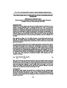
Fig. 1. Chemical Structures of Natural trans-Crotonin and trans-Dehydrocrotonin
treated with crotonin at 100 and 250 mg/kg or between the crotonin 50 mg/kg group and the control. So, we selected a fixed oral dose of 100 mg/kg for all assays because this dose also was the lowest effective dose that presented antiulcerogenic activity. In this study, crotonin significantly protected the gastric mucosa against injury induced by ethanol. These results indicate that crotonin may enhance gastric-mucosal defensive factors. The diterpene crotonin inhibits HCl/ ethanol gastric-lesion formation (51%) slightly better than DHC (48%) at the same dose. Hypothermic-restraint stress ulcers have been widely used experimentally for the evaluation of anti-ulcer activity in rats because of data reproducibility.27) Stress-induced ulcers are probably mediated by the release of several biogenic amines and metabolites of arachidonic acid by lipoxygenase with enhancement of acid secretion and reduction of mucous production.28,29) Disturbances of gastricmucosal microcirculation and abnormal motility also have been considered to be the pathogenic mechanisms responsible for stress-induced gastric-mucosal lesions.30) Our data showed that pre-treatment with crotonin (100 mg/kg) significantly protected the gastric mucosa against hypothermic restraint-stress induced ulcers in mice as well as cimetidine at the same dose. In this assay, crotonin showed a more potent inhibition of gastric-lesion formation (72%) when compared to DHC (67%). NSAID like indomethacin reduce the gastric cyclo-oxygenase pathway of arachidonic acid metabolism, inhibiting the overproduction of leukotrienes and other products of the 5-lipoxygenase pathway.12) These agents also break the mucosal barrier, provoke an increase in gastric-mucosal permeability to H1 and Na1 ions, a drop in the transmucosal potential difference and also induce the formation of erosions and ulcers.31,32) There is mounting evidence that an increase of certain endogenous PG can enhance gastric-mucosal resistance against ulcerogenic agents such as anti-inflammatory agents.33) In this assay, crotonin was able to produce a significant reduction of the gastric mucosal damage induced by indomethacin, again indicating a probable local increase in the synthesis of PGs or of the 5-lipoxygenase inhibitory effect. When compared to DHC in the same model, crotonin presents marked decrease in gastric haemorrhage. In the following section we present the biochemical results obtained after submitting mice to pylorus ligature. Mice were orally pretreated with crotonin, DHC or cimetidine and another group was pre-treated by the intraduodenal route with the same drugs. Pre-treatment with cimetidine provoked changes in the acidity and volume of gastric juice. Crotonin only provoked a marked increase in the volume of gastric
456
juice. But the intraduodenal administration of crotonin was effective in reducing gastric acidity. Moreover it seems that crotonin exerts a kind of cytoprotective action mainly by the systemic route because it is not present when administered orally, but only when administered intraduodenally. Thus its protective effect does not depend on contact of the drug with the gastric mucosa. On the other hand, DHC did not induce any alteration in gastric volume in Shay mice but modified the pH and total acid concentration of gastric juice when administered by the intraduodenal route.3) The primary therapeutic approach of anti-ulcer agents involves the maintenance of a delicate balance of factors controlling the synthesis, secretion and breakdown of its proteins, glycoproteins and lipid components,34) so as to strengthen the mucosal integrity. Amongst the mechanisms by which crotonin could provide gastroprotection in various models, the involvement of a mucosal-adaptation process by means of an increase in the activity of defense mechanisms such as mucous secretion is probable. But pre-treatment with crotonin or DHC was not able to increase the free gastric mucous in pylorus-ligated mice. However, further experiments are required to elucidate the exact mechanism of anti-ulcerogenic activity of this compound. It is not possible to discount the possibility of the involvement or regulation of leukotriene release in the gastric cytoprotection of crotonin. Therefore, on the basis of the present results, we can attribute to crotonin, in addition to DHC and essential oil,35) a part of the antiulcerogenic action of traditional Croton cajucara preparations. Furthermore, the very similar pattern of antiulcerogenic activity of both diterpenes shows that this action does not depend on the a ,b -unsaturated ketone group present in DHC.
Vol. 25, No. 4 5) 6)
7)
8) 9) 10) 11) 12) 13) 14) 15) 16) 17) 18) 19)
20) 21) 22) 23) 24) 25) 26) 27) 28)
Acknowledgements The authors are grateful to FAPESP, CAPES and CNPq for financial support.
29) 30)
REFERENCES 1)
Di Stasi L. C., Santos E. M. C., Moreira dos Santos C., Hiruma C. A., “Plantas Medicinais da Amazônia,” Editora UNESP, São Paulo, 1989, pp.127—128. 2) Souza Brito A. R. M., Nunes D. S., Ciência e Cultura, 49, 402—408 (1997). 3) Souza Brito A. R. M., Rodríguez J. A., Hiruma-Lima C. A., Haun M., Nunes D. S., Planta Med., 64, 126—129 (1998). 4) Hiruma-Lima C. A., Spadari-Bratfisch R. C., Grassi Kassisse D. M., Souza Brito A. R. M., Planta Med., 65, 325—330 (1999).
31) 32)
33) 34) 35)
Rodriguez J. A., Haun M., Pharmacology and Toxicology, 65, 1—5 (1999). Hiruma-Lima C. A., Gracioso J. S., Toma W., Almeida A. B. A., Paula A. C. B., Brasil D. S. B., Muller A. H., Souza Brito A. R. M., Phytomedicine, 8, 94—100 (2001). Hiruma-Lima C. A., Gracioso J. S., Toma W., Paula A. C. B., Almeida A. B. A., Brasil D. S. B., Muller A. H., Souza Brito A. R. M., Biol. Pharm. Bull. 23, 1465—1469 (2000). Olfert E. D., Cross B. M., McWilliam A. A., “Canadian Council on Animal Care,” Ottawa, Ontario, 1, 1993, pp. 1—213. Itokawa H., Ichihara Y., Kojima H., Watanabe K., Takeya K., Phytochemistry, 28, 1667—1669 (1989). Mizui T., Doteuchi M., Jpn. J. Pharmacol., 33, 939—945 (1983). Szelenyi I., Thiemer K., Arch. Toxicol., 41, 99—105 (1978). Rainsford K. D., J. Pharm. Pharmacol., 39, 669—672 (1978). Levine R. J., “Peptic Ulcer,” ed. by Pfeiffer, C. J., Munksgaard, Copenhagen, 1971, pp. 92—97. Shay H., Komarov S. A., Fels S. S., Meranze D., Gruenstein M., Siplet H., Gastroenterol., 5, 43—61 (1945). Bolton J. P., Palmer D., Cohen M., Digest. Diseases, 23, 359—364 (1978). Sun S. B., Matsumoto T., Yamada H., J. Pharm. Pharmacol., 43, 699—704 (1991). Wallace J. L. Granger D. N., FASEB J., 10, 731—740 (1996). Weir D. G., Br. Med. J., 296, 195—200 (1988). Lewis D. A., Hanson P. J., “Progress in Medicinal Chemistry,” ed. by Ellis G. P., West G. B., Elsevier Science Publishers, Amsterdam, 1991, pp. 201—231. Alkofahi A., Atta A. H., J. Ethnopharmacology, 67, 341—345 (1999). Kitazawa E., Sato A., Takahashi H., Kuwano H., Ogiso A., Chem. Pharm. Bull., 28, 227—234 (1980). Desai J. K., Parmar N. S., Agents Actions, 42, 149—152 (1994). Sarosiek J., Slomiany B. L., Kojima K., Swierczec J., Slomiany A., Konturek S. J., J. Appl. Biochem., 5, 429—436 (1984). Guth P. H., Paulsen G., Nagata H., Gastroenterol., 87, 1083—1090 (1984). Szabo S., Scand. J. Gastroenterol., 22, 21—28 (1987). Parnaham M. J., Brune K., Agents Actions, 21, 232—234 (1987). Murakami M., Lam S. K., Inada M., Miyake T., Gastroenterol., 88, 660—665 (1985). Kitagawa H., Fujiwara M., Osumi Y., Gastroenterol., 77, 298—302 (1979). Koo M. W. L., Ogle C. W., Cho C. H., Pharmacology, 32, 326—334 (1986). Sato N., Kawano S., Tsuji S., Ogihara T., Yamada S., Scand. J. Gastroenterol., 30, 14—20 (1995). Whittle B. J., Gastroenterol., 80, 94—98 (1981). Droy-Lefaix M. T., “Prostaglandins: Biology and Chemistry of Prostaglandins and Related Eicosanoids,” ed. by Curtis-Prior P. B., Churchill Livingtone, New York, 1988, pp. 345—360. Wallace J. L., Whittle B. J., Eur. J. Pharmacol., 115, 45—51 (1985). Bilski J., Sarosiek J., Murty V. L. N., Aono M., Moriga M., Slomiany A., Slomiany B. L., Biochem. Pharmacol., 36, 4059—4065 (1987). Hiruma-Lima C. A., Gracioso J. S., Nunes D. S., Souza Brito A. R. M., J. Pharm. Pharmacol., 51, 341—346 (1999).