A"ilrAi nLri", Facultid de Bioquimica, Quimica y Farmacict, [Jnit'ersidad Nrtc:ional de. Tuclmin, Chacabuco i1t' mOO - San Miguel de Tucuntdn' Argentina.
MODELS OF CYTOPLASMIC ARCHITECTURE AND FUNCTION M.A. AON, S. CORTASSA UNT)' Instituto superior de Investigacianes Biol6gicas (INSIBIO' CONICET de Quitnica BioL6gica "Dr' Instituto and Nutrici6tt La de Bioquimictt de Departamento A"ilrAi nLri", Facultid de Bioquimica, Quimica y Farmacict, [Jnit'ersidad Nrtc:ional de Tuclmin, Chacabuco i1t' mOO - San Miguel de Tucuntdn' Argentina A. CACERES
Instituto de Investigaci6n M,dica
X.
tl
Systems,
\ ork, 1985)
,,Mercedes y
Martin Ferreyra,,, 5000 C6rdoba,
Argentina
Introduction
In the last two decades, the ideas concerning the organization and functional first pr"p".,i", the cytoplasm of cel1s have grown around two main concepts. The whereas "f crystal2'3; protein organized tiiough one is that the cytopiasm is a crowdedl gene expression the second one aeats with the fact that metabolism and the that organization4-6. by influenced profoundly but are not indiflerent machinery ------n"g-airg cellular protein;, at least one third of them take part in the and nucleus4' The macromolecuiar networts and laminae of the cytoplasm of RNA4'7replication and transcription of DNA and the pro.".ring ond translation l;'*Jo"ytor;s and secretion, occur deeply associar'ed with reorganization of nuclear 2'4' 1 0'
and cytoplasmic macromolecular' lattices their dynamics have Intimate association of enzymes and ultrastructure as well as
Several workers treen considered of utmost importance in metabolic regulation' with actin, its properties binding enzyme of elucidation the before were interested in r
in enzyrne kinetic properties, the inreraction between enzymes consecutive in times "; aggregates an'd ii. inlluence upon transition supramolecular Similarly, studies have be-en-reported for interaction of sreps3,1"l"-1{. .ui^f,ri. gty"otytl" enzymes with tubulin or microtubul"tl5'16 and their effect on enzyme
;if;;i
988)
L1n"6.t6'l7'18. NY,
1990).
that rnetabolism Overa1l, experimental evidence increasingly^f?YqT^tl" idea these insoluble that un6 scaffolds3'11'19-22 occurs strongiy associated to the cellular reactions3'5chemical of dynamics the matrices and their assembly strongly affect 6,20,21,24-25.
2. Modelling functional global properties of cells: coherence r
995).
rnechanisms of
alld transduction
cytopl asm2-3'2-5 ' At present, within the framework of the "structured view" of the These are the function' cell concerning model.s distinguishable two ih"i" u." at least ,'tensegrity"2l and the "fractal percolation"2J rnodels. We will compare two aspects
195
196
of these models: the mechanism of coherence and how is this coherence transduld.
into functional proPerties. By mechanism of coherence we mean how is effected the passage from microscopic to macroscopic order in cell function, i.e. how the activity of millim of molecules or architecture of supramolecular stluctures, become synchronized L space and time in an apparently "purposeful", functional, way. Transduction de,ab with the mechanisms through which cells transduce environmental stimuli (e-8. hormones, neurotransmitters) into differential gene expression, metabolism and cellular energetics. Both aspects represent tightly linked concepts since th mechanisms of coherence play a central role in the cellular machinery of transduction. On this basis let us briefly compare the two models.
22 Th The ctr is orga percola quantifi
parcrn
cell's
a
clusteil
rocess
Inl
becomr
cluster,
2.1 The tensegrity model.
instabi
For Ingber'r ,ro6"121'24, coherence is given by changes in the "tensiond
pathwa
integration" of components of the cytoskeleton and its transduction is essentially conformationul2l'?1 .In essence, the tensegrity model proposes that the entirc cytoskeleton responds to stress as a single, tensionally integrated (tensegrit-v) siructure. Experimental evidence in support of this model came from experiments of stress-strain relation measured with magnetic microbeads attached to the surface of living endothelial cells27. The mechanical stress applied to the surface of cells with a twisting device showed that the cytoskeleton response to the applied stress was r property of the integrated system. The partial disruption of the cytoskeleton with depolymerizing agents such as cytochalasin D or nocodazole did not completely suppress the cytoskeleton stiffening, i.e. a large resistance to mechanical deformatim was evident at high levels of applied stress27. Using a tensegrity model to interpret
fansdu be achi
the data, the stiffness-stress (force) linear response ofthe cytoskeleton
occupy occupit
ofliving cells
could be simulated and it was further shown that, when increasing the force, thc mechanically interdependent structural elements realranged without topologicd
disruption or loss
of tensional continuity. Thus, a molecular continuum of
mechanically interdependent struts and tensile elements structurally _stabilize by
global rearrangement rather than local deformation in response to stress2l'24. In this light, the cytoskeleton response to applied stress appeared as a property of the integrated system and not a iharacteristic of any individual par?T ' According to the tensegrity model, coherence would be given by the use of tensegrity architecture by cells. In this way, a mechanism is provided to distribute mechanical stresses to regulatory molecules and enzymes that are immobilized on or interact with cytoskeletal and nuclear scaffotds and thereby to integrate cell structure and function. Mechanical stress in turn, is transduced into biochemical function through conformational changes, i.e. a mechano-chemical transduction2l.
topolog
and
en
dimens
2.3 (Mt Anothe
properl macron
regulari
of intra
suggesl
volumc cytopla
what tI nonider
3.
Hr meche
The vie
fractal
l
self-ass
This ef
the pF,
197
2.2
rce transduc€..
The
fractal percolation model.
fror.
The conception of the cellular cytoplasm as a fractal proposes that the cytoskeleton
ity of million.
is organized according to the principles of fractal geometry. A random fractal percolation ciusters - has been hypothesized as a plausible model on the basis of
passage
vnchronized r:
sduction L1
stimuli
dea'..
quantification of the fractal dimension in micrograp\t23,28 . Iteration of an invariant pattern that spans several length scales might describe the complex forms shown by cell's architecture23. From a dynamic perspective, a main feature of percolation clusters is given by the existetce of apercolation threshold fbr which a percoiation process undergoes a transition from local to global connectedness 23,29. In the fractal model, coherence arises when the dynamics of cellular processes become unstable at sites in the cytoplasmic lattice be.longing to the percolation cluster, i.e. further from the "percolation threshold". Transduction occurs because
(e.g
etabolism an-
:pts since th: machinery t
.
instability is triggered by environmental stimuli acting through multiple the "tension.,. n is essentiail" lhat the entir;
'd
(tensegritr
experiments ' r the surface ,
pathways22-23,30. Brlrunced catalytic performance may be one of the outcomes of transduction. In heterogeneous fractal media such enhanced catalytic performance may be achieved through two mechanisms: (l) segregation of reactants in disjointed fractal topologies; (ii) increase of the encounter probability between substrate or effector,
.
and enzyme or target, by their coexistence in the same (2D) topological
:
dimension23.
;e of cells rl'ii:. ed stress was ., .oskeleton wii:: not completel\ .'al deformatior.
,del to interprc. n of living cell. g the force, th.' rut topologica.
continuum o: l1y stabilize b," .rr21,24. 6 61',r.
nroperty of thc -r
by the use o: rid to distribut.. rnobilized on t,: rte cell structur!'
:mical functior:
2.
3 ( M acro)Molecular
Crowding
Another view of cytoplasmic structure emphasizes the "macromolecular crowding" properties of the ground plan of living cells. Cells contain a large variety of macromolecules in their cytoplasm, largely proteins, which taken together will occupy a substantial fraction of the total cytoplasmic volume, called yolume occupied or crowded1,31-32. Following changes in cell volume during osmotic regulation, molecular crotvding and confinement directly affect the form and function of intracellular macromolecules. From data obtained in red blood cells, it has been suggested that cells may use macromolecular crowding as a means of sensing their volume; what appears to be cell volume regulation is really a regulation of cytoplasmic macromolecular crowding33-34. 4ppu.ently, protein concentration is what the cells seek to maintain to approach an optimal degree of cytoplasmic nonideality33.
3. Heterogeneous distribution of cytoskeletal proteins as mechanism of metabolic fluxes modulation
a
,p21.
The view ofthe cytoplasm as a crowded object is not excluded by either tensegrity or fractal percolation models. For instance, crowding may alter the average degree of self-association of an enzyme, in turn changing its average catalytic activiry1,6,31. This effect was shown to be specifically induced by microtubular protein or actin in the pyruvate kinase/lactate dehydrogenase enzymatic cotpl.e22,35. Furthermore, the
198
supramoleculat organization of microtubular protein could entrain the dynamics c:
enzymatic reactions through mechanisms of self-association-dissociation of enzyme. toward high-er or lower oligomeric forms with higher or lower levels of activir\ respecti vely6
We have
a
enzyme ma1' bt depending on *
location35. -{c
postulated to el rearrange their i:
reactions takrng This is a ri suggests that th
and that its (sup neuronal polant
of distinct cyto;
these cells to cr
spatio-tempora)
polarity, form
ar
attributing a m cytoskeletal prc by the dynamics eplightened. Ac change their dis the cell cycle-de well studied39-r
continuously ass or rearrange it-s ,
A predictic:
geometry or arc ps. In percolatic at the threshold when a locaL in: whole cellular.fi'
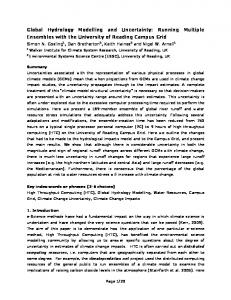
4. A Figure 1. Heterogeneous distribution of anti-tubulin and anti-actin immunofluorescent staining at the cellular Ievel.
Micrographs showing the distribution of microtubules (A) or microfilaments (B) in astrocytes. Microtubules and microfilaments extends throughout the cell cytoplasm.
Microtubules were labeled with an antibody against tyrosinated o-tubulin, and microfilaments were labeled with fluorescein-phalloidin. Note the straight actin bundles
radiating from the nuclear p.eriphery alternating with dots, and the regi"onalized staining adjacent to the plasma membrane. The tubulin lattice appears as an inlricated meshwork of fibers highly dense around the nuclear periphery.
rheologir
Tensegrity and different precon,
assembly is evid principles of s1 s form. There exist. polymers behar e assembled and d The tensegri
ofindependent
s'
Cytoskeleton
st
199
ie dynamics ol lrn of enzymes
:Is of activitl,
We have also presented experimental evidence that suggests that the same enzyme may be differentially modulated at different locations in the cytoplasm depending on the cytoskeletal protein (and their associated proteins) present at that
location35. According to this proposal, a precise biochemical coupling can
be
postulated to exist between the highly sophisticated mechanisms by which ceils realrange their (supra)molecular architecture (Fig. 1) and the dynamics of biochemical reactions taking place concomitantly. This is a relevant matter since the information presently availabie strongly suggests that the cytoskeleton is one of the endogenous determinants of cell form, and that its (supra)molecular asymmetries are involved in the development of, e.g. neuronal polarity36-38. Furthermore, recently it has become apparent the existence of distinct cytoplasmic domains in neurons intimately associated with the ability of these cells to create microtubule subsets that differ in molecular composition, and spatio-temporal distribution3T-38. Thus, a clear link is established between cell polarity, form and the underlying dynamics of celluiar biochemistry, by the proposal attributing a modulatory role to the heterogeneous cytoplasmic distribution of cytoskeletal proteins35 (Fig. 1). Considering the entrainment of enzymatic reactions by the dynamics of the cellular architecture, a whole set of observations are distinctly enlightened. Actin and tubulin as major structural proteins in all eukaryote cells, change their distribution along the cell cycle. In the budding yeast Saccharomyces, the cell cycle-dependent patterns of microtubules and microfilaments localization are well studied39-40. Reported experimental data show that cytoskeleton components continuously assemble/disassemble along the length of the axon of cultured neurons or rearrange its spatial distribution in response to growth 1u.1o..10,41-43. A prediction of the fractal model of cytoplasm is that a ce1l by regulating its geometry or arohitecture may in turn regulate the level of its percolation threshold, p6. In percolation lattices, transitions between local to global connectedness happen
I
at the threshold level of occupancy of the lattice29. Different p. would determine when a local instability in the dynamics of an enzymatic reaction may extend to the whole cellulctr field if it spreads in a domain belonging to the spanning cluster.
4. A rheological view of tensegrity and fractal
nd anti-actin :ilaments (B) in ce1l cytoplasm.
cr-tubulin,
and :ht actin bundles onaiized staining rcated meshwork
models
Tensegrity and fractal models do not exclude each other although they belong to different preconceptionr44. A structural preconception based on geometry and selfassembly is evident in the tensegrity model whilst the fractal model follows kinetic principles of systems organization away from equilibrium for generating pattern and form.
There exists compelling rheological evidence which shows that cytoplasmic polymers behave as a reversible non-covalent gel network which is constantly being assembled and disassem61"6,25,45 -46 . The tensegrity model describes cytoplasm as a prestressed molecular continuum of independent struts and tensile elements (see section above: The tensegrity model).
Cytoskeleton stiffness and apparent viscosity change
in parallel following
2N application of mechanical stress because of the postulated molecular continuum. Shifts in cytoplasmic rheology, i.e. viscosity, result from filament alignment or interfilamental friction. Altering the global architecture of the cell, changes the force balance of the cytoskeleton that in turn elicits changes of its stiffness and apparent viscosity4T.
The supramolecular network of the cytoplasm effects rheological shifts involving sol-gel transitions. The connectivity of such a network would certainly depend on the concentration of assembling filaments, the strength of their interaction, or the number of cross-linkers, e.g. MAPs46. Besides, geometrical arrangements certainly influence the connectivity of the gel network. As already stressed (see: The fractal percolation model), a cornmon ingredient in these systems
disassemble48-49. 6 the lattice, transitions
threshold of the frac physiological prooes which cytoskeleton la
proposed that the pal achieved through sell an analogy between t of spatio-temporal sq organization3o.
is that at a given value of a characteristic parameter, a macroscopic connected cluster forms in the system, e.g. a cluster of connected polymer molecules results in gel formation. When the latter happens, the physical properties of the system can change
drastically. In the case of sol-gel transitions, the polymer solution has liquid properties before the gel forms, that is, a well-defined value of the viscosity, whereas in the gel phase, the viscosity is practically infinite and the system has well-defined elastic properties. The critical parameter at which these transitions happen is the
percolation threshold, that for sol-gel transitions may be the concentration of polymers in solution. Summarizing, a clear link exists between the behavior of a fluid in a percolation supramolecular network, and the rheological behavior of the molecular network itself. Otherwise stated, at the percolation threshold, pc, not only the dynamics of the (bio)chemical reactions have the possibility to reach a global influence, but also the rheological character of the polymer network, i.e. may effect a sol-gel transition. This is utmost important since it integrates in a unified and coherent scheme, rheological and biochemical evidence with the self-organizing properties of non linear kinetic schemes. The latter is a potential source of self-emergent spatiotemporal behaviors, i.e. bistability, oscillations, thresholds, chaos or waves of chemicals. In the following we will show the possibility of the existence of such phenomena in the coupling of two dynamical subsystems such as the assemblydisassembly of microtubular protein and the kinetics of an enzymatic reaction.
5. Dynamic organization of biochemical reactions and
cytoskeleton
components The more adequate models for cellular functional coherence will be those ones in which s-tructure and dynamics are combined. In the way to shape those models we should not miss that cellular processes, scale in space and time, from the conformational change in the tx-helix of a protein up to cell division. By the latter we mean that processes such as enzymatic reactions or cell division occur within
characteristic spatial coordinates and when perturbed they
will relax with a
characteristic relaxation time30. We have proposed that microtubular protein which organizes as fractal percolation 1ugi"."22'28 exhibits self-organization in the spatiotemporal "window" (micrometers and minutes) in which microtubules assemble-
Figure 2.
Schemr
polymerization-dcp
kinetics.
PK in the presence ofl MTP polymerization< (Cp) or non-polymeriz The dashed arrows int either the tetrameric
In order to test
tr
if
the dynamics of en assembly-disassembl
ordinary differential
model which links ft The equations which the kinetic behavior t
201
. geometrical
disassemble48-49. 1n percolation processes, at the threshold level of occupancy of the lattice, transitions between local to global connectedness happen. The percolation threshold of the fractai cytostructure may be related to the scaling exhibited by physiological processes at the 'transition p,oint' in the spatio-temporal domain for which cytoskeleton lattices self-organiz"6,30. p.o* these consideratlons, it has been proposed that the passage from microscopic to macroscopic functional coherence achieved through self-organization of cytoskeleton components, allows to postulate an analogy between the concepts of self-similarity in fractal cytostructures and that of spatio-temporal scaling exhibited by physiological processes at distinct levels of
k. As alreadr
organization3o.
rr contlnuum. alignment or rges the forcc and apparent
ogical shifts ruld certainlr rgth of their
s^ ssY
these system: nected cluster
results in
ge1
'm can chang.-
rn has liquid osity, whereas s well-defined happen is the
rcentration
o1
r a percolation
rular network .'dynamics
..,1/., ,9[
o1'
rence, but alscr
-gel transition. erent scheme. pcrties of non
ergent spatioi or waves of .tence of such the assemblyeaction.
cytoskeleton
V
,*o
ffi
I I
ttruo
T
I
---:-_>
.E-\-
V m ffif ffi
PKr
MAps
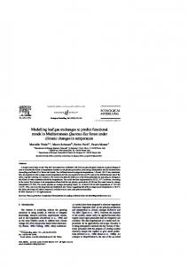
Figure 2. scheme of the model which Iinks microtutrular protein polymerization-depolymerization dynamics and pyruvate kinase (PK) kinetics.
PK in the presence of MTP, may exist as a tetramer (PK1) or pentamer (PKp). The cycie of MTp polymerization-clepolymerization assumes that tubulin may exist as polymerized (cp) or non-polymerized, bound to GTP (C1), or non-polymerized bound to GDP (CD). The dashed arrows indicate that polymerized or depolymerized forms of MTP stabilize either the tetrameric or pentameric forms of PK (Reproduced from Aon et a1., 1995).
: those ones in ,,se models we
me, from the n. By the latter n occur within
i relax with a : protein which n in the spatioLrles assemble-
In order to test if self-organization of microtubular protein (MTP) could entrain the dynamics of enzymatic reactions, we modelled the coupling between the
assembly-disassembly of MTP and pyruvate kinase (PK) through a system of four ordinary differential equations (ODEs) (Eq. 1-a). Figure 2 shows a scheme of the model which links the dynamics of assembly-disassernbly of MTP and PK kinetics' The equations which describe the activity of PK (Eq. 5-7) were obtained by fitting the kinetic behavior of the enzyme with respect to the concentration of its substrate
202
phosphoenolpyruvate (PEP), in the presence of either polymerized or non polymerized MTP6. The enzyme, in the presence of MTP, may exist as a tetram€r polymeric staus iTy-o. p"ntu*er (P); the proportion of each form depending on the 1). The MAPs of Eq. the r.h.s. term at (through the 3rd or GTP MAPs of-MTir, bound to the microtubular lattice appears to promote the formation of the P form through the first term at the r.h.s. of Eq. l; the VPK*ry of the P form being larger than that of the T one6. The equations that describe the MTP polymerization-depolymerization assumes
one of three forms, i.e. either polymerized' or nonpolymerized bound to GTP; or non-polymerized bound to GDP50. A conservation equation relates the three forms of MTP (Eq. 8). The polymerization term of the state variable Cp is autocatalytic with n1 as the nuclei size (n1=2)' The coupling between the kinetic behavior of PK and the polymeric status of MTP is realized through the term of pentamer formation, PKp, which is proportional to the amount of polymerized MTP, Cp, in Eq. 1 and 4. The mathematical formulation of the model is as follows:
that tubulin may exist
k2 @K1- tPl) Cp -
H=0, -
Hf
in
k:
[P] - k4 tPltGTPl
vpr - k1 GEPg - IPEPI)
= - kP
cl1 c1+ ksco
(1)
(z)
The parameters GI and PEP, as well i inputs of the mod
polymerization an
concentration are si The existence and in the activitY system is bistable, or non-polymerizc value of the total depending on the systems are revers
happen. In these c previous branch o previous historY (
jump, the state va indefinitely on the
situation. These rt phenomenon of dY in the sense that sl sbte41,48,51-52. Figure 3 emPl
cells, i.e. the
[GTPI - ku
cr (GrPo - IGTPI)
(3)
cha
variation of the tt This results in a' stable and unstabk
3). In fact, the va (4)
Y=kpcllcr-kapcp vfrff vPK=
-:-
rrenrn
(s)
Ki + tPEPln
ufff=t[u**(ril* -,T*
several physiologit
For instance, actil phosphoinositides affect Kpol. Likev By inspecting
transitions in PK
amount of polY polymerization,
)*t
(6)
irreversible chang state namely aggn
In summarY, t
n=4+ IBL PKt
(7)
Co=CD+C1+CP
(8)
bistability or mult
and a biochemical Kpol. These two I
through this the This reinforces t
1
components maY
I
203
rized or non t as a tetramer rlymeric statu: l). The MAPs of the P fornr m being larger zation assumes rized, or non-
\
conservation on term of the The coupling ITP is realized L to the amount :.rulation
of
the
(1)
The parameters GTP6 and PEP. which represent the total intracellular pools of GTP and PEP, as well as the amount of intracellular tubulin, Cs, are main biological inputs of the model. Essential experimental results such as the kinetics of MTP polymerization and the increase in PK-_catalyzed fluxes as a function of MTP concentration are simulated by the model35'
The existence of coupled-irreversible transitions in the MTP polymeric status and in rhe activity of PK, is a main result obtained with the model (Fig. 3). The system is bistable, i.e. it shows two possible steady states of activity (polymerized or non-polymerized MTP, and a fully active or half-active enzyme) for the same value of the total MTP concentration, Co, and the rate of polymerization, Kpol, depending on the sense of variation of the latter. Generally speaking, bistable ,yit"*r are reversible although under some conditions irreversible transitions may happen. In these cases the system once passed a limit point can not return to the pr"uio.rr branch of steady states. The irreversible jump occurs depending on the previous history of the system (e.g. the direction of change of Kpol). After the jump, the state variables, e.g. the non polymerized or polymerized MTP, remain inaefinit"ty on the steady branch without possibility of coming back to the previous situation. these results may also provide a putative explanation to the well known phenomenon of dynamic instability shown by microtubules either in vitro or in vivo in the sense that shrinking microtubules seldom undergo a transition to the growing state4 1 ,48 ,51-52
(2)
?l)
(3)
.
Figure 3 emphasizes the multidimensional character of functional regulation in cells, i.e. the changes in steady state values of PEP versus the simultaneous variation of the total amount of MTP, Ce, and its rate of polymerization, Kpo1.
This results in a 'line of bifurcation points', i.e. joining the points that limit the stable and unstable branches of steady states in the individual bifurcation plots (Fig.
3). In fact, the variation of the parameter Kpol, may result from the 'lumping' of (4)
(5)
(6)
several physiological processes which, in turn, entrain ditferent functional prop^erIies. For instance, actin or tubulin polymerization are sensitive to cytoplasmic Ca2+ and phosphoinositides 1eve1s53 and pH changes54. These physiological variables will urc.t f,ot Likewise, Cs may be modulated by changes in gene expression55-56. By inspecting Fig. 3, as the amount of protein increases either thc irreversibie transiiions'in ef aciivlty (displayed by the level of the substrate PEP) or the
amount of polymerized protein, Cp (not shown) occur at lower rates of polymerization, Kpol. The transitions in MTP polymeric status also entrain irreversible changes in the catalytic properties of PK as well as of its conforrnational state namely aggregation as a pentamer (Fig. 2)'
tn
(7)
(8)
ru-*uli
.ri" hur" been able to show that a selt'-organized phenomenon, i.e.
bistability o. ,rultipl" stationary states, may occur in the coupled dynamics of MTP
C6 and a biochemical ieaction as a function of two relevant cellular parameters, of MTP and lattice the Kpol. These two parameters in turn regulate the geometry of and
p23'28'29' through this the percolation threshold, ps, and the fractal dimension, This ieinforces the idea that polymerization-depolymerization of cytoskeleton
.o.npon"n,, may
be involved in-the passage from microscopic to macroscopic order
2M at the cellular level. Besides, the existence of abrupt transitions at limit points in tb dynamic behavior of MTP polymeric status together with pK activity, furthcr suggests a link between ps and self-organization of cytoskeleton components, end their involvement in the synchronization of cellular activities further apart in th cytoplasm of living cells.
6. Concluding
r
How does curren
metaphors of the c of cells?
According to
t
local cytoskeleton
remodelling mol
0.75
cytoskeleton, and
unstable architectu the rearrangement coherence results l local (metabolic) h
0.7 z,
E
0.6s
o o
The view
FI
0.6
assembly-disassem inducer of chang
0.55
supramolecular or feasible physicoch global behavior in
0.5
ps, the cytoplasn
&
K"o, (mM'2
subsystems. Struc subtly modulated b messenger levels, r change the ps. Thr
s-1)
Figure 3 A 3-dimensional representation of the qualitative dynamic properties of two coupled systems. The study of the qualitative dynamic behavior of a model based on experimental data6 was performed with the program-AUTO. The stability properties as well as the different type of steady states (ss) exhibited by the model are represented as a function of two bifuicition parameters of physiological relevance such as the rate of polymerization of MTp, Kpol, and the total amount of MTP species, co, present in the system. The steady state value! of phosphoenolpyruvate (PEP), the substrate of PK, were plotted. The steady values of state variables represented by the system of Eq. l-4 were computed with AUTO. The bifurcation behavior presented stable (-) and unstable (- - -) branches of steady states with limit points. The diagrams were obtained with the following parameter values:_k1 = 1.0 s-1, kZ = tO mM-l s-1, k3 = 0.05 s-1, k4 6.62 = mM-l s-1, kf = 3.0 mM-l s-1, kb = 2.5 tO-3 mU-l s-1, kdp = 2.5 lO-3 r-1, n1 = 2, Ks 0.6 mM, PEP' =
l0 mM,
PK1 = g.g1 mM,
t-', uTu* = 14 s-1. The shaded
em,
GTP. = 1.0 mM, [GTP] = 0.9 mM,
areas represent the domain (or'surface')
,ilu*
=
architectural arran! regions can give r functional propertir
exffa-cellular envirr Overall, we ha
not a unique, 'ro) manifested at sever Even all metaphors such as the 'cell-as-r
parallel distributed organization to giv think at the coexistr which implies the
cr
,t Acknowledgeme
of stable behavior
of the system separated by a 'gap' of unstable comportment. The parameter values of the subsystem which describes assembly-disassembly of MTP were taken from Carlier et
We are indebted to
a1.,50. Those parameter values corresponding to
and Consejo de Inv and AC are career ir
et
a1.,6.
PK kinetics were obtained from Cortassa
computing facilities
205
6. Concluding remarks: models and metaphors of the cell
ut points in thc
:tivity, further
How does current models or views of the cytoplasm of cells, fit into current metaphors of the cells? What does this mean for modelling of functional properties of cells? According to the tensegrity model, global changes in tissue structure alter the local cytoskeleton force balance hence eliciting an integrated cellular response. The
'mponents, and rer apart in the
remodelling molecular cascade would result from tension moiding of the cytoskeleton, and internal oscillatory transformations between locally stable and
-.@"4ffi
unstable architectural states. The larger the magnitude of the applied stress, the larger
the rearrangement of the cytoskeleton matrix. In this scheme, the mechanism of
coherence results from the passage between global cytoskeieton reorganization to local (metabolic) behavior, rather than the inverse. The view emerging from the fractal percolation model suggests that the assembly-disassembly of cytoskeleton components act as a macroscopic, coherent,
w
rtive
inducer of changes
dynamic
nental data6 wa: different type oi : two bifurcation r of MTP, Kpol
iv
state values o;
ri' Eq. 1-4
were
and unstable
(-
-
btained with the 5 s-i, k4 = 0.01
,-1, n1 = 2, Ks =
nM, vpax =
in
systemic properties such as metabolic fluxes. The
supramolecular organtzation of the cytoskeleton as a percolation lattice adds a feasible physicochemical mechanism of coherence when the passage from local to global behavior in the cytoplasm is effected at the percolation threshold, p6. Below pq, the cytoplasm would behave as loosely coupled, independent clynamical subsystems. Structural microscopic features comprising molecular interactions subtly modulated by relevant physiological parameters such as pH, Ca2+ or second messenger levels, determine macroscopic geometrical characters of the lattice that change the ps. The dynamics of chemical reactions can be either entrained by the architectural arrangements of cytoskeleton components, or at unstable parametric regions can give rise to self-organized behavior whose emergence induce new functional properties to cells, i.e. adaptive conduct ensuing changes of the intra- or extra-cellular environments. Overall, we have the impression, as well as other researchers5T-58, that there is not a unique, 'royal', way to model systems whose functional properties are manifested at several simultaneous levels of organization such as cellular systems30. Even all metaphors, with the appropriate biological constraints, will have to be used such as the 'cell-as-machine', 'cell-as-society', 'cell-as-text' or'cell-as-field' exhibiting parallel distributed processing5S. Instead of a single model spanning all levels of organization to give a unified account of the whole complexity, one approach is to think at the coexistence of a 'chain of models' linking adjacent levels of organization which implies the coexistence of explanations at several levels.
2,s
Acknowledgements
,f stable behavior 'ter values of the
from Carlier et ed from Cortassa
We are indebted to Centro de C51cu1o de la Universidad Nacional de Tucumdn for computing facilities. This work was supported by grants from Fundaci6n Antorchas and Consejo de Investigaciones de la Universidad Nacional de Tucum6n. and AC are career investigators from CONICET, Argentina.
MAA,
SC
206
References
44. L.G. Harrison,
1. A.P. Minton, Mol. CeIl. Biochem.55, 119 (1983). 2. A. Fulton, Cell30, 345 (1982). 3. J.S. Clegg, Am. J. Physiol.246, P.l33 (1984). 4. S. Penman et al., Cold Spring Harbor Symp. Quant. Biol. 1013 (1981). 5. K.J. Pienta and C.N. Hoover, J. Cell. Biochem.55, 16 (1994). 6. S. Cortassa et al., J. Cell. Biochem.55, 120 (1994). 7. R. van Driel et al., l. Cell. Biochem.47,311(1991). 8. K.A. Suprenant, Cell Motil. Cytoskel.25, | (1993). 9. A. Fulton, J. Cell. Biochem. 52, 148 (1993). 10. N. HirokawainThe Neuronal Cytoskeleton, (Wiley-Liss, New york, 1991). 11. C. Masters, J. Cell Biol. 99, 222 (1984). 12. F. Clarke et al., in Regulation of carbohydrate metabolism, ed. R Beitner (CRC Press, Boca Raton,
13. 14. 15. 16. 17. 18. 19.
1
985).
M.A. Luther and J.C. Lee, J. Biol. Chem.26l, 1753 (1986). T. Keleti and J. Ovadi, Curr. Top. Cell. Regul.29,1 (1988). H.R. Knull and J. L. Walsh, Curr. Top. Cell. Regul.33, 15 (1992). J. Ov6di and F. Orosz, Curr. Top. Cell. Regul.33,105 (1992). A. Lehotzky et al., J. Biol. Chem.268,10888 (1993). P. Marmlllot et al., Arch. Biochem. Biophys.3l5, 461 (1994).
G.R. Welch, Prog. Biophys. Molec. Biol.32, 103 (1977). J.S. Clegg, Curr. Top. Cell. Regul.33,3 (1992). D.E. Ingber, Cell 7 5 , 1249 (1993). S. Cortassa and M.A. Aon, Cell Biol. Internat.18,68l (1994). M.A. Aon and S. Cortassa, FEBS Lett.344,1 (1994). 24. D.E.Ingber et al., Int. Rev. Cytol. 150, 173 (1994). 25. K. Luby-Phelps et al., Ann. Rev. Biophys. Biophys. Chem.17,369 (1988). 27. N. Wang et al., Science 260, 1124 (1993). 28. C. Raboullle et al., J. Biomol. Struct. & Dynamics 9, 1013 (1992). 29. LFeder, Fractals, (Plenum Press, New Vort, teSA;. 30. M.A. Aon and S. Cortassa, MoL Cell. Biochem.l20, 1 (1993). 31. A.P. Minton, Biophys. "/. 63, 1090 (1992). 20. 2l . 22. 23.
32. M.M. Garner and M.B. Bwg, Am. J. Physiol. 266 (Cell Physiol. 35), CBlj (tee4). 33. J.C. Parker, Am. J. Physiol.265, C1191 (1993). 34. D. Haussinger et al., Am. J. Physiol. (Endocrinol. Metab.) 30, E343 (1994). 35. M.A. Aon et al., J. Cell. Biochem.59 (in the press) (1995). 36. F. Solomon, J. Cell Biol.90, 547 (1981). 31. A. Ciceres et al., Dev. Brain Res. 13, 314 (1984). 38. A. Ferreira and A. C6ceres, Dev. Brain Res.49,205 (1989) . 39. D.J. Lew and S.I. Reed, J. Cell 8io1.120,1305 (1993). 40. J. Chant, Trends in Genet. 10,328 (1994). 41. E.M. Tanaka and M.W. Kirschner, J. Cell Biol.l15,345 (1991). 42. G. Morfini et al., J. Neurosc. Res. 39, 219 (1994). 43.M. DiTella et al., Cell Motil. Cytoskel.29,Ill (1994) .
45. K. Luby-Phelp 46. G. Forgacs anr 47. N. Wang and I
48. M. Kirschner e 49. H.P. Erickson (1e92).
50. M.F. Carlier et 51. T. Mitchison a: 52. T. Horio and F 53. P.A. Janmey, ,
54. K.A. Suprenan 55. 56. 57. 58.
A. Ben-Ze'ev, D.W. Clevelan, P.S. Churchlan
R.C. Paton, .Bl
I
20-7
44.L.G. Harrison, J. theor. Biol.125,369 (1981).
r
98 1).
York, 1991). R Beitner (CRC
eez).
'. 369 (1988). qq?\ ).
vsiol. 35), C871 E343 (t994).
)1)
45. K. Luby-Pheips et al., J. Cell Biol. 102,2015 (1986). 46. G. Forgacs and S.A. Newman, Int. Rev. Cytol.l50, 139 (1994). 47. N. Wang and D.E. Ingber, Biophys. J.66,2181 (1994). 48. M. Kirschner and T. Mitchinson, CelL 45,329 (1986). 49.H.P. Erickson and E. T. O'Brien, Annu. Rev. Biophys. Biomol. Struct 21,145 (r9e2). 50. M.F. Carlier et al., Proc. Natl. Acad. Sci. USA84,5251 (1987). 51. T. Mitchison and M. Kirschner, Nature 312,231 (1984). 52. T. Horio and H. Hotani, Nature 32L,605 (1986). 53. P.A. Janmey, Ann. Rev. Physiol.56,169 (1994). 54. K.A. Suprenant, Cell Motil. Cytoskel.19,20l (1991). 55. A. Ben-Ze'ev, Trends Biochem. Sci. 475 (1986). 56. D.W. Cleveland, Trends Biochem. Scl. 13, 339 (1988). 57. P.S. Churchland and T.J. Sejnowski, Science242,141(1988). 58. R.C. Paton, Biosystems 29, 63 (1993).
[otnputation in
[ellular
and
Molecular Biological SUstetns
Roy Cuthbertson Depf Human Anafomy & Cell Biology, Univ. Liverpod
Mike Holcombe Depl Compufer Science, Uni. Sheffield
Roy Polon Dept Computer Science, Uni, Liverpool
\P
y;',',1 :,: ::j::,:: ;?,
n
"
d. n . H. n g K. n s
Pttblislud by World Scientific Publishing Co. Pte. Ltd. PO
IISA
Box 128, Farrer Road, Singapore 912805
ofice:
Suite
lB,
1060 Main Street, River Edge' NJ 07661
ItK office: 57 Shelton Street, Covent Garden, London WC2H 9HE
T
COMPUTATION IN CELLT]LAR AND MOLECULAR BIOLOGICAL SYSTEMS Selected Papers from IPCAT 95 Copyright O 1996 by World Scientific Publishing Co. Pte. Ltd. All rights resemed.. This booh or parts thereof, may not be reproduced. in any fonn or by any means, electronic or mechanical, including photocopying, recording or any information storage and retrieval system now lotown or to be invented, witlaut witten permission from tlu Publisher.
For photocopying of material in this volume, please pay a copying fee through the CopyrightClearanceCenter,Inc.,222 RosewoodDrive, Danvers,MA01923, USA. Inthis case permission to photocopy is not required from the publisher.
rsBN 981-02-2878-3
This book is printed on acid-free paper.
Printed in Sing-apore by UtoPrint
1
Contents Dedication Preface
vll
Introduction
1
Information Processing in Cells and Tissues R. Paton and M. Holcombe
Information Processing and Signalling Processes
l.
Interplay of Cell-Cell Signalling and Multicellular Morphogenesis during Dictyostelium Aggregation T. Hdfer and P. K" Maini
-1
+.
5
Concerted Regulation of Cyclic Adenosine Monophosphate by Calmodulin/Calcium Complex and Dopamine: A Kinetic Modelling Approach R. Kiitter D. Schirok and K. Zilles
15
29
A Role of Ca2+lcalmodulin-Dependent Protein Kinase II in the Induction of Long-term Potentiation: Switching Characteristics accordin gto Caz+ Signalling and Digitization of Graded Information into Binary Information H. Okamoto and K. Ichikawa
43
Mathematical Aspects of Signal-Processing via G-Proteins J. Nauroschat and U. an der Heiden
57
Oscillations, Positive Feedback and the Flow of Biological Information K. ^S. R. Cuthbertson A Data and Knowledge Base for Cell Signalling Igarashi, T. Kaminuma and Y. Nadaoka
T.
Networks
69
77
xii
8.
Modelling the Emergence of coregulated Proteins in Biological Regulation Networks E. Chiva and P. Thrroux
g.
Specrfying Logical Agents in Cellular Hierarchies R. Paton, G. Stanifud and G. Kendall Processing using Artificial Neural Nets M. Bogdan, A. Babanine, t. Kaniecki aildW' Rosenstiel
10. Nerve Signal
,*. 9l
12.
105
tzt
Dynamical Modd
Elementary Modes of Functioning in Biochemical Netrvorks S. Schuster C. Hilgetag, J. H' Woods and D' A' Fell
151
The Cell as a Problem-Solving "Engine" P. C.
21. ComPlexlhi R. Thonas
22. Reconstnrctl .PathoPhYsiol
M. R. BoY$ r67
23.
?/1. T\eEffecrst Magoinrdeo Hormone
195
Cellular Structures P. Hartmann
t7. Genomic Cellular Automata Transposed on Silicon: Experiments in SYnthetic Life P. Marclul, P. Nussbaum, C. Piguet, S' Durand, D' Mange' E. Sanclwz A. Snuffer and G. Tbmpesti
h
J. P. A. Fowt
M. A. Aon, S. Cortassa and A. Cdceres 16. Models of Information Processing within Inhomogeneous
Roles of tbel T. R. ChtrY
183
Marijuan
15. Models of Cytoplasmic Architecture and Function
20. ParameterEsl S.IYengar
A. Bell and M. Holcombe
14.
19. Bioenergetbs
Stein'sModd
137
Processing
Cc
M.-W.IIo
Systems: Computer Simulation Study P. Mendes, P. Mannillot, J-E Hervagault aild G' R' Welch
13. Computational Models of Cellular
as EnergY
K. Matsutto
Information Processing and Cellular Systems ll.Long-RangeMetabolicsigm[inginHeterogeneousCatalytic
go6"slyelrH
25. AModelof, E.
2W
A. StcPlu
26.
TheEffectr MicroParai M. J. Hdd
n.
Onthe Sim
223
G.tuiic*
xiii
18. 91
Cohesive Interaction in Biomolecules and their Organizations as Energy Consumers
237
K. Matsuno 105
19. Bioenergetics
and Biocommunication
251
I
M.-W. Ho
I
,
121
20.
I
Parameter Estimation for a Diffusion Approximation to Stein's Model
265
S.Iyengar Dynamical Models of Cellular Systems and Information Processing
! t37
21. Complex Trajectories
from Simple Feedback Circuits
28t
R. Thomas 151
22.
Reconstruction of Excitable Tissue Physiology and Pathophysiology from Cellular Models M. R. Boyett, A. V. Holden and H. Zhang
287
t67 /.3.
Roles of the Ion Channels in Reentrant Cardiac Arrhythmia T. R. Chay
301
r83 24. The Effects of Random Variation in Stimulation Timing and
Magnitude on Excitable and Oscillatory Forms of Hormone Pulse Generator Model J. P. A. Foweraker and D. Brown
19-5
209
a
Luteinizing
25. A Model of Pituitary Release of Growth Hormone E. A. Stephens, D. Brown, G. Leng and R. G. Smith 26.
223 27.
The Effect of the Ernbryonic Bottleneck on Vertical Microparasite Transmission M. J. Hatcher A. M. Dunn and C. Tofts On the Simulation of Health with the Tissue Automat G. Z,ajicek
315
329
339
353