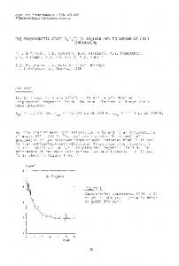
16. - 18. 10. 2013, Brno, Czech Republic, EU
2D SEPARATION USING PARAMAGNETIC MICROPARTICLES FOR SARCOSINE ISOLATION – POSSIBLE WAY OF PROSATE CARCINOMA DIAGNOSIS Natalia CERNEI1,2, Zbynek HEGER1,2, Jaromir GUMULEC2,3, Pavel KOPEL1,2, Ondrej ZITKA1,2, Michal MASARIK 2,3, Vojtech ADAM1,2, and Rene KIZEK1,2* 1 Mendel
University in Brno, Brno, Czech Republic, EU University of Technology, Brno, Czech Republic, EU 3 Masaryk University, Brno, Czech Republic, EU *
[email protected]
2 Brno
Abstract Prostate cancer (CaP) is the second leading cause of male cancer-related deaths in the Europe. Currently does not exist reliable non-invasive biomarker for determination of cancer of prostate. Simple metabolites such as amino acids would be able to serve as the new non-invasive biomarkers for determination of aggressive types of CaP. One of them – sarcosine, nowadays abundantly discussed CaP marker arouses much attention. Sarcosine, also known as N-methylglycine is a non-protein amino acid forming during glycine synthesis and degradation as an intermediate product. It was shown that sarcosine is excreted in urine of patients suffering from aggressive forms of CaP in low amounts in an intact form. Hence we aimed to synthesize paramagnetic microparticles able to isolate and immobilize sarcosine from urine and thus preconcentrate it for subsequent analysis on ion-exchange liquid chromatography with VIS detector. We modified titanium isopropox as a carrier for NH3 functional groups providing binding with sarcosine and nanomaghemite (γ-Fe2O3) providing perfect paramagnetic properties. Our paramagnetic microparticles are able to establish a binding with sarcosine and have potential to improve sarcosine isolation and preconcentration from urine of patients suffering from carcinoma of prostate and in future can serve for application in biosensors. Keywords: Ion Exchange Chromatography; Maghemite Nanoparticles; Prostate Cancer; Sarcosine; Amino Acids 1.
INTRODUCTION
Nowadays there is not a complex test for prostate carcinoma (CaP) stage identification and diagnosis. Screening tests include examination per rectum with tandem determination of prostatic specific antigen (PSA) level in blood serum [16] and subsequent biopsy of prostate [15]. These procedures are painful and stressful for patients. Hence non-invasive markers of CaP useful for earlier diagnosis are studied nowadays. As the new potential biomarkers found in urine alfa-metyl CoA-racemosis (AMACR) [1], PCA3 (prostatic antigen 3) [6, 12], annexin A3 [1] and sarcosine [13] were described. Sarcosine, also known as Nmethylglycine is colourless, non-toxic and solid amino acid well soluble in water and was firstly isolated and named by the German chemist Justus von Liebig in 1847. Increased interest about sarcosine as a secondary urine metabolite is dated from 2009, when Sreekumar and colleagues published their study providing new information about possible application of sarcosine as a potential prostate carcinoma marker with ability to determinate the aggressive types of tumours [4, 13]. Sarcosine is not present in urine of health people, therefore, false positivity is minimized [2, 5], but application of sarcosine as a marker is still under discussions, because there can be found some publications refuting the connection between sarcosine and prostate tumours [9, 10]. Currently many methods for sarcosine determination are developed. Most of them are based on chromatography (GC, LC, HPLC) [8] linked with mass spectrometry (MS). Because sarcosine occurs in urine in low levels we decided to use modified paramagnetic microparticles able to bind sarcosine and thus imobilize and preconcentrate it for subsequent analysis. Surface of PMPs has been modified by
16. - 18. 10. 2013, Brno, Czech Republic, EU
nanomaghemite (γ-Fe2O3) which is gaining great interest in analytical chemistry for its small size and high surface area providing better kinetics as well as the possibility of manipulation under the influence of an external magnetic field [7]. In our study we decided to use ion exchange chromatography with post-column ninhydrin derivatization and VIS detector for sarcosine determination according to our previous studies [2, 3, 5]. 2.
MATERIALS AND METHODS
2.1
Chemicals
Sarcosine as well as other chemicals were obtained from Sigma Aldrich (St. Louis, MO, USA) unless noted otherwise. Dilution buffer for preparing of samples was composed of 0.10 g of N3Na, 11.5 g of NaCl and 14 g of citric acid per 1 L H2O. For post-column derivatization was used ninhydrin in methylcelosolve (m/v) (Ingos, Prague, Czech Republic) and SnCl2 used as a reduction agent. 2.2
Preparation of paramagnetic microparticles
Maghemite nanoparticles were prepared by sodium borohydride (NaBH 4) reduction of iron chloride (FeCl3.6H2O) according to Magro et al. and Prucek et al [11, 14]. 10 g of FeCl3 • 6H2O was dissolved in 800 mL of water. Briefly, 2 g of FeCl3·6H2O was dissolved in 80 mL of MiliQ water and a solution of 0.2 g of NaBH4 in ammonia (10 mL, 3.5 % m/v) was poured into the first solution with vigorous stirring. The obtained solution was heated at boiling temperature for 2 h. After cooling and standing for 2 h at room temperature, the obtained magnetic nanoparticles were separated by external magnetic field and washed several times with water. The nanoparticles, prepared as described above were suspended in 80 mL of water and Ti(isopropox)4 was added. Resulting product was stirred on Biosan OS-10 through whole night. Then microparticles were separated by external magnetic field, washed with MiliQ water and finally dried at 40 °C. 2.3
SECM characterization of paramagnetic microparticles
Scanning electrochemical microscope consisted of 10 mm measuring platinum disc probe electrode with potential of 0.2 V. Another platinum disc electrode with O-ring as conducting substrate used potential of 0.3 V. During scanning, the particles were attached on the substrate platinum electrode by magnetic force from neodyme magnet, situated below the electrode. Platinum measuring electrode was moving from 150 µm above the surface. The mixture consisted of 5 % ferrocene in methanol mixed in ratio 1:1 with 0.05 % KCl in water (v/v). Measuring was performed in Teflon cell with volume of 1.5 mL according to the following parameters: amperometric mode, vertical scan was carried out in area 500 × 500 µm with rate 30 µm.s -1. 2.4
X-ray fluorescence analysis of paramagnetic microparticles
XRF element analysis of PMPs was carried out on Xepos (SPECTRO analytical instruments GmbH, Kleve, Germany) fitted with three detectors: Barkla scatter – aluminium oxide, Barkla scatter – HOPG and Compton/secondary molybdenum respectively. Analyses were conducted in Turbo Quant cuvette method of measuring. Analysis parameters were set to – measurement duration: 300 seconds, tube voltage from 24.81 to 47.72 kV, tube current from 0.55 to 1.0 mA, with zero peak at 5000 cps and vacuum switched off. 2.5
Sample preparation
PMPs stock solution consisted of 40 mg PMPs in 1,000 µL of water with ACS purity was sonicated with ultrasonic needle homogenizer SONOPULS mini20 (Bandelin electronic, Berlin, Germany) at 55% power for 2 minutes. Then, diluted 50 µL of PMPs were mixed together with 200 µL of PBS prior to further experiments. Workflow process consisted of three times washing of PMPs with PBS buffer and three times washing of PMPs with Britton-Robinson buffer. As the following step, the incubation was carried out according to conditions from our preliminary study [17]. For subsequent incubation analysed amino acids were diluted in ACS water for final concentration 100 µg.mL-1. After incubation another three washing steps
16. - 18. 10. 2013, Brno, Czech Republic, EU
with Britton-Robinson buffer were included. Thereafter eluate was discarded with the permanent magnet and PMPs with sarcosine bound were dissolved in 3 M HCl. Through this, the particles were prepared for the evaporation by nitrogen evaporator Ultravap 96 with spiral needles (Porvair Sciences limited, Leatherhead, United Kingdom). Subsequent analysis using automatic amino acid analyser was performed after resuspension of evaporated retentate with dilution buffer. 2.6
Ion-exchange liquid chromatography
An AAA 400 (Ingos, Prague, Czech Republic) liquid chromatography apparatus was used for determination of sarcosine. The system consists of a glassy filling chromatographic column and steel precolumn, two chromatographic pumps for transport of elution buffers and derivatization reagent, cooled carousel for 25 test tubes of 1.5 – 2.0 mL volume, dosing valve, heat reactor, VIS detector and cooled chamber for derivatization reagent. Volume of injected sample was 200 µL with an accuracy of application RSD of about 1 %. A twochannel VIS detector with a 5 µL flow volume cuvette operated at wavelengths of 440 and 570 nm. 3.
RESULTS AND DISCUSSION
Our major idea was an isolation and 2D separation of the low mass molecules, able to serve as the potential CaP biomarkers, based on the adsorption on the paramagnetic microparticles with various modifications and subsequent ion-exchange liquid chromatography. We modified titanium isopropox with nanomaghemite and these PMPs showed the best properties for binding of sarcosine (recovery 34.5 %) (Fig 1.A). Our PMPs showed also relatively good selectivity which is probably based on principles of workflow process used for deprivation of undesired impurities from beads. Britton-Robinson buffer with pH 2 causes sarcosine protonation which leads into a positive charging of its molecules due to its pI = 6.2 [3]. Interaction between positively charged molecule and surface of PMP provides the forming of binding between them. These interactions depend on isoelectric points of amino acids which are in this manner behaving as the ionexchangers. Other amino acids (or peptides in case of GSH and GSSG) showed also relative good recoveries (serine recovery 24.6 %, GSSG 21.8 %, leucine 16.2 %, glutamic acid 15.1 %, GSH 10.9 %, cysteine 6.2 %, proline 2.9 % and glycine 0.41 % respectively); nevertheless their amounts in urine are not so high to interfere during sarcosine binding to PMPs. The largest problem may be expected at glycine, but its ability to form binding with PMPs is very weak as well as at proline. Moreover our second separation step – IELC eliminates the influence of interferents at minimum.
16. - 18. 10. 2013, Brno, Czech Republic, EU
Fig. 1 Characterization of paramagnetic microparticles for sarcosine binding. (A) Expression of PMPs ability to bind different amino acids bound to them by the constant workflow process. Sarcosine shows highest yields and thus PMPs have highest specificity to establish a binding with it. (B) XRF element analysis, expressing the PMPs elemental composition. (C) SECM 3D imaging characterization of PMP surface without sarcosine bound, (D) SECM 3D imaging characterization of PMP surface with sarcosine bound showing increase of relative current response caused by sarcosine binding. Scans were obtained on scanning electrochemical microscope. After confirmation of ability to bind sarcosine we carried out a characterization of PMPs. First characterization step was X-ray fluorescence analysis providing information about element composition of microparticles. In Fig 1.B can be seen that as the most abundant elements were determined titanium (Ti represented in 25.05 %) and iron (Fe represented in 14.28 %). This information was not surprising for us, because we used titanium isopropox as a carrier and maghemite as provider of paramagnetic properties, but confirmed us that process of PMPs synthesis worked well. In addition for further PMPs characterization we carried out scanning electrochemical microscope analysis for recognition of PMPs surface relative current response changes in dependence on sarcosine binding. In Fig. 1C can be seen a 3D image, expressing a relative current response of paramagnetic microparticle surface without sarcosine bound. Comparing with Fig. 1D considerable changes of surface current response can be observed. Relative current response after establishing of sarcosine binding is higher than before binding. This obvious difference (approximately 20 pA) is indicating that the sarcosine binding leads to a change of PMPs attributes. As was mentioned above sarcosine gets protonated under influence of acidic pH maintained by Britton-Robinson buffer used during workflow process. Positively charged molecule increases current response of paramagnetic particle. Hence we received a confirmation that our paramagnetic microparticles bind sarcosine properly and are usable for its isolation and preconcentration from urine of patients suffering from prostate carcinoma.
16. - 18. 10. 2013, Brno, Czech Republic, EU
4.
CONCLUSION
In this study we synthesize new paramagnetic microparticles able to establish binding with sarcosine – currently discussed biomarker of prostate carcinoma. Nowadays used tests include examination per rectum, determination of prostate-specific antigen (PSA) levels and transrectal sonography with a biopsy of prostate tissue. Due to the non-specifity of PSA and biopsy soreness, approaches using paramagnetic microparticles for sarcosine isolation for subsequent analyses can serve as a rapid, cheap and painless method. Paramagnetic microparticles may be used also as a part of biosensors for screening diagnostic and thus improve the detection of tumor growth at the earliest possible stages guaranteeing better results in the treatment. ACKNOWLEDGMENT Financial support by Research centrum SIX CZ.1.05/2.1.00/03.0072 is highly acknowledged. Special thanks are dedicated to Michal Zurek for perfect technical assistance. LITERATURE [1]
[1] CAO, D. L.; YE, D. W.; ZHANG, H. L.; ZHU, Y.; WANG, Y. X.; YAO, X. D. A Multiplex Model of Combining Gene-Based, Protein-Based, and Metabolite-Based With Positive and Negative Markers in Urine for the Early Diagnosis of Prostate Cancer. Prostate, 2011, roč. 71. č. 7, s. 700-710.
[2]
[2] CERNEI, N.; MASARIK, M.; GUMULEC, J.; ZITKA, O.; BABULA, P.; KIZEK, R. Determination of sarcosine as possible tumour marker of prostate tumours. J. Biochem. Techol., 2010, roč. 2. č. 5, s. S7-S8.
[3]
[3] CERNEI, N.; ZITKA, O.; RYVOLOVA, M.; ADAM, V.; MASARIK, M.; HUBALEK, J.; KIZEK, R. Spectrometric and Electrochemical Analysis of Sarcosine as a Potential Prostate Carcinoma Marker. International Journal of Electrochemical Science, 2012, roč. 7. č. 5, s. 4286-4301.
[4]
[4] CERNEI, N.; ZITKA, O.; SKALICKOVA, S.; GUMULEC, J.; MASARIK, M.; HRABEC, R.; ADAM, V.; KIZEK, R. Sarcosine in urine of patients with prostate carcinome (in Czech). Praktický lékař, 2012, roč. 92. č., s. 444-448.
[5]
[5] CERNEI, N.; ZITKA, O.; SKALICKOVA, S.; GUMULEC, J.; SZTALMACHOVA, M.; RODRIGO, M. A. M.; SOCHOR, J.; MASARIK, M.; ADAM, V.; HUBALEK, J.; TRNKOVA, L.; KRUSEOVA, J.; ECKSCHLAGER, T.; KIZEK, R. Effect of sarcosine on antioxidant parameters and metallothionein content in the PC-3 prostate cancer cell line. Oncology Reports, 2013, roč. 29. č. 6, s. 2459-2466.
[6]
[6] CRAWFORD, E. D.; ROVE, K. O.; TRABULSI, E. J.; QIAN, J. Q.; DREWNOWSKA, K. P.; KAMINETSKY, J. C.; HUISMAN, T. K.; BILOWUS, M. L.; FREEDMAN, S. J.; GLOVER, W. L.; BOSTWICK, D. G. Diagnostic Performance of PCA3 to Detect Prostate Cancer in Men with Increased Prostate Specific Antigen: A Prospective Study of 1,962 Cases. Journal of Urology, 2012, roč. 188. č. 5, s. 1726-1731.
[7]
[7] GIAKISIKLI, G.; ANTHEMIDIS, A. N. Automated magnetic sorbent extraction based on octadecylsilane functionalized maghemite magnetic particles in a sequential injection system coupled with electrothermal atomic absorption spectrometry for metal determination. Talanta, 2013, roč. 110. č., s. 229-235.
[8]
[8] CHANG, H. H.; CHEN, B. Y.; WU, C. Y.; TSAO, Z. J.; CHEN, Y. Y.; CHANG, C. P.; YANG, C. R.; LIN, D. P. C. Hedgehog overexpression leads to the formation of prostate cancer stem cells with metastatic property irrespective of androgen receptor expression in the mouse model. Journal of Biomedical Science, 2011, roč. 18. č., s.
[9]
[9] ISSAQ, H. J.; VEENSTRA, T. D. Is sarcosine a biomarker for prostate cancer? Journal of Separation Science, 2011, roč. 34. č. 24, s. 3619-3621.
[10]
[10] JENTZMIK, F.; STEPHAN, C.; MILLER, K.; SCHRADER, M.; ERBERSDOBLER, A.; KRISTIANSEN, G.; LEIN, M.; JUNG, K. Sarcosine in Urine after Digital Rectal Examination Fails as a Marker in Prostate Cancer Detection and Identification of Aggressive Tumours. European Urology, 2010, roč. 58. č. 1, s. 12-18.
[11]
[11] MAGRO, M.; SINIGAGLIA, G.; NODARI, L.; TUCEK, J.; POLAKOVA, K.; MARUSAK, Z.; CARDILLO, S.; SALVIULO, G.; RUSSO, U.; STEVANATO, R.; ZBORIL, R.; VIANELLO, F. Charge binding of rhodamine derivative to OH- stabilized nanomaghemite: Universal nanocarrier for construction of magnetofluorescent biosensors. Acta Biomaterialia, 2012, roč. 8. č. 6, s. 2068-2076.
16. - 18. 10. 2013, Brno, Czech Republic, EU
[12]
[12] MOROTE, J.; RIGAU, M.; GARCIA, M.; MIR, C.; BALLESTEROS, C.; PLANAS, J.; RAVENTOS, C. X.; PLACER, J.; DE TORRES, I. M.; REVENTOS, J.; DOLL, A. Behavior of the PCA3 gene in the urine of men with high grade prostatic intraepithelial neoplasia. World Journal of Urology, 2010, roč. 28. č. 6, s. 677-680.
[13]
[13] SREEKUMAR, A.; POISSON, L. M.; RAJENDIRAN, T. M.; KHAN, A. P.; CAO, Q.; YU, J. D.; LAXMAN, B.; MEHRA, R.; LONIGRO, R. J.; LI, Y.; NYATI, M. K.; AHSAN, A.; KALYANA-SUNDARAM, S.; HAN, B.; CAO, X. H.; BYUN, J.; OMENN, G. S.; GHOSH, D.; PENNATHUR, S.; ALEXANDER, D. C.; BERGER, A.; SHUSTER, J. R.; WEI, J. T.; VARAMBALLY, S.; BEECHER, C.; CHINNAIYAN, A. M. Metabolomic profiles delineate potential role for sarcosine in prostate cancer progression. Nature, 2009, roč. 457. č. 7231, s. 910-914.
[14]
[14] TUCEK, J.; ZBORIL, R.; PETRIDIS, D. Maghemite nanoparticles by view of Mossbauer spectroscopy. Journal of Nanoscience and Nanotechnology, 2006, roč. 6. č. 4, s. 926-947.
[15]
[15] VAN VUGT, H. A.; ROOBOL, M. J.; BUSSTRA, M.; KIL, P.; OOMENS, E. H.; DE JONG, I. J.; BANGMA, C. H.; STEYERBERG, E. W.; KORFAGE, I. Compliance with biopsy recommendations of a prostate cancer risk calculator. Bju International, 2012, roč. 109. č. 10, s. 1480-1488.
[16]
[16] VICKERS, A. J.; LILJA, H. We Need a Better Marker for Prostate Cancer. How About Renaming PSA? Urology, 2012, roč. 79. č. 2, s. 254-255.
[17]
[17] ZITKA, O.; CERNEI, N.; HEGER, Z.; MATOUSEK, M.; KOPEL, P.; KYNICKY, J.; MASARIK, M.; KIZEK, R.; ADAM, V. Microfluidic chip coupled with modified paramagnetic particles for sarcosine isolation in urine. ELECTROPHORESIS, 2013, roč. č., s. n/a-n/a.