www.nature.com/scientificreports
OPEN
Received: 22 May 2017 Accepted: 22 September 2017 Published: xx xx xxxx
A comparative study of machine learning methods for time-to-event survival data for radiomics risk modelling Stefan Leger1,2, Alex Zwanenburg 1,2,8,9, Karoline Pilz1,2,8,10, Fabian Lohaus1,2,8,10, Annett Linge 1,2,8,10, Klaus Zöphel12,13, Jörg Kotzerke12,13, Andreas Schreiber14, Inge Tinhofer3,15, Volker Budach3,15, Ali Sak 4,16, Martin Stuschke4,16, Panagiotis Balermpas5,17, Claus Rödel5,17, Ute Ganswindt18,19,20, Claus Belka6,18,19,20, Steffi Pigorsch6,21, Stephanie E. Combs6,21,22, David Mönnich7,23, Daniel Zips7,23, Mechthild Krause1,2,8,10,11, Michael Baumann1,2,8,9,10,11, Esther G. C. Troost1,2,8,10,11, Steffen Löck1,2,10 & Christian Richter1,2,10,11 Radiomics applies machine learning algorithms to quantitative imaging data to characterise the tumour phenotype and predict clinical outcome. For the development of radiomics risk models, a variety of different algorithms is available and it is not clear which one gives optimal results. Therefore, we assessed the performance of 11 machine learning algorithms combined with 12 feature selection methods by the concordance index (C-Index), to predict loco-regional tumour control (LRC) and overall survival for patients with head and neck squamous cell carcinoma. The considered algorithms are able to deal with continuous time-to-event survival data. Feature selection and model building were performed on a multicentre cohort (213 patients) and validated using an independent cohort (80 patients). We found several combinations of machine learning algorithms and feature selection methods which 1 OncoRay - National Center for Radiation Research in Oncology, Faculty of Medicine and University Hospital Carl Gustav Carus, Technische Universität Dresden, Helmholtz-Zentrum Dresden – Rossendorf, Dresden, Germany. 2 German Cancer Research Center (DKFZ), Heidelberg and German Cancer Consortium (DKTK) partner site Dresden, Dresden, Germany. 3German Cancer Research Center (DKFZ), Heidelberg and German Cancer Consortium (DKTK) partner site Berlin, Berlin, Germany. 4German Cancer Research Center (DKFZ), Heidelberg and German Cancer Consortium (DKTK) partner site Essen, Essen, Germany. 5German Cancer Research Center (DKFZ), Heidelberg and German Cancer Consortium (DKTK) partner site Frankfurt, Frankfurt, Germany. 6German Cancer Research Center (DKFZ), Heidelberg and German Cancer Consortium (DKTK) partner site Munich, Munich, Germany. 7German Cancer Research Center (DKFZ), Heidelberg and German Cancer Consortium (DKTK) partner site Tübingen, Tübingen, Germany. 8National Center for Tumor Diseases (NCT), partner site Dresden, Dresden, Germany. 9 German Cancer Research Center (DKFZ), Heidelberg, Germany. 10Department of Radiotherapy and Radiation Oncology, Faculty of Medicine and University Hospital Carl Gustav Carus, Technische Universität Dresden, Dresden, Germany. 11Helmholtz-Zentrum Dresden – Rossendorf, Institute of Radiooncology - OncoRay, Dresden, Germany. 12 Department of Nuclear Medicine, Faculty of Medicine and University Hospital Carl Gustav Carus, Technische Universität Dresden, Dresden, Germany. 13Helmholtz-Zentrum Dresden-Rossendorf, PET Center, Institute of Radiopharmaceutical Cancer Research, Dresden, Germany. 14Clinic of Radiation Oncology, Teaching Hospital Dresden - Friedrichstadt, Technische Universität Dresden, Dresden, Germany. 15Department of Radiooncology and Radiotherapy, Charité University Hospital, Berlin, Germany. 16Department of Radiotherapy, Medical Faculty, University of Duisburg-Essen, Essen, Germany. 17Department of Radiotherapy and Oncology, Goethe-University Frankfurt, Frankfurt, Germany. 18Heidelberg Ion Therapy Center (HIT), Department of Radiation Oncology, University of Heidelberg Medical School, Heidelberg, Germany. 19Department of Radiation Oncology, Ludwig-MaximiliansUniversität, Munich, Germany. 20Clinical Cooperation Group, Personalized Radiotherapy in Head and Neck Cancer, Helmholtz Zentrum, Munich, Germany. 21Department of Radiation Oncology, Technische Universität München, München, Germany. 22Institute of Innovative Radiotherapy (iRT), Helmholtz Zentrum München, Oberschleißheim, Germany. 23Department of Radiation Oncology, Faculty of Medicine and University Hospital Tübingen, Eberhard Karls Universität Tübingen, Tübingen, Germany. Steffen Löck and Christian Richter jointly supervised this work. Correspondence and requests for materials should be addressed to S.L. (email:
[email protected])
SCIENTIFIC RePorTS | 7: 13206 | DOI:10.1038/s41598-017-13448-3
1
www.nature.com/scientificreports/ achieve similar results, e.g., MSR-RF: C-Index = 0.71 and BT-COX: C-Index = 0.70 in combination with Spearman feature selection. Using the best performing models, patients were stratified into groups of low and high risk of recurrence. Significant differences in LRC were obtained between both groups on the validation cohort. Based on the presented analysis, we identified a subset of algorithms which should be considered in future radiomics studies to develop stable and clinically relevant predictive models for time-to-event endpoints. In the era of patient specific cancer therapy, radiomics is a new and promising field in radiation oncology1. Radiomics aims to predict patient specific outcomes based on high-throughput analysis and mining of advanced imaging biomarkers by machine learning algorithms. It has shown promising results in several studies on lung, head and neck, breast as well as brain tumours2–7. In radiomics, feature selection is used to identify prognostic biomarkers (signature) and to reduce the dimensionality of the feature space8. Machine learning algorithms subsequently use the signature to construct predictive models by learning the decision boundaries of the underlying data distribution. A variety of feature selection methods and machine learning approaches exist. However, most radiomics studies only consider the combination of one feature selection with one learning algorithm. For instance, L. van Dijk et al.9 used the Pearson correlation coefficient to identify relevant image features in combination with the Lasso regularisation to develop a multivariable logistic regression model. In contrast, Kickingereder et al.3, used a supervised principal component analysis based on coefficients of the Cox regression model to develop the radiomics signature in combination with a multivariate Cox regression model for prediction of survival. To date it is not clear whether these methodological choices led to models with the highest prognostic accuracy. Therefore, a systemic evaluation to identify a set of suitable feature selection methods and learning algorithms is a critical step to develop clinically relevant radiomics risk models. Thus far, only few studies have performed such an evaluation. Recently, Parmar et al.10,11, investigated different algorithms in two different studies for patients with non-small cell lung (NSCLC) cancer and locally advanced head and neck squamous cell carcinoma (HNSCC). However, in these studies the outcome of interest, overall survival (OS), was transformed to a binary endpoint. While dichotomisation of the endpoint is a method for stratifying patient groups, it incurs the risk of biasing prediction accuracy12. Therefore we avoid dichotomisation of continuous time-to-event data, and instead base patient stratification on the predicted risk. In the present study we systematically assessed 11 machine learning algorithms and 12 feature selection methods for the prediction of continuous time-to-event data. Pre-treatment computed tomography (CT) scans were recorded in 293 HNSCC patients from a multicentre cohort. The patients were divided into strictly separated exploratory (n = 213) and validation (n = 80) cohorts. We then used the CT images of the exploratory cohort to build risk models for loco-regional tumour control (LRC) and overall survival (OS). Subsequently, we assessed both, predictive performance of the models and patient risk stratification, for each combination of feature selection and learning algorithm on the validation cohort. Furthermore we assessed the robustness of the selected signatures for each feature selection method using the intra-class correlation coefficient (ICC)12 calculated for rotated and translated images of the exploratory cohort. In addition we evaluated the predictive performance of our models to the radiomics signature previously defined by Aerts et al.5. The evaluations above led to the identification of a subset of useful feature selection and learning algorithms for time-to-event survival data.
Material and Methods
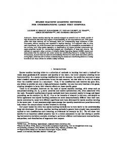
Patient cohorts. In this study, two cohorts with a total of 293 patients from different institutions were included. All patients suffered from histologically confirmed loco-regionally advanced HNSCC and received primary radiochemotherapy. Patients were allocated to an exploratory and validation cohort with a ratio of 2:1 based on the different included studies rather than on the treatment places. The exploratory cohort included 213 patients. 152 of the 213 patients were treated in one of the seven partner sites of the German Cancer Consortium Radiation Oncology Group (DKTK-ROG)13 between 2005 and 2011. The remaining 61 patients of the exploratory cohort were treated at the University Hospital Dresden (UKD, Germany) between 1999 and 2006. The validation cohort consisted of 80 patients. 50 of the 80 patients were treated within a prospective clinical trial [NCT00180180, ref.14] at the UKD between 2006 and 2012. The remaining 30 patients were treated at the UKD and the Radiotherapy Center Dresden-Friedrichstadt (RCDF) between 2005 and 2009. The clinical characteristics of both cohorts are summarised in Supplement A. Ethical approval for the multicentre retrospective analyses of clinical and imaging data was obtained from the Ethics Committee at the Technische Universität Dresden, Germany, EK177042017. All analyses were carried out in accordance with the relevant guidelines and regulations. Informed consent was obtained from all patients. Image pre-processing and feature extraction. Figure 1 illustrates the image pre-processing and feature extraction workflow. Prior to analysing the pre-treatment computed tomography (CT) scans without contrast agent, the gross tumour volume (GTV) was manually delineated taking into account patient examination and the findings of additional imaging modalities. Voxels in each CT image volume were re-sampled to an isotropic voxel size of 1.0 × 1.0 × 1.0 mm3 to correct for different voxel spacings and slice thicknesses between different centres5. Subsequently, the GTV was re-segmented to cover only soft tissue voxels between −150 and 180 Hounsfield units, removing voxels containing air and bone. Spatial filtering was applied to the base image to quantify additional image characteristics such as edges and blobs. We performed a stationary coiflet-1 wavelet transformation along the three spatial dimensions which produced eight transformed images in addition to the base image. A mean
SCIENTIFIC RePorTS | 7: 13206 | DOI:10.1038/s41598-017-13448-3
2
www.nature.com/scientificreports/
Figure 1. Illustration of image pre-processing and feature extraction.
Laplacian of Gaussian (LoG) image of five different kernel widths (0.5 mm, 1.0 mm, 2.0 mm 3.0 mm, 5.0 mm, respectively, ref.15) was generated from the base CT image as a further image volume. In each image set 18 statistical, 18 morphological, 30 histogram-based and 95 texture features were extracted from the GTV, leading to 1610 features in total. The following texture matrices were used: grey-level co-occurrence (GLCM)16, grey-level run length (GRLM)17,18, neighbourhood grey tone difference (NGTDM)19, grey-level size zone (GLZSM)20, grey-level distance zone (GLDZM)21 and neighbourhood grey level dependence (NGLDM)22 matrix. All features were calculated using a volumetric approach, and not by slice. Mathematical descriptions of all features are published in ref.23. The GTV was discretised using 64 quantization levels before calculation of texture matrices and the intensity histogram24,25. GLCM and GLRLM-based features were first calculated for each of the 13 different spatial directions and subsequently averaged. All features were normalised on the exploratory cohort using z-score normalisation. The resulting scale and shift constants were applied to the independent validation cohort. For image pre-processing and feature extraction we developed in-house software based on Python 2.7 (Python Software Foundation).
Feature selection methods and machine learning algorithms. In the present study different feature selection methods were considered: (I) correlation-based methods: Pearson, Spearman; (II) feature selection algorithms based on mutual information optimisation: mutual information maximisation (MIM), mutual information feature selection (MIFS), minimum redundancy maximum relevance (MRMR); and (III) model-based approaches: a univariate (uni)- and a multivariate (multi)-Cox-regression model, a random forest minimal depth (RF-MD), a random forest variable importance (RF-VI), a random forest based on maximally selected rank statistics variable importance (MSR-RFVI) and a random forest based on permutation variable importance (PVI-RF). Additionally, we selected features at random (RAND) and performed no feature selection (None). The comparison of different machine learning algorithms included the following non-parametric models: (I) the Cox model, the NET-Cox method with lasso and elastic-net regularisation; (II) models based on boosting trees (BT): BT-Cox, BT-CIndex; (III) boosting gradient linear models (BGLM): BGLM-Cox, BGLM-CIndex; and (IV) random forest based methods: random survival forest (RSF), random forest using maximally selected rank statistics (MSR-RF). Furthermore we investigated the following full-parametric models (V): survival regression (Survival-Regression) and models based on the Weibull distribution: BT- and BGLM-Weibull. A short description of all of these methods can be found in Supplement B. All feature selection methods and machine learning algorithms assessed here are able to handle continuous time-to-event data. Radiomics modelling framework. A radiomics modelling framework (RMF) was developed to create radiomics signatures, to optimise the hyper-parameters of machine learning algorithms, to train predictive models, and subsequently determine the predictive performance of the models as well as to perform the Kaplan-Meier survival analyses on validation data. Figure 2 shows the RMF and its four major processing steps: (I) feature selection, (II) hyper-parameter selection, (III) model building and (IV) model validation. The RMF was developed in-house using R 3.3.226. Feature selection. After feature extraction, feature clustering was performed on the exploratory cohort to
obtain an initial non-redundant set of biomarkers27. Highly correlated imaging biomarkers (Spearman correlation coefficient (SCC) >0.90) were clustered using hierarchical clustering28. The resulting clusters were represented by a meta-feature calculated by averaging over all features within the cluster. Negatively correlated features were inverted before averaging. A total of 229 non-singular clusters were created. The same clusters and meta-features were also generated for the validation cohort. After clustering, the feature set of the exploratory cohort was used to identify the most relevant features using feature selection algorithms. Feature selection was repeated n = 100 times using n bootstrap samples (i.e., 0.632 bootstrap method with replacement) of the exploratory cohort to ensure the selection of stable features. Feature selection ranks each feature according to a score, which depends on the method used. The top 20 best ranking SCIENTIFIC RePorTS | 7: 13206 | DOI:10.1038/s41598-017-13448-3
3
www.nature.com/scientificreports/
Figure 2. Illustration of the major radiomics processing chain within the radiomics modelling framework (RMF). (I) feature clustering and selection to identify prognostic biomarkers, (II) automatic hyper-parameter optimisation Θ for each model using a 2-fold cross validation with 40 times repetitions based on the exploratory cohort, (III) model building and (IV) model validation were performed.
features were selected from each bootstrap sample. We subsequently aggregated the selected features j over the bootstraps by calculating an importance score Ij, defined as Ij =
∑ ni =1 R ij occ j2
,
(1)
where Rij defines the rank within the i-th bootstrap sample and occj the frequency of occurrence of feature j over all bootstrap samples. The feature rank aggregation score is based on the enhanced Borda score29, with the difference being that feature occurrence receives a greater weight.
Hyper-parameter optimisation. After feature selection and rank aggregation, hyper-parameters of the
machine learning algorithms, such as signature size or algorithm-specific settings were optimised for each combination of feature selection and machine learning algorithm. A major objective of hyper-parameter tuning is to limit model overfitting. Overfitting would otherwise lead to poor predictive performance on unseen data. The individual hyper-parameter set Ω of each learning algorithm A was tuned by using an internal 2-fold cross validation scheme which was repeated 40 times (nRep = 40) based on the exploratory cohort. Hyper-parameter optimisation was performed using a grid search through a pre-defined hyper-parameter space. The objective of the hyper-parameter optimisation is to minimise a loss function L(Y, X, A) over the internal training and validation folds, X and Y, respectively, by a trained learning algorithm A to obtain an optimal set of parameters Ω*:
(
)
Ω⁎ = argmin L Y valid , X train , A γ (X train) , γ∈Ω
(2)
L(Y , X , A) = γ1 + γ2 + γ3
(3)
1−α 1−α − − γ1 = max 1, 1 , 2 (μvalid − α)2 (μtrain − α)
(4)
with
SCIENTIFIC RePorTS | 7: 13206 | DOI:10.1038/s41598-017-13448-3
4
www.nature.com/scientificreports/
1 − 1, γ2 = 50 2 (1 − μ bal) 1000 µ train > α γ3 = 0
∪ µvalid < α
or otherwise
µ train < α
(5)
∪ µvalid > α .
(6)
Here, μtrain and μvalid are the average prediction accuracy of the internal training and validation folds and μbal is the difference in average prediction accuracy between training and validation folds. The correction factor α = 0.5 represents the performance of a random experiment. The terms γ1, γ2 define penalties for the training-test error to minimise both test error and differences between training and test error. This leads to hyper-parameter sets where train and test error are more similar (balanced), which may increase model generalisability. The penalty term γ3 accounts for the discordance in train and test errors, to avoid selecting hyper-parameter sets where the predictions on the training set were concordant with the outcome, yet discordant on the test set.
Model building and validation. Model training was performed m = 100 times using bootstrap samples
(i.e., 0.632 bootstrap method with replacement) of the exploratory cohort for each combination of feature selection method and machine learning algorithm. The learning algorithms were trained on the generated bootstrap samples based on the top rank features as well as the optimised hyper-parameter set. Afterwards, an ensemble prediction30 was made by averaging the predicted risk scores for each model using data of the independent validation cohort. The ensemble model performance on the validation cohort was assessed using the concordance index (C-Index)31,32. The C-Index is a generalisation of the area under the curve for continuous time-to-event survival data. C-Index = 0.5 describes a random prediction whereas a perfectly predicting model has C-Index = 1.0.
Clinical endpoints and statistical analysis. The clinical endpoints LRC and OS were calculated from the first day of radiochemotherapy to the date of event or censoring. The number of events for LRC and OS were 86 and 120 for the exploratory cohort, and 26 and 51 for the validation cohort, respectively. In the present study, four analyses were performed: (I) the predictive performance of all combinations of feature selection methods and machine learning algorithms was evaluated based on the validation C-Index of the ensemble of models. (II) The median and standard deviation of the validation C-Indices of a feature selection method over all machine learning algorithms and vice versa was assessed to measure the variance induced by the respective algorithms. (III) The robustness of the radiomic signatures was assessed by applying different image rotations (±2°, ±6°, ±10°) and translations in x-y-direction (0.25 mm, 0.75 mm) for all combinations to the exploratory cohort and subsequently calculating the ICC for each feature selected by the various feature selection methods. (IV) Patients were stratified into a low and high risk group based on the predicted risk of the radiomics models. The cut-off value used for stratification was selected by using 1000 bootstrap samples based on the exploratory cohort. The fraction of significant stratification results (power) was calculated for each cut-off, leading to the optimal value which has the largest power33. Cut-off values were applied to the validation cohort unchanged. Survival curves were estimated by the Kaplan-Meier method and compared by log-rank tests. Finally, (V) validation performance for both endpoints was determined for the external signature, obtained by Aerts et al.5. In addition, we performed a statistical test to compare the C-Indices of the best performing models for LRC using the R package “compareC”34. Two-sided tests were applied and p-values