https://jap.or.kr
J Adv Prosthodont 2018;10:395-400
https://doi.org/10.4047/jap.2018.10.6.395
A pilot study using machine learning methods about factors influencing prognosis of dental implants Seung-Ryong Ha1a, Hyun Sung Park2a, Eung-Hee Kim3, Hong-Ki Kim4, Jin-Yong Yang5, Junyoung Heo6, In-Sung Luke Yeo7* Department of Prosthodontics, Dankook University College of Dentistry Jukjeon Dental Hospital, Yongin, Republic of Korea Private Practice, The Seoul Dental Clinic, Seongnam, Republic of Korea 3 Biomedical Knowledge Engineering Lab., Seoul National University School of Dentistry, Seoul, Republic of Korea 4 Dental Research Institute, Seoul National University School of Dentistry, Seoul, Republic of Korea 5 Private Practice, Yang’s Dental Clinic, Seoul, Republic of Korea 6 Department of IT Engineering, Hansung University, Seoul, Republic of Korea 7 Department of Prosthodontics and Dental Research Institute, Seoul National University School of Dentistry, Seoul, Republic of Korea 1 2
PURPOSE. This study tried to find the most significant factors predicting implant prognosis using machine learning methods. MATERIALS AND METHODS. The data used in this study was based on a systematic search of chart files at Seoul National University Bundang Hospital for one year. In this period, oral and maxillofacial surgeons inserted 667 implants in 198 patients after consultation with a prosthodontist. The traditional statistical methods were inappropriate in this study, which analyzed the data of a small sample size to find a factor affecting the prognosis. The machine learning methods were used in this study, since these methods have analyzing power for a small sample size and are able to find a new factor that has been unknown to have an effect on the result. A decision tree model and a support vector machine were used for the analysis. RESULTS. The results identified mesio-distal position of the inserted implant as the most significant factor determining its prognosis. Both of the machine learning methods, the decision tree model and support vector machine, yielded the similar results. CONCLUSION. Dental clinicians should be careful in locating implants in the patient’s mouths, especially mesio-distally, to minimize the negative complications against implant survival. [ J Adv Prosthodont 2018;10:395-400]
KEYWORDS: Machine learning; Decision tree; Support vector machine; Dental implant; Implant prognosis
Introduction Corresponding author: In-Sung Luke Yeo Department of Prosthodontics and Dental Research Institute, Seoul National University School of Dentistry, 101 Daehak-ro, Jongno-gu, Seoul 03080, Republic of Korea Tel. +82220722661: e-mail,
[email protected] Received May 2, 2017 / Last Revision September 12, 2017 / Accepted September 17, 2017 © 2018 The Korean Academy of Prosthodontics This is an Open Access article distributed under the terms of the Creative Commons Attribution Non-Commercial License (http://creativecommons. org/licenses/by-nc/3.0) which permits unrestricted non-commercial use, distribution, and reproduction in any medium, provided the original work is properly cited. These authors equally contributed to this work. This work was supported by the Technology Innovative Program funded by the Ministry of Trade, Industry & Energy (MI, Korea) (Grant No. 10052753).
a
pISSN 2005-7806, eISSN 2005-7814
Prosthodontic restoration plans to guide the locations of dental implants are utterly important.1 Inadequate implant placement makes the restorative procedure difficult. Fabricating prosthesis is sometimes impossible for a misplaced implant, and removing the implant is considered.2 The prosthodontic procedures without the removal of implant lead to undesired outcomes biomechanically and aesthetically. There are many possible reasons for implant misplacement, one of which appears to be that the precise implant placement directly depends on the amount of bone at the planned site.3 There are several studies trying to find some factors affecting implant prognosis and survival.4-6 Such studies generally used the logistic regression methods or other traditional tools to infer the factors statistically.7 Regression modThe Journal of Advanced Prosthodontics
395
J Adv Prosthodont 2018;10:395-400
els evaluate the influences of the factors on the implant survival, including the age, installed site, and bone quality, which are the variables already planned to be investigated. However, modern data mining tools including machine learning methods are considered necessary in analyzing the effects of the various variables associated with the implant surgery, which include mesio-distal position, bucco-lingual angulation, and the depth of an implant. Two advantages of these machine learning methods are to precisely analyze the data of a small sample size and to detect a new factor affecting the results.8 Those two features are the requirements that many clinical studies need in the field of dentistry, the sample size of which is usually small. The decision tree, one of the machine learning methods, is often used for classification and prediction and consists of a root node on the top, internal nodes for grouping criteria, links for nodes, and leaves as the final classification using a recursive partitioning method. A decision tree is appropriate for discovering patterns from binomial data with a learning binomial function.9 Many researchers in medical science are currently applying the decision tree model in diverse fields.10,11 The support vector machine is a supervised learning method used to determine the decision surface with the best classification of data.12 This method showed at least equal or better performance to that of other classification methods such as Bayesian classifier or artificial neural network.13 Medical researchers use support vector machines in various research areas, such as diagnosing Alzheimer’s disease through single-photon emission computed tomography image classification, discriminating breast cancer patients from a control group based on nucleosides in urine samples, and prognosis of drugs for heart failure patients.14-16 However, support vector machines have not been used in the field of prosthodontics. This study tried to find the determinant location factors of an inserted implant, which influences implant survival or complication. Additionally, both the decision tree and the support vector machine investigated whether there was a new factor affecting the implant prognosis.
Materials and Methods The data were collected from the retrospective chart review and filtered very carefully at at Seoul National University Bundang Hospital for one year (IRB No. B-0602-030-016). Subjects included in the analysis were partially edentulous patients who wanted their consecutively missing teeth to be restored by implants, having the implants placed without surgical stents. The patients having well-controlled hypertension or diabetes were included. However, smokers and patients with any other systemic disease that was likely to compromise implant survival were excluded.17,18 After consulting with a prosthodontist about implant location, all operations included in this study were performed by a maxillofacial surgeon with the same surgical protocol. The surgeon placed 667 implants in the mouths 396
of 198 patients according to the anatomic guide structures. Two dental implant systems (Osstem, Osstem Co., Busan, Korea, and Implantium, Dentium Co., Yongin, Korea) were used. All subsequent prosthodontic procedures were similarly performed by a prosthodontist. The problems that were confronted during the prosthodontic procedure were assessed and the final prostheses were evaluated by the prosthodontist. A year after prosthodontic restoration, the prosthodontist evaluated the implant’s location, features, and biomechanical aspects with relation to the outcome. The prosthodontic evaluations were categorized into cases according to the chart records. If a patient had several implants, more than one case could exist. The authors processed the descriptive evaluations into several features designated in nominal form for the analysis. During this process, some cases that lacked sufficient information for evaluation were excluded. Therefore, a total of 53 patients and 59 cases were analyzed, and the machine learning method was applied because of the small sample size. The features consisted of explanatory variables and output variables. The explanatory variables were divided into controllable variables, which could be managed by dentists, and host variables (Table 1 and Table 2). As explained above, these variables had nominal values. In Table 1, for example, the ‘immediate implantation’ variable has ‘Yes’ or ‘No’, and the other variables have ‘Adequate’ or ‘Inadequate’. In Table 2, the ‘site of placement’ variable has ‘Maxilla’, ‘Mandible’, or ‘Both’, and the ‘posterior site of placement’ variable has ‘premolar’, ‘molar’, or ‘both’. There were two kinds of output variables: the first was the survival of the implant, evaluated as whether the implant was osseointegrated or not; and the second was complications of the implant, evaluated as any discomfort or defect despite of adequate function with osseointegration (Table 3). The result variable has one of ‘Survival’ and ‘Osseo-disintegration’. The complication variable has one of ‘None’, ‘Thread/Implant exposure’, ‘Screw loosening’, ‘Loading problem’, and ‘Others’. All the features had nominal values in this study. Therefore, we selected a decision tree model among many available machine learning methods. Also, we used the support vector machine since this method is widely utilized in various fields including medical science. The authors trained and tested the decision tree using the collected data. Learning and classification of the factors affecting survival and complication were accomplished for the decision tree model analysis, using WEKA (Waikato Environment for Knowledge Analysis, University of Waikato, Hamilton, New Zealand) - Java based machine learning software. All possible combinations of features were made for the support vector machine. For each combination of features, cross validation was performed on the support vector machine. Linear function was used for classification by the support vector machine. The evaluation of support vector machine classification by machine learning was carried out by leaveon-out-cross-validation (LOOCV), which classified one sample with machine learning that was trained using the
A pilot study using machine learning methods about factors influencing prognosis of dental implants
Table 1. Controllable variables Variables
Description
Immediate implantation (Yes or No)
Whether implant is placed immediately after tooth extraction
Implant insertion depth (Adequate or Inadequate)
Inadequate if there was less than 5 mm between the implant top and the functional cusp tip of the occluding tooth
Bucco-lingual angulation (Adequate or Inadequate)
Inadequate when the screw hole of the straight, not angulated, abutment appeared on the buccal or lingual surface of the implant-supported crown
Mesio-distal position for restoration (Adequate or Inadequate)
Inadequate when the distance between two implants was less than 3 mm or the distance between the implant and the adjacent tooth was less than 1.5 mm
Implant length (Adequate or Inadequate)
Inadequate when the length of implant was shorter than 7 mm19
Crown-root ratio (Adequate or Inadequate)
Inadequate if the ratio was less than 1:1.520
Table 2. Host variables Variables
Description
Site of placement (Mx, Mn, or Both)
Placement of implants in the maxilla, mandible, or both jaws
Site of placement in jaw (Ant, Post, or Both)
Placement of implants at the anterior (central incisor to canine), posterior (first premolar to second molar), or both sides of the jaw
Posterior site of placement (Premolar, Molar, or Both)
Placement of implants at the premolar, molar, or both sites of the posterior area
Bone sufficiency (Adequate or Inadequate)
Extent in bone quantity: ‘Adequate’ if the bone width is larger than the implant diameter, and the bone height is greater than 5 mm
Soft tissue problem (Yes or No)
Whether soft tissue has any problem at the time of implant placement
Bone graft (Yes or No)
Whether the bone graft procedure was performed or not
Bone graft resorption (Yes or No)
Whether resorption occurs at the time of placing the implant-supported restoration
Table 3. Output variables Variables
Classification
Result
Survival Osseo-disintegration
Implant placement fails with osseo-disintegration
Complication
None
Implant functions without complication
Thread/Fixture exposure
Implant functions with thread/fixture exposure
Screw loosening
Implant functions with screw loosening
Loading problem
Implant is loaded over 30 degrees to the implant long-axis
Others
Other complications
other 58 samples. The accuracy, defined as true positive and negative over the whole sample, was calculated with the confusion matrix.
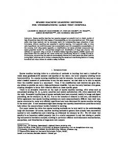
Results The decision tree model for implant survival is presented in Fig. 1. The mesio-distal position was the most significant factor determining implant prognosis (accuracy = 0.93).
Descriptions Implant functions well
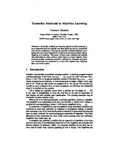
The decision tree model for complications of implant is shown in Fig. 2. Thread/fixture exposure appeared when the implant was placed with bone graft even when the implants were placed adequately (accuracy = 0.64). Support vector machine with LOOCV - survival measure of implant is presented in Table 4. Support vector machine with LOOCV - complication measure of implant is shown in Table 5.
The Journal of Advanced Prosthodontics
397
J Adv Prosthodont 2018;10:395-400
Fig. 1. The decision tree model for implant survival. The mesio-distal position is the most significant factor determining implant prognosis (accuracy = 0.93).
Fig. 2. The decision tree model for complications of implant. Thread/fixture exposure appeared when the implant was placed with the bone graft even when the implants were placed adequately (accuracy = 0.64).
Discussion First, both the decision tree model and support vector machine clearly identified ‘mesio-distal position’ as the most important factor during implant treatment. Both complication and survival of implant were associated with this factor in the decision tree model and most sets of the support vector machine also included this factor. The decision tree model showed that the implant functions successfully without osseo-disintegration when placed appropriately in the mesio-distal position. Even when this condition was inappropriate, there was no problem unless 398
the placement was in the anterior part of jaw. According to features of the decision tree model, the mesio-distal position variables potentially affected the survival of the implant to a greater extent because they were located in the root of the tree (Fig. 1, accuracy = 0.93). Regarding complications, noticeable thread exposure appeared when the implant was placed with bone graft. Thread/fixture exposure occurred even when the implants were placed with the appropriate bucco-lingual angulation and mesio-distal position in the maxilla and mandible. This result indicated that there was thread exposure even with adequate bucco-lingual angulation of the implant axis. In
A pilot study using machine learning methods about factors influencing prognosis of dental implants
Table 4. Support vector machine with LOOCV - survival measure of implant (top 5 results) Factors
Accuracy
Site of placement Site of placement in jaw Posterior site of placement Mesio-distal position for restoration
0.95
Site of placement in jaw Implant insertion depth Bucco-lingual angulation Mesio-distal position for restoration
0.95
Site of placement Site of placement in jaw Posterior site of placement Bone sufficiency Mesio-distal position for restoration
0.95
Site of placement Site of placement in jaw Posterior site of placement Soft tissue problem Mesio-distal position for restoration
0.95
Site of placement Site of placement in jaw Posterior site of placement Mesio-distal position for restoration Crown-root ratio
0.95
Table 5. Support vector machine with LOOCV - complication measure of implant (top 5 results) Factors
Accuracy
Site of placement in jaw Bone graft Bone graft resorption Immediate implantation Bucco-lingual angulation Mesio-distal position for restoration
0.75
Site of placement in jaw Bone graft Bone graft resorption Bucco-lingual angulation Mesio-distal position for restoration Implant length
0.75
Site of placement in jaw Bone graft Bone graft resorption Immediate implantation Bucco-lingual angulation Mesio-distal position for restoration Implant length
0.75
Site of placement in jaw Bone graft Bucco-lingual angulation Mesio-distal position for restoration
0.73
Site of placement in jaw Bone graft Bone graft resorption Bucco-lingual angulation Mesio-distal position for restoration
0.73
addition, loading problems occurred if the implant was placed without bone graft, inappropriate bucco-lingual angulation, and appropriate mesio-distal position. This loading problem led to non-axial loading on the implant (Fig. 2, accuracy = 0.64). Support vector machine with LOOCV showed outstanding performance in linear kernel function (Table 4, Table 5). In the survival measure, two sets with only four features were sufficient to classify the result of the implant with high accuracy (0.95). One of the sets consisted of the following features: site of placement-maxilla or mandible; site of placement in jaw-anterior, posterior, or both; posterior site of placement; and mesio-distal position for restoration. The other set included the following: site of placement in jaw-anterior, posterior, or both; implant insertion depth; bucco-lingual angulation; and mesio-distal position for restoration. Interestingly, all of the sets had one common feature, mesio-distal position for restoration (Table 4). As Bryant (1998) found, most sets showed that the site of placement, whether it is on the maxilla or mandible, was important.4 Regarding complication of implants, the support vector machine indicated that six factors were important in order to predict the prognosis with the best accuracy (0.746). All sets commonly included the following four factors: site of placement in jaw-anterior, posterior, or both; bone graft; bucco-lingual angulation; and mesio-distal position for restoration. The other factors in these sets were bone graft resorption, immediate implantation, and implant length. In the present study, the machine learning method was selected for analysis.9,19 This term was first used in the literature by Samuel, who proposed a learning game through alternating features and weighted values.20 Thereafter, the machine learning method integrated with computation theory was studied as a new research area in the 1980s. Document classification, information searching, and various other areas utilize machine learning methods.21,22 Many practitioners have realized how agonizing it is to encounter unexpected situations during implant placement. We also know how important it is to reach consensus between surgical operation and biomechanical prosthetic rehabilitation. Knowledge of the factors primarily considered during the operation would be very valuable for functional rehabilitation. Several factors have been proposed by other researchers, including quality and quantity of bone, history of trauma to the region, proximity of important structures (sinus, inferior alveolar nerve), need for bone grafting, degree of arterial blood supply, and rate of tissue healing.6 For implant fixture, a wider diameter and long length are known to be important factors for the survival of the implant.23,24 The shape of dental implants has been one of the most contested aspects and may have an effect on implant biomechanics.25 It has been demonstrated that bone loss of 1.5 to 2 mm apical to the implant-abutment junction would occur after uncovering surgery.26 Moreover, it has been documented that this bone loss could result in less inter-implant crestal bone loss if inter-implant distance is greater than 3 mm.27 The Journal of Advanced Prosthodontics
399
J Adv Prosthodont 2018;10:395-400
Therefore, it has been suggested that two implants must be spaced more than 3 mm apart.27 It is commonly observed that it is more difficult to increase inter-implant distance at the anterior than posterior. This results from differences in mesio-distal width of the crowns at the anterior and posterior. Therefore, the residual bone must be carefully studied to place implant fixtures in mesio-distally correct positions for anterior implants. It will ensure at least 3 mm of the inter-implant distance. Moreover, it may indicate that wider diameter implants may be of limited use in the anterior.
Conclusion Adequate mesio-distal positioning of the implant significantly affected the implant prognosis. Thus, site of placement was the most critical feature in this pilot study, as also indicated in other research. The small sample size demonstrated the strength of the machine learning method. However, traditional statistical tools should be applied with large samples to verify our conclusion.
ORCID Seung-Ryong Ha https://orcid.org/0000-0003-4176-6865 Hong-Ki Kim https://orcid.org/0000-0002-2610-4321 Jin-Yong Yang https://orcid.org/0000-0002-6731-1992 Junyoung Heo https://orcid.org/0000-0001-6407-6678 In-Sung Luke Yeo https://orcid.org/0000-0002-6780-2601
References 1. Academy of Osseointegration. 2010 Guidelines of the Academy of Osseointegration for the provision of dental implants and associated patient care. Int J Oral Maxillofac Implants 2010;25:620-7. 2. Kohner JS. Implant team: problems and solutions with osseointegrated implants. Pract Periodontics Aesthet Dent 1992;4:27-32. 3. Garber DA, Belser UC. Restoration-driven implant placement with restoration-generated site development. Compend Contin Educ Dent 1995;16:796, 798-802, 804. 4. Bryant SR. The effects of age, jaw site, and bone condition on oral implant outcomes. J Prosthodont 1998;11:470-90. 5. Omran MT, Miley DD, McLeod DE, Garcia MN. Retrospective assessment of survival rate for short endosseous dental implants. Implant Dent 2015;24:185-91. 6. Tolstunov L. Implant zones of the jaws: implant location and related success rate. J Oral Implantol 2007;33:211-20. 7. Walton JN, MacEntee MI. Choosing or refusing oral implants: a prospective study of edentulous volunteers for a clinical trial. Int J Prosthodont 2005;18:483-8. 8. Furey TS, Cristianini N, Duffy N, Bednarski DW, Schummer M, Haussler D. Support vector machine classification and validation of cancer tissue samples using microarray expression data. Bioinformatics 2000;16:906-14. 9. Mitchell TM. Machine Learning. New York: McGraw-Hill; 1997. p. 414. 10. Gambhir SS, Hoh CK, Phelps ME, Madar I, Maddahi J.
400
Decision tree sensitivity analysis for cost-effectiveness of FDG-PET in the staging and management of non-small-cell lung carcinoma. J Nucl Med 1996;37:1428-36. 11. Nakano Y, Takeshita T, Kamio N, Shiota S, Shibata Y, Suzuki N, Yoneda M, Hirofuji T, Yamashita Y. Supervised machine learning-based classification of oral malodor based on the microbiota in saliva samples. Artif Intell Med 2014;60:97-101. 12. Ward JJ, McGuffin LJ, Buxton BF, Jones DT. Secondary structure prediction with support vector machines. Bioinformatics 2003;19:1650-5. 13. Chen J, Xing Y, Xi G, Chen J, Yi J, Zhao D, Wang J. A comparison of four data mining models: Bayes, neural network, SVM and decision trees in identifying syndromes in coronary heart disease. Advances in Neural Networks-ISNN 2007: Springer; 2007. p. 1274-9. 14. Bullinger D, Fröhlich H, Klaus F, Neubauer H, Frickenschmidt A, Henneges C, Zell A, Laufer S, Gleiter CH, Liebich H, Kammerer B. Bioinformatical evaluation of modified nucleosides as biomedical markers in diagnosis of breast cancer. Anal Chim Acta. 2008;618:29-34. 15. Fung G, Stoeckel J. SVM feature selection for classification of SPECT images of Alzheimer’s disease using spatial information. Knowl Inf Syst 2007;11:243-58. 16. Son YJ, Kim HG, Kim EH, Choi S, Lee SK. Application of support vector machine for prediction of medication adherence in heart failure patients. Healthc Inform Res 2010;16:253-9. 17. Hwang D, Wang HL. Medical contraindications to implant therapy: part I: absolute contraindications. Implant Dent 2006;15:353-60. 18. Hwang D, Wang HL. Medical contraindications to implant therapy: Part II: Relative contraindications. Implant Dent 2007;16:13-23. 19. Michalski RSE, Carbonell JGE, Mitchell TME. Machine learning: an artificial intelligence approach. New York: Springer-Verlag; 1984. 20. Samuel AL. Some studies in machine learning using the game of checkers. IBM J Res Dev 2000;44:206-26. 21. Freitag D. Machine learning for information extraction in informal domains. Mach Learn 2000;39:169-202. 22. Sebastiani F. Machine learning in automated text categorization. Acm Comput Surv 2002;34:1-47. 23. Ivanoff CJ, Sennerby L, Johansson C, Rangert B, Lekholm U. Influence of implant diameters on the integration of screw implants. An experimental study in rabbits. Int J Oral Maxillofac Surg 1997;26:141-8. 24. Winkler S, Morris HF, Ochi S. Implant survival to 36 months as related to length and diameter. Ann Periodontol 2000;5:22-31. 25. Lin S, Shi S, LeGeros RZ, LeGeros JP. Three-dimensional finite element analyses of four designs of a high-strength silicon nitride implant. Implant Dent 2000;9:53-60. 26. Hermann JS, Cochran DL, Nummikoski PV, Buser D. Crestal bone changes around titanium implants. A radiographic evaluation of unloaded nonsubmerged and submerged implants in the canine mandible. J Periodontol 1997;68:1117-30. 27. Tarnow DP, Cho SC, Wallace SS. The effect of inter-implant distance on the height of inter-implant bone crest. J Periodontol 2000;71:546-9.