Printed in the Netherlands. PLSO 9555. A comparison between minirhizotron and monolith sampling methods for measuring root growth of maize (Zea mays L.).
Plant and Soil 147: 127-134, 1992. © 1992 KluwerAcademic Publishers. Printedin the Netherlands.
PLSO 9555
A comparison between minirhizotron and monolith sampling methods for measuring root growth of maize (Zea mays L.) H O O S H A N G MAJDI 1, ALVIN J.M. SMUCKER 2 and HANS PERSSON 1 ~Department of Ecology and Environmental Research, Swedish University of Agricultural Sciences, P.O. Box 7072, S-75007, Uppsala, Sweden and 2Department of Crop and Soil Sciences, Michigan State University, East Lansing, MI 488242, USA Received 1 April 1992. Revised August 1992
Key words:
image processing, methods, maize, minirhizotron, Zea mays L.
Abstract
Transparent plastic minirhizotron tubes have been used to evaluate spatial and temporal growth activities of plant root systems. Root number was estimated from video recordings of roots intersecting minirhizotron tubes and of washed roots extracted from monoliths of the same soil profiles at the physiological maturity stage of a maize (Zea mays L.) crop. Root length was measured by the line intercept (LI) and computer image processing (CIP) methods from the monolith samples. There was a slight significant correlation (r -- 0.28, p < 0.005) between the number of roots measured by minirhizotron and root lengths measured by the LI method, however, no correlation was found with the CIP method. Using a single regression line, root number was underestimated by the minirhizotron method at depths between 0-7.6 cm. A correlation was found between root length estimated by LI and CIP. The slope of estimated RLD was significant with depth for these two methods. Root length density (RLD) measured by CIP showed a more erratic decline with distance from the plant row and soil surface than the LI method.
Introduction
The need for more accurate and reproducible methods to quantify root growth and activity has led to the development of several new approaches in recent years. The minirhizotron tube and associated micro video camera technology reported by Upchurch and Ritchie (1983) is one new and promising method. The monolith sampling method has furthermore been improved by separating the roots from soil by hydropneumatic elutriation (Smucker et al., 1982), and then using either the line intercept (LI) method or video recording of the roots followed by computer image processing (CIP) for measuring root length. Minirhizotrons can be used to visually observe
and photograph plant roots intersecting plastic tubes. This method has among other things been used to measure root numbers, branching frequencies as well as mesofaunal activity in the rhizosphere (Box et al., 1989; Ferguson and Smucker, 1989; Snider et al., 1990). Its main advantage is that production, development, ageing and mortality of roots can be followed continuously throughout the year at the same place in the soil profile. However, it is not clear how the plastic tubes affect the root growth observed. Sampling of roots in field soils by the hand auger or the monolith sampling method provides washed root samples from which the root surface area, biomass, necromass, width, length and other morphological variables can be estimated
128
Majdi et al.
(Smucker et al., 1982, 1987; Srivastava et al., 1982; Vogt and Persson, 1991). Roots, rhizosphere and bulk soil samples can also be collected for chemical analyses. All these methods of root investigations have their specific applications to the plant-root-soil interface and are, except for the minirhizotron method, very time-consuming. The purpose of this paper is a comparison of measurements of similar root systems by minirhizotron and monolith sampling methods.
Material and methods
This study was carried out at the Kellog Biological Station in Michigan. The soil type is a highly stratified glacial Kalamazoo loam soil (fine loamy, mixed, mesic, typic Hapludalf). Root growth was studied in four different treatments: conventional tillage, no tillage and fertilizer treatments of 0 and 150kg N ha -I during one year of the project. The experimental design was replicated 4 times. Each plot was 35 x 28 m in size. Maize (Zea mays L.) was planted on May 10. The monolith samples were taken in early September and minirhizotron observations were completed in late August 1987. The data derived from all replicates was considered as a general sample without regarding treatments. Statistical analyses were carried out using the SAS, GLM procedure (Student's t-test with accounting for missing values).
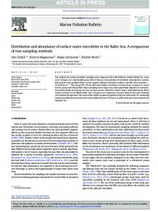
Minirhizotron measurements In each plot, three minirhizotron tubes (183 cm length, 5.1cm i.d., 5.7cm o.d., made from butyrate) were installed into the soil beneath the plants. The tubes intersected the soil at a depth equivalent to position 7 in sample profile A (Fig. 1). Profile B, with the same subsample dimensions as profile A, was sampled between the plant rows at 22.8 to 38.0cm from the plant row. This double-profile sampling scheme provided data for root distribution patterns for one half the distance between the plant rows (Fig. 1). Soil cores (5.5 cm in dimeter) were removed at an angle of 45 ° to the soil surface, to depths of
119 cm using a Giddings hydraulic power auger soil sampler. The machined cutting points of the probe, which replaced conventional cutting points, resulted in compressing of the displaced soil toward the interior of the sampling tube (Box et al., 1990). Soil on the walls of the hole which had been oriented by the sampling probe, was removed by using a 5.5 cm fine circular steel brush. Transparent minirhizotron tubes were manually forced into the holes. After installation, the 15 cm of the tube which remained above ground was painted black and then white to exclude light and reduce solar warming. The bottom and top openings of each tube were sealed by rubber stoppers. Tubes were labelled with an identification number and an index reference notch was installed 5 cm from the top end. After inserting the tubes in the holes, the soil surface was compressed around them to reduce water and light penetration along the tube surface. On each measurement occasion a Circon color video camera, was lowered into each minirhizotron tube by a square aluminium handle, containing registration holes 1.2 cm from center to center as described by Ferguson and Smucker (1989). Camera depth was controlled to ---0.1 mm by a plastic drawer-carriage wheel which stopped at each registration hole in the handle. Four incandescent light bulbs, 3 watts each, placed around the lens, provided uniform, diffuse light at each 'window' of the minirhizotron tube. Computer generated bar codes were recorded on video tapes in the field to identify the date, time, minirhizotron number and depth to the deepest root. Root images were video recorded on standard 1.9cm VHS tapes and catalogued for further analysis in the laboratory. Root counts were measured from the videotape by using one monitor for displaying each recorded image and another monitor for registration of the root count, depth, time and date of recording. Two large profiles were extracted in the area of each minirhizotron tube (Fig. 1). The monoliths were extracted by the profile sampling technique described by Srivastava et al. (1982). Each profile was fractionated into 18 subsamples (each 439cm3), labeled and stored at 3°C.
Minirhizotron and monolith sampling methods
Top view
Front vie~
o
15.2 cm
I
I X×
3
2
:
6
5
•
5
6
9
8
'~
8
9
X
22.8 cm
X X
12 11
,,10 11 12
([~)
15 14
i3114115 I
1~ 17
;x
I I i
x
B
:
X
B
129
A
L-.J
×
(
)
7.6 c m
0
A b,
x ~x
,
_
_
5.6 cm
X
©
Minirhizotron tube
Side view
"""'i""
~1~ ",, -,.
Fig. 1. Diagram of relative locations of the monolith profile samples (A and B) and the minirhizotron tubes with respect to the plant row.
130
Majdi et al.
Computer image processing
Results and discussion
Roots were washed from the soil by the hydropneumatic elutriation method (cf Smucker et al., 1982). Washed roots were processed immediately or stored in plastic bags containing 100-200 mL 10-15% methanol solution and stored at 3°C. Roots were dyed with Malachite green oxalate 24-48 h before video recording, by injecting 510mL of 1% dye into the plastic storage bag. Stained roots were rinsed with distilled water on an ultra fine (25 txm) nylon screen and placed uniformly into a clear glass tray where the roots were covered with a thin water film of approximately 1 mm above the root samples. The glass tray was illuminated from below by a translucent light table and video recorded by a computer controlled robotic camera which recorded 64% of the area on a VHS video tape (Smucker, 1990). The video images were transferred to the Vicom image analysis system (Smucker et al., 1987; Smucker, 1990) and digitized for measurement of root length and diameter classes. Video recorded root images (64 per tray samples from each 439cm 3 of soil) were analyzed by the Vicom pipeline and parallel image processor. Processing time for each image varied between from 1.6 to 2.5 minutes, depending on the quantities of non-root residues in each sample. The amount of residue in each sample ranged from zero, for the deepest samples to 17% for the surface samples. The Vicom computer algorithms are designed to measure root lengths and diameters with a 3% error for roots ranging in diameter from 0 to 3 mm when residue contents are less than 20%. Samples with more than 20% residues result in greater errors.
Figure 2 indicates a correlation between the LI and CIP methods of estimating root length (r = 0.782, p