Iran. J. Radiat. Res., 2012; 10(1): 43-51
A Comparison of dosimetric parameters between IAEA TRS-398, AAPM TG-51 protocols and Monte-Carlo simulation S.R.M. Mahdavi1*, M. Mahdavi2, H. Alijanzadeh2, M. Zabihzadeh3, A. Mostaar4 1Department
of Medical Physics, Tehran University of Medical Sciences, Tehran, Iran of Physics, Faculty of Basic Sciences, Mazandaran University, Babolsar, Iran 3Department of Medical Physics, Joundishapour University of Medical Sciences, Ahvaz, Iran 4Department of Medical Physics, Faculty of Medicine, Shahid Beheshti University of Medical Sciences, Tehran, Iran 2Department
Background: Two protocols of AAPM TG-51 and IAEA TRS-398 were compared followed by a measurement and Monte Carlo simulation of beam quality correction factor, KQ, AAPM TG-51 and IAEA TRS-398 protocols were compared for the absorbed dose to water for DW, and KQ parameters. Materials and Methods: Dose measurements by either protocols were performed with cylindrical and plane parallel chambers for 6 and 18 MV photons, and 6, 9, 12, 15, 18 MeV electron clinical beams were traced to the calibration factor of Iranian secondary standard dosimetry laboratory. MCNP-4C simulation of depth doses, beam profiles and KQ factors were validated typically for 18 MV and 12 MeV beams by experimental measurements. Results: The differences between simulation and measurements were 0.07% for beam profile, -2.60% and 1.19% for 12 MeV build up and linear portion of the depth dose curve, respectively. The figures of merit for 18 MV were about -4.17%, 1.62% and 0.38%. The differences of KQ's between simulation and measurement of 12 MeV, and 18 MV beams for TG-51 were -0.194% and 0.169%, and for TRS-398, they were about -0.465% and 0.097%, respectively. Conclusion: These differences between the two dosimetry protocols (IAEA TRS-398 & AAPM TG-51) from the point of absolute dosimetry were not significant at least when they were used under the same calibration procedure. The good agreement between Monte Carlo and measurement may also be even more important regarding the contribution into the development of radiotherapy treatment planning system, based on Monte Carlo procedures. Iran. J. Radiat. Res., 2012; 10(1): 4351
Keywords: Clinical dosimetry, TRS-398, AAPM TG-51, Monte Carlo simulation.
INTRODUCTION Higher precision of dose delivery is the first approach for the development of a
dosimetry protocol. However, two criteria should be considered for the selection of a clinical dosimetry protocol. First a calibration dosimetry method normally traceable to regional standard dosimetry laboratory and, secondly to be practical and easy to use in clinic; so, that radiotherapy physicists will spend a little time for absolute dosimetry of various beams (1-3). Air kerma calibration factor In Iran is provided by secondary standard dosimetry laboratory (SSDL) for 60Co beam quality. Radiotherapy departments are applying the ion chamber calibration factor to different photon and electron beams qualities. On the other hand, the success of radiotherapy depends on the absorbed dose within the target volume with no more than ± 5 % uncertainty (1). Since it is possible to delineate the target and other critical structures, using sophisticated diagnostic imaging modalities, there is a need to evaluate the absorbed dose accurately to maximize the target dose and minimize the normal tissue dose. However, it requires the measurement procedures in calibration laboratories as much possible as to be comparable to the user condition. Different studies have investigated the correspondence of the two ionizing radiation dosimetry protocols of American Association of Physicists in Medicine Task Group-51 *Corresponding author: Dr. Seied Rabi Mehdi Mahdavi, Department of Medical Physics, Tehran University of Medical Sciences, Tehran, Iran. Fax: +98 21 88058647 E-mail:
[email protected]
S.R.M. Mahdavi, M. Mahdavi, H. Alijanzadeh, et al.
(AAPM TG-51) and International Atomic Energy Agency Technical Report Series-398 (IAEA TRS-398) through measurement by cylindrical and plane-parallel ionization chambers (4,5). In this study, we have studied the differences of the absorbed dose to water and the beam quality factor of these two protocols with calibration factors traced to the regional (SSDL). The formalism and dosimetry procedures in the new TG-51 and International Atomic Energy IAEA TRS-398 protocols were based on the use of an ion chamber with a 60Co absorbed-dose to water calibration factor, N , and the beam quality correction factor, KQ, for the user beam (6). However, the compatibilities of the measurements and Monte Carlo (MC) simulation of depth doses (DD's), as well as beam profiles of 18 MV and 12 MeV radiations were analyzed typically for upcoming projects in which we would propose the comparison of the measurement by protocols with exact simulation of dose distribution for even more precise dosimetry. 60 Co D ,W
MATERIALS AND METHODS IAEA TRS-398 and the AAPM TG-51 have published different protocols for the calibration and/or measurement of clinical beams. These protocols are based on the use of an ionization chamber in terms of absorbed dose to water in standard laboratories and reference beam quality. Absorbed doses to water and beam quality factors were measured and then their ratios of AAPM TG-51 / IAEA TRS-398 were calculated. Measurements were performed within a computer-control scanner water tank of 40 × 40 × 40 cm3 (MP2 beam analyzer, PTW Freiburg, Germany). For central axis depth dose, the measurements were performed with a PTW Markus plane parallel chamber and a 0.6 cm3 PTW 30001 cylindrical chamber; both chambers were connected to a PTW Unidos E electrometer. The reference setup corresponded to a 10 × 10 cm2 field 44
Iran. J. Radiat. Res., Vol. 10 No. 1, June 2012
size and SSD = 100 cm. The scanning system had a position accuracy of ≤ 1 mm and a reproducibility of ≤ 0.1 mm. Measurements were made in 6 and 18 MV photons, as well as 6, 9, 12, 15 and 18 MeV electron beams. In this work, the MCNP-4C code (7) was used to run the 18 MV photon and 12 MeV electron beams spectra from the head of a Varian Clinac 2100 C/D linear accelerator to obtain the correspondence of dosimetric properties (e.g. depth doses and beam profiles). The simulated models included the bremsstrahlung target, the primary collimator, the vacuum window, the flattening filter, the monitor ion chamber, the mirror, the scattering foil and applicator (in the case of 12 MeV), as well as the upper and lower jaws. Beam monitoring chamber (more details reported by Duzenli et al. 1993) (8) and flattening filter (only in the case of 18 MV) were accurately modeled due to the fact they were the main sources of contaminating electrons. For the electron beam, the target was not present, scattering foil replaced the flattening filter and the primary collimator was also omitted from the electron beam simulations since it did not influence the beam significantly. For electron beams the applicator and a field defining insert in its bottom scraper was also included. This detailed description of the geometry required for the accurate simulation was provided by the manufacturer. The exact energy and radial spread of the hitting electrons to the target were unknown and must have been obtained by calibrating each spectral distribution against the corresponding depth dose curve and profiles. It should be noted that the central axis -depth dose curves have been dependent to the hitting electron energy while the dose profiles (especially for larger field sizes) were more affected by the radial spread of these electrons. The range of the primary mean electron energy was ranged from 17.7 to 18.4 MeV. The final incident electrons had a Gaussian energy distribution with a
Comparison between IAEA TRS-398 and AAPM TG-51
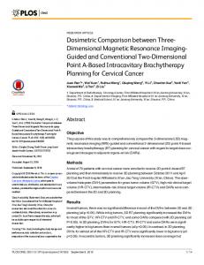
full width at half maximum (FWHM) of 1 MeV and centered at 18.2 MeV for the 18 MV photon beam. The electron beam radial intensity distribution was also set to be a Gaussian with the FWHM of 1.4 mm. A study reported by Ding et al. 1995 (9) showed that there has been little difference in the depth dose when using incident electrons which were either mono-energetic or having symmetric energy spectra. The incident electron energy on the exit window is usually higher than the nominal beam energy. For 12 MeV, we started the simulation by selecting incident electron energy to match with measured value of R50 for the 10 × 10 cm2 applicator. The model fine-tuning process resulted in peak energy of 12.25 MeV for 12 MeV electron beam. The FWHM was set to 0.103 cm. The number of electrons in the primary beam was set to 108. The cutoff energies of electrons and photons were set to 100 KeV and 10 KeV respectively. No photon interaction forcing and no Rayleigh scattering were used. The CPU used for the simulation was a Pentium IV with 2.5 GHz processors. Such simulations can also model the interactions which electrons undergo within the treatment head of the linear accelerator, allowing the dose at each point in the tissue to be broken down into several components, including that from contaminant photons ( 1 0 ) . The maximum statistical uncertainties of the results were about 2 % and 3% at the deeper depth (20 cm) of DD, and with more distance from the central axis (15 cm), respectively. For depth dose calculations in water phantom, a cylinder with a radius of one-tenth the size of the open field size was defined and divided into scoring cells with 2 mm height along the beam central axis. For beam profile calculations the primary cylinder was located at considered depth vertically to the beam central axis with the radius of 2 mm. Therefore, the dose resolution was 2 mm in this study. The set up depicted in figure 1, is the simulated geometry for the Varian 2100C/D linac and water phantom.
Figure 1. Schematic representation of the Monte Carlo models for the Varian 2100C/D linear accelerator geometry in photon mode.
To compare the simulation and measurement data of DD's and beam profiles, the average percent of difference was estimated through equation 1 (11). For this purpose, a FORTRAN program was released which can find point to point difference in percent and then average them out for the range of measured depth on the depth dose and/or beam profile: Average difference% = [(calculation - measurement)/measurement] (1) × 100 Dosimetry Formalism The dosimetry system was calibrated by SSDL of Iran at a reference condition in a 60Co gamma-ray beam. To measure the absorbed dose to water, the calibration factor, ND,W, was obtained to be 1.33 and 0.05335 Gy/nC for plane-parallel and cylindrical chambers, respectively. According to TRS-398, the absorbed dose to water at the reference depth, Zref, in water for a reference beam of quality Q0 (60Co) is equal to: D W , Q = M Q N D ,W , Q 0 K Q , Q 0
(2)
Where, MQ is the reading of the dosimeter under the reference condition which Iran. J. Radiat. Res., Vol. 10, No. 1, June 2012
45
S.R.M. Mahdavi, M. Mahdavi, H. Alijanzadeh, et al.
should be corrected to influence quantities such as polarity and recombination effects and ND,W,Q0 is the calibration factor in terms of absorbed dose to water of the dosimeter obtained from a standard laboratory at the reference beam quality, (12). When a dosimeter is used in a beam quality Q different from that used for calibration, Q0, the absorbed dose to water has to be corrected for the beam quality factor, which corrects the effect of the difference between the reference beam quality Q0 and the actual user quality Q (13). The TG-51 protocol provides a formulation at beam quality Q and for a chamber calibrated at 60Co gamma-rays energy that the absorbed dose to water at the reference depth in a beam of quality Q is obtained:
D WQ = MK
60
Q
N D ,WCo
(3)
where KQ converts the absorbed dose to water calibration factor for the 60Co beam, N instead of the calibration factor of an arbitrary beam of quality Q (14). For electron beams, KQ is written as a product of three factors; 60 Co D ,W
K Q = PgrQ K R′ 50 K ecal
(4)
where K´R50, Kecal and PgrQ are the electron quality conversion factor, photon-electron conversion factor and gradient correction factor, respectively (14). In calibration process, influencing quantities should be properly corrected. They are the quantities not being considered in the measurement, but yet influencing the quantity under measurement. These might be different in nature such as pressure, temperature and polarization voltage. Also, they may also arise from the dosimeter and / or the radiation field (e.g. beam quality, dose rate, field size, depth in a phantom) (13).
RESULTS The findings on quality correction factors and absorbed dose to water for 6 and 18 MV photon beams of Varian 2100 C/D 46
Iran. J. Radiat. Res., Vol. 10 No. 1, June 2012
accelerator are given in table 1. The TG-51/ TRS-398 value for the absorbed dose to water at field size of 10×10 cm2 were obtained to be 0.994 and 0.995 for 6 and 18 MV photons (SD4 gr/Cm2 but in TG -51, it is possible to calculate it for 2< R50