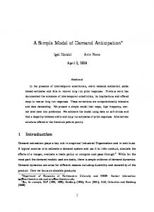
Oct 9, 2006 - Abstract. Some visual search tasks require to memorize the location of stimuli that have been previously scanned. Considerations about the ...
A Computational Model of Spatial Memory Anticipation during Visual Search
arXiv:cs/0610041v1 [cs.NE] 9 Oct 2006
J´er´emy Fix and Julien Vitay and Nicolas P. Rougier Loria, Campus Scientifique, BP239 54506 Vandoeuvre-les-Nancy, France
Abstract. Some visual search tasks require to memorize the location of stimuli that have been previously scanned. Considerations about the eye movements raise the question of how we are able to maintain a coherent memory, despite the frequent drastically changes in the perception. In this article, we present a computational model that is able to anticipate the consequences of the eye movements on the visual perception in order to update a spatial memory.
1
Introduction
While the notion of anticipation has been known for quite a long time in both psychology, biology or physics domains, it remains difficult to agree on a standard definition that can account for its multiple facets. For example, in [1], the author proposes an analogy between motor control and kalman filters where a controller is supposed to produce a signal that is sent to both the plant to control and to the emulator that is then able to produce a prediction of the behavior. In [2], the author refutes this standard definition of anticipatory systems as being based on a predictive model of the system itself and its environment. However, even if there does not exist such a general definition, there is a large consensus on the fundamental role played by anticipation in behavior. Someone that would have been deprived from any anticipation abilities would be severely impaired in its everyday life, from both a perception and action point of view. Of course, the deprivation of any anticipatory capabilities does not need to be so radical and we can also imagine a lighter impairment of the system. For instance, let us simply consider the inability to anticipate changes in the visual information resulting from an eye saccade. This anticipation is known to be largely based on unconscious mechanisms that provide us with a feeling of stability while the whole retina is submerged by different information at each saccade : producing a saccade results in a complete change in the visual perception of the outer world. If a system is unable to anticipate its own saccadic movements, it cannot pretend to obtain a coherent view of the world: each image would be totally uncorrelated from the others. One stimulus being at one location before a saccade could not be identified easily at being the same stimulus at another location after the saccade. The aim of this paper is to precisely pinpoint the importance of this visual
anticipation in establishing a coherent view of the environment and to propose a computational model that rely on anticipation to efficiently scan a visual scene. After a quick review of the literature demonstrating that visual anticipation is a critical part of the visual system, we introduce a simple experiment of visual search and explain how the model we propose can solve the task by using both anticipation and a dynamic model of working memory.
2
Visual search
Visual search is a cognitive task that most generally involves an active scan of a visual scene for finding one or several given targets among distractors. It is deeply anchored in most animal behaviors, from a predator looking for a prey in the environment, to the prey looking for a safe place to avoid being seen by the predator. Psychological experiments may be less ecological and may propose for example to find a given letter among an array of other letters, measuring the efficiency of the visual search in terms of reaction time (the average time to find the target given the experimental paradigm). In the early eighties, [3] suggested that the brain actually extracts some basic features from the visual field in order to perform the search. Among these basic features that have been recently reviewed by [4], one can find features such as color, shape, motion or curvature. Finding a target is then equivalent to finding the conjunction of features (that may be unique) that best describ the target. In this sense, [3] distinguished two main paradigms (a more tempered point of view can be found in [5]). Feature search refers to a search where the target differs from distractors against exactly one feature. Conjunction search refers to a search where the target differs from distractors against two or more features. What characterizes best the feature search is a constant search time that does not depend on the number of distractors. The target is sufficiently different from the distractors to pop out. However, in the case of conjunction search, the time to find the target seems to be tightly linked to the number of distractors that share at least one feature with the target (cf. Fig. 1). These observations lead to the question of how a visual stimulus could be represented in the brain. In [6], the authors proposed that the visual perception relies on two separated pathways: one would be dedicated to the extraction of features independently on their spatial positions (the so-called What pathways) while the other would only extract stimuli position without any information regarding feature properties (the so-called Where pathway). In this article, we don’t deal with the high-level processing of the visual input (the What pathway) nor with the difficult problem of the communication between the two pathways known as the binding problem and only consider a spatial representation of the visual input, filled by computing basic filters.
Fig. 1. Feature search can be performed very quickly as illustrated on the left part of the figure; the disc shape literally pops out from the scene. However, as illustrated on the right part of the figure, if the stimuli share at least two features, the pop out effect is suppressed. Hence, finding the disc shape with the stripes going from up-left to down-right requires an active scan of the visual scene.
2.1
Saccadic eye movements
The eye movements may have different behavioral goals, leading to five different categories of movements : saccades, vestibulo-ocular reflex, optokinetic reflex, smooth-pursuit and vergence. However, in this article we will only focus on saccades (for a detailed study of eye movements, see [7], [8]). Saccades are fast and frequent eye movements that move quickly the eye from the current point of gaze to a new location in order to center a visual stimulus on the fovea, a small area on the retina where the resolution is at its highest. The velocity of the eyes depends on the amplitude of the movement and can be reached up to 700 degrees per second at a frequency of 3 Hz. The question we would like to address is how the brain may give the illusion of a stable visual space while the visual perception is drastically modified every 200 ms. While the debate to decide whether or not the brain is blind during a saccade has not been settled ([9], [10]), the coherence between the perception before and after a saccade cannot be established accurately solely based on perception. One solution is to consider that the brain may use an efferent copy of the voluntary eye movement to remap the representation it has built of the visual world. Several studies shed light on pre-saccadic activities in areas such as V4 and LIP where the locations of relevant stimuli are supposed to be represented. In [11], the authors suggest that “the presaccadic enhancement exhibited by V4 neurons [...] provides a mechanism by which a clear perception of the saccade goal can be maintained during the execution of the saccade, perhaps for the purpose of establishing continuity across eye movements”. In [12], the authors review evidences that LIP neurons, whose receptive field will land on a previously stimulated screen
location after a saccade, are excited even if the stimulus disappears during the saccade. 2.2
Visual attention
The capacity to focus on a given stimulus of the visual scene is tightly linked to visual attention that has been defined as the capacity to concentrate cognitive ressources on a restricted subset of sensory information ([13]). In the context of visual attention, only a small subset of the retina information is available at any given time to elaborate motor plans or cognitive reasoning (cf. change blindness experiments presented in [14], [15]). The selection of a target for an eye movement is then closely related to the notion of spatial attention ([16]) that is classically divided into two types: overt attention which involves a saccade to center an object on the fovea and covert attention in which no eye movement is initiated. These two types of spatial attention were first supposed to be independent ([17]) but recent studies such as the premotor theory of attention proposed in [18] (see also [19], [20], [21]) consider that covert and overt attention rely on the same neural structures but movement is inhibited in covert attention. 2.3
Computational models
Over the past few years, several attempts at modeling visual attention have been engaged ([22], [23], [24], [25], [26]). The basic idea behind most of those models is to find a way to select interesting locations in the visual space giving their behavioral relevance and whether or not they have been already focused. The two central notions in this context have been proposed by [22] and [27]: – saliency map – inhibition of return (IOR). The saliency map is a single spatial map, in retinotopic coordinates, where all the available visual information converge in order to obtain a unified representation of stimuli, according to their behavioral relevances. A winner-take-all algorithm can be easily used to find what is the most salient stimulus within the visual scene which is identified as the attentional point of focus. However, in order to be able to go to the next stimuli, it is important to bias the winner-take-all algorithm in such a way that it prevents returning to an already focused stimulus. The goal of the inhibition of return mechanism is precisely to feed the saliency map with such a bias. The idea is to have another neural map that records focused stimuli and inhibits the corresponding locations in the saliency map. Since an already focused stimulus is actively inhibited by this map, it cannot pretend to win the winner-take-all competition, even if it is the most salient. The existence of a single saliency map is still not proved. In [26] the author proposes a more distributed representation of these relevances, clearly dividing the what and the where pathways stated before, and where spatial competition
occurs in a motor map instead of a perceptive one. The related model exhibits good performances regarding visual search task in natural scene, but is restricted to covert attention. Therefore, authors do not take into account eye movements and the visual scene is supposed to remain stable: scanning is done without any saccade. During the rest of this article, we will stick to the saliency map hypothesis, even if controverted, in order to illustrate the anticipatory mechanism.
3 3.1
A model of visual search with overt attention Experiment
In order to accurately evaluate the model, we setup a simple experimental framework where some identical stimuli are drawn on a blackboard and are observed by a camera. The task is to successively focus (i.e. center) each one of the stimuli without focusing twice on any of them. We estimate the performance of the model in terms of how many times a stimulus has been focused. Hence, the point is not to analyze the strategy of deciding which stimulus has to be focused next (see [28,29] for details on this matter). In the context of the proposed model, the strategy is simply to go from the most salient stimulus to the least salient one, and to randomly pick one stimulus if the remaining ones are equally salient. Figure 2 illustrates an experiment composed of four identical stimuli where the visual scan path has been materialized. The effect of making a saccade from one stimulus to another is shown and underlines the difficulty (for a computational model) of identifying a stimulus before and after a saccade. Each one of the stimulus being identical to the others, it is impossible to perform an identification based solely on features. 3.2
Model
The model is based on three distinct mechanisms (cf. Fig. 3 for a schematic view of the model). The first one is a competition mechanism that involves potential targets represented in a saliency map that were previously computed according to visual input. Second, to be able to focus only once on each stimulus, the locations of the scanned targets are stored in a memory map using retinotopic coordinates. Finally, since we are considering overt attention, the model is required to produce a camera movement, centering the target onto the fovea, used to update the working memory. This third mechanism works in conjunction with two inputs: current memory and parameters of the next saccade. This allows the model to compute quite accurately a prediction of the future state of the visual space, restricted to the targets that have been already memorized. A different version of this model, without the anticipatory mechanism can be found in [30]. Moreover, the model uses the computational paradigm of two dimensional discrete neural fields (the mathematical basis of this paradigm can be found in [31] for the one dimensional case, extended to a two dimensional study in [32]).
1
1
2
1
2 4
4
3
3
2 4 3
1
1
2
2 4
4
3
3
Fig. 2. When scanning a visual scene, going for example from stimulus 1 to stimulus 4, as illustrated on the left of the figure, the image received on the retina is radically changed when each stimulus is centered on the retina, as illustrated on the right of the figure. The difficulty in this situation is to be able to remember which stimulus has already been centered in order to center another one. The figures on the stimuli are shown only for explanation purpose and do not appear on the screen; all the stimuli are identical.
Update Saliency map
Working memory
Gating Current memory
Competition Focus
Prepared saccade
Prediction
Memory anticipation
Fig. 3. Schematic view of the architecture of the model. The image captured by the camera is filtered and represented in the saliency map. This information feeds two pathways : one to the memory and one to the focus map. A competition in the focus map leads to the most salient location that is the target for the next saccade. The anticipation circuit predicts the future state of the memory with its current content and the programmed saccade.
The model consists of six n×n maps of units, characterized by their position in a map, denoted x ∈ [1..n]2 and their activity as a function of their position and time, denoted u(x,t). The basic dynamic equation that follows the activity of a unit at position x, depends on its input, computed as a weighted sum over input units, and on an weighted influence of the lateral units in the same map. Equation (1) is the equation proposed in [31], discretized in space, where M is the set of the lateral units, M ′ the set of the input units, wM (x − x ′ ) the lateral connection weight function, and s(x, y) the afferent connection weight function. Usually, the weighting functions s(x, y) and wM (x−x ′ ) are chosen as a Gaussian or as a difference of Gaussians, as given by (2).
τ.
X X ∂u(x, t) = −u(x, t) + wM (x − x ′ )u(x ′ , t) + s(x, y).u(y, t) ∂t ′ M
s(x, y) = C.e wM (x − x ′ ) = A.e
kx−yk2 c2
(1)
M
with C, c ∈ IR∗+
kx−x ′ k2 a2
− B.e
kx−x ′ k2 b2
with A, B, a, b ∈ IR∗+
(2)
where u(x, t) is the activity of the unit at the location x in a map M, u(x ’, t) the activity of the unit at the location x ’ in the same map, u(y,t) the activity of the unit at the location y in a map M ’, different from M and τ is a given parameter that defines the temporal dynamics . A unit whose activity satisfies (1) will be called a sigma unit in the following. We also introduce sigma-pi units ([33]) whose activity satisfies (3). While in (1) the input of a unit is computed as a sum of activities, in (3), the input of the unit is computed as a sum of product of activities.
τ.
X X Y ∂u(x, t) = −u(x, t) + wM (x − x ′ )u(x ′ , t) + wi . u(y, t) ∂t ′ M
i∈I
(3)
y∈Mi
In the following, we denote I(x, t) the input of the unit x, at time t, that can be written as :
I(x, t) =
X M
I(x, t) =
s(x, y).u(y, t) for sigma units
′
X i∈I
wi .
Y y∈Mi
u(y, t) for sigma-pi units
(4) (5)
′
We will now describe briefly how the different maps interact. Since the scope of this article is the anticipation mechanism, the description of the saliency map, the focus map and the working memory will not be accurate but a more detailed explanation, with the appropriate dynamical equations, can be found in [30].
Saliency map The saliency map is updated by convolving the image captured with the camera of the robot used for the simulation with gaussian filters. The stimuli we use are easily discriminable from the background on the basis of the color information. This computation leads to a representation of the visual stimuli with gaussian patterns of activity in a single saliency map. We point out again that this is one of our working hypothesis, detailed in section 2.3. Focus Units in the focus map have direct excitatory feedforward inputs from the saliency map. The lateral connections are locally excitatory and widely inhibitory so that a competition between the units within the map leads to the emergence of only one stimulus in the focus map. This stimulus is the next target to focus and the movement to perform to center it on the fovea is decoded from this map. Working memory Once a stimulus has appeared within the focus map and because it is also present in the saliency map, it emerges immediately within the working memory. Both excitations from the focus map and the saliency map (at a same location) are necessary for the emergence of the stimulus in the working memory area. If the focused stimulus changes, it will not be present anymore in the focus map such that an additional mechanism is needed to maintain it in the memory. It is not shown on the schematic illustration 3 but the memory consists in two maps that share excitatory connections in the two ways : the first map excites the second and the second excites the first, weighted so that the excitation is limited in space. Memory anticipation The memory anticipation mechanism aims at predicting what should be the state of the working memory, after an eye movement needed to center the stimulus in the focus map, before the movement is initiated. The sigma-pi units in the anticipation map has two inputs : the activity of the units of the focus map and the activity of the units of the working memory. If we denote wm(x,t) the activity of the unit x of the working memory at time t, and f(x,t) the activity of the unit x of the focus map at time t, we define the input I(x) of the unit x in the anticipation map as : X I(x, t) = β. wm(y, t).f (y − x, t) (6) y∈IR2 The input of each unit in the anticipation map is computed as a convolution product of the working memory and the focus, centered on its coordinates. To make (6) clearer, the condition of the sum is weaker than the one that should be used : since the input maps are discrete sets of units, the two vectors y and y-x mustn’t exceed the size of the maps. From (3) and (6), the activity of the units in the anticipation map, without lateral connections, satisfies (7). τ.
X ∂u(x, t) = −u(x, t) + β. wm(y, t).f (y − x, t) ∂t y∈IR2
(7)
Then, the shape of activity in the anticipation map converges to the convolution product of the working memory and the focus map. Since the activity in the focus map has a gaussian shape and the working memory can be written as a sum of gaussian functions, the convolution product of the working memory and the focus map leads to an activity profile that is the profile in the working memory translated by the vector represented in the focus map. This profile is the prediction of the future state of the working memory and is then used to slightly excite the working memory. After the eye movement and when the saliency map is updated, the previously scanned stimuli emerge in the working memory as a result of the conjunction of the visual stimuli in the saliency map and the prediction of the working memory; This is the same mechanism than the one used when a stimulus emerges in the working memory owing to the conjunction of the activity in the saliency map and the focus map. 3.3
Simulation and results
The visual environment consists in three identical stimuli that the robot is expected to scan successively exactly once. A stimulus is easily discriminable from the background, namely a green lemon on a white table. A complete activation sequence of the different maps is illustrated on Fig. 4. The saliency map is filled by convolving the image captured from the camera by a green filter in HSV coordinates such that it leads to three distinct stimuli. At the beginning of the simulation (Fig. 4a), only one of the three stimuli emerges in the focus map, thanks to the strong lateral competition that occurs within this map. This stimulus, present both in the focus map and in the saliency map, emerges in the working memory. The activation within the anticipation map reflects what should be the state of the saliency map, restricted to the stimuli that are in the working memory, after the movement that brings the focused one in the center of the visual field. During the eye movement (Fig. 4b), no visual information is available and the parameter τ in 1 and 7 is adjusted so that only the units in the anticipation map remain active, whereas the activity of the others tends to zero. After the eye movement and as soon as the saliency map is fed with the new visual input, the working memory is updated thanks to the excitation from both saliency and anticipation map at a same location : the prediction of the state of the visual memory is compared with the current visual information. A new target can now be elicited in the focus map thanks to a switch mechanism similar to that described in [30].
4
Discussion
We have presented a computational model of visual memory anticipation that is able to ensure the coherence of the visual world despite abrupt changes in the perception that occur after each eye movement. The prediction of the future state of the visual memory enriches the perception of the visual world in order to avoid focusing twice a same stimulus. As we explained previously, saccades
thal_wm
wm
anticipation
focus
input
thal_wm
wm
wm
focus
input
a)
thal_wm
anticipation
anticipation
focus
input
wm
thal_wm
wm
anticipation
focus
input
b)
anticipation
focus
input
e)
thal_wm
wm
thal_wm
wm
anticipation
focus
input
c)
anticipation
focus
input
f)
thal_wm
d)
thal_wm
wm
anticipation
focus
input
g)
h)
Fig. 4. A sequence of evolution of the model during an overt visual scan trial. a) One of the three stimuli emerges in the focus map and the anticipation’s units predict the future state of the visual memory (the maps wm and thal wm). b) During the execution of the saccade, only the units in the anticipation map remain active. c) The focused stimulus emerge in the memory since it is both in the saliency map and the anticipation map at the same location. d) A new target to focus is elicited. e) The future state of the memory is anticipated. f) The saccade is executed and only the prediction remains. g) The two already focused stimuli emerge in the memory. h) The attentional focus lands on the last target.
are generally too fast and it is impossible, even in the case we were not blind during eye movements, to continuously update a visual memory. An efferent copy of the eye movement is used to establish the missing link between the pre and post-saccadic perceptions. This mechanism is clearly an extension of visual attention models that have been presented in section 2.3 and where the visual world is purely static. The question of learning the underlying transformation of the anticipatory mechanism, namely the convolution product of the focus map and the working memory, remains open and still studied. We did implement a learning mechanism, under restrictions and strong hypotheses, that relies heavily on the difference between the pre-saccadic prediction and the post-saccadic actual perception. This self generated signal is able to measure to what extent the predicition is correct or not. Hence, it is quite easy to modify weights accordingly. The main difficulty during learning remains the sampling distribution of examples within the input space which is a well known problem in information and learning theory. Without an additional motivational system that could bias examples according to a given task, it is quite unrealistic to rely on a regular distribution of examples.
References 1. Grush, R.: The emulation theory of representation : motor control, imagery and perception. Behavioral and brain sciences 27 (2004) 377–442 2. Riegler, A.: The role of anticipation in cognition. Computing Anticipatory Systems: CASYS 2000 - Fourth International Conference 573 (2001) 534–541 3. Treisman, A., Gelade, G.: A feature-integration theory of attention. Cognitive Psychology 12(1) (1980) 97–136 4. Wolfe, J.: Visual search. In: Attention, University College London Press (1998) 5. Duncan, J., Humphreys, G.: Visual search and stimulus similarity. Psychological Review 96(3) (1989) 433–458 6. Goodale, M., Milner, A.: Seperate visual pathways for perception and action. Trends in Neurosciences 15(1) (1992) 20–25 7. Leigh, R., Zee, D.: The neurology of eye movements, 3rd edition. (1999) 8. Carpenter, R.: Movements of the eyes, 2nd edition. (1988) 9. Kleiser, R., Seitz, R., Krekelberg, B.: Neural correlates of saccadic suppression in humans. Current Biology 14 (2004) 386–390 10. Ross, J., Morrone, C., Goldberg, M., Burr, D.: Changes in visual perception at the time of saccades. Trends in Neurosciences 24(2) (2001) 113–121 11. Moore, T., Tolias, A., Schiller, P.: Visual representations during saccadic eye movements. Neurobiology 95(15) (1998) 8981–8984 12. Merriam, E., Colby, C.: Active vision in parietal and extrastriate cortex. The Neuroscientist 11(5) (2005) 484–493 13. James, W.: The principles of psychology. (1890) 14. O’Regan, Noe: A sensorimotor account of vision and visual consciouness. Behavioral and Brain Sciences 24 (2001) 939–1031
15. Simons, J.: Current approaches to change blindness. Visual Cognition 7(1–2–3) (2000) 1–15 16. Moore, T., Fallah, M.: Control of eye movements and spatial attention. PNAS 98(3) (2001) 1273–1276 17. Posner, M., Petersen, S.: The attentional system of the human brain. Annual Review of Neurosciences 13 (1990) 25–42 18. Rizzolatti, G., Riggio, L., Dascola, I., Ulmita, C.: Reorienting attention accross the horizontal and vertical meridians. Neuropsychologia 25 (1987) 31–40 19. Chelazzi, L., Miller, E., Duncan, J., Desimone, R.: A neural basis for visual search in inferior temporal cortex. Nature 363 (1993) 345–347 20. Kowler, E., Andersen, E., Dosher, B., Blaser, E.: The role of attention in the programming of saccade. Vision Research 35 (1995) 1897–1916 21. Craighero, L., Fadiga, L., Rizzolatti, G., Umilta, C.: Action for perception : a motor-visual attentional effect. Journal of Experimental Psychology 25 (1999) 1673–1692 22. Koch, C., Ullman, S.: Shifts in selective visual attention : Towards the underlying neural circuitry. Human Neurobiology 4(4) (1985) 219–227 23. Tsotsos, J., Culhane, S., Lai, W., Davis, N.: Modeling visual attention via selective tuning. Artificial Intelligence 78 (1995) 507–545 24. Wolfe, J.: Visual attention. In: Seeing : Handbook of Perception and Cognition, 2nd ed., De Valois KK (2000) 335–386 25. Itti, L., Koch, C.: Computational modeling of visual attention. Nature Reviews Neuroscience 2(3) (2001) 194–203 26. Hamker, F.: A dynamic model of how feature cues guide spatial attention. Vision Research 44 (2004) 501–521 27. Posner, M., Cohen, Y.: Components of visual orienting. (1984) 531–556 28. Findlay, J., Brown, V.: Eye scanning of multi-element displays: I. scanpath planning. Vision Research 46(1–2) (2006a) 179–195 29. Findlay, J., Brown, V.: Eye scanning of multi-element displays: Ii. saccade planning. Vision Research 46(1–2) (2006b) 216–227 30. Vitay, J., Rougier, N.: Using neural dynamics to switch attention. In: International Joint Conference on Neural Networks, IJCNN (2005) 31. Amari, S.: Dynamical study of formation of cortical maps. Biological Cybernetics 27 (1977) 77–87 32. Taylor, J.: Neural bubble dynamics in two dimensions. Biological Cybernetics 80 (1999) 5167–5174 33. Rumelhart, D., Hinton, G., McClelland, J.: A general framework for parallel distributed processing. In: Parallel Distributed Processing, Vol. 1, MIT Press (1987)