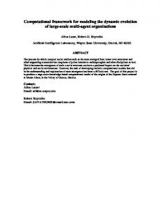
88
INTERNATIONAL JOURNAL OF COMPUTATIONAL COGNITION (HTTP://WWW.YANGSKY.COM/YANGIJCC.HTM), VOL. 3, NO. 3, SEPTEMBER 2005
A Framework for the Computational Approach to Cellular Metabolism Supporting Neuronal Activity (Invited Paper) Alfredo Pereira Jr and Carlos Lungarzo
Abstract— Computational neuroscience has focused mainly on electrical properties of the neuron membrane, related to computations performed by neural networks. In this paper we develop a framework for the inclusion of metabolism in a computational model of the single cortical pyramidal neuron. Two mechanisms control the activity of the neuron’s metabolic networks: calcium ions entering chemically gated/voltage-dependent channels, and metabotropic receptors binding to G-proteins. These pathways converge to the activity of the kinase family of proteins that controls the phosphorylation of other enzymes and activates gene expression, then promoting feedback on both metabolism and membrane states. The activity of the kinase family of proteins supports the functional specialization of cells. Besides firing and non-firing, neuronal computational models account for sub-threshold states of the neuronal membrane, related to the metabolic setting and tuning of functional specialization c 2004mechanisms by means of kinase activity. Copyright ° 2005 Yang’s Scientific Research Institute, LLC. All rights reserved. Index Terms— Biocomputation, Kinase family, feature detection, metabolic networks, signal-transduction pathways.
I. I NTRODUCTION
I
N THE current Proteomics era, special attention has been directed to the identification of the pool of proteins present in each kind of tissue. Estimated to be around 15.000 for each tissue, proteins are responsible for the biological functions carried by these tissues, organs and body systems. Many proteins and their respective functions have been discovered and proposed in recent years, opening new and exciting possibilities for the scientific explanation of cognitive, motivational and emotional brain processes. Metabolic networks are also known in the literature as Signal-Transduction Pathways (STPs). They include membrane receptors, other proteins present in the cytoplasm and molecular signals (ions, hormones). Although the mapping of neuronal proteins is still incomplete, the available data is sufficient to begin a systematic effort towards understanding the role of metabolic processes in neuronal function. Manuscript received October 13, 2004; revised October 14, 2004. Alfredo Pereira Jr, UNESP - Botucatu - S˜ao Paulo - Brasil. Carlos Lungarzo, UNICAMP – Campinas - S˜ao Paulo - Brasil. Email:
[email protected](A. Pereira Jr),
[email protected](C. Lungarzo) Acknowledgements: CNPQ (Brasil) for financial support to the research and Juliana Lima for the drawings. Publisher Item Identifier S 1542-5908(05)10311-X/$20.00 c Copyright °2004-2005 Yang’s Scientific Research Institute, LLC. All rights reserved. The online version posted on October 15, 2004 at http://www.YangSky.com/ijcc33.htm
In spite of the tremendous variety of proteins, they can be classified in classes or families, according to common functional characteristics. Comparing their characteristics, some redundant patterns are apparent, allowing conjectures about possible computational mechanisms. Current attempts to understand neuronal function have not focused on metabolic processes (an important exception is Rocha, 1997), in spite of the importance of metabolism in the measurement of brain function by means of techniques as functional Magnetic Resonance Imaging (fMRI) and microdialysis. Computational models of brain function have focused on other aspects and levels of information processing, as patterns of electrical activity and cognitive representation. On the other hand, molecular models of biological function, as the classical cellular automata developed by Stuart Kaufmann, describe the self-regulation of gene expression, without considering the interaction between genes, metabolism and environment. Several input lines, from genes and environmental factors, converge to the cytoplasm, an interaction space in the cell, where they undergo transformations, generating several outputs as the production of new proteins. As the inputs are not initially correlated and obey different rules, the biological machine cannot be identified with the classical computational concept of a Turing machine. A current alternative to classical computation has been the Connectionist approach, focusing on the pattern of connections between multiple nodes in a distributed processing network. In neuroscience, the pattern of connections is assumed to correspond to cognitive representations defined as vectors in a multidimensional space (Churchland, 1986; Churchland and Sejnowski, 1993). However, the advances brought by this approach have not been sufficient to incorporate cellular metabolism in the picture. Neural networks usually assume that neuronal activity reduces to receiving and sending discrete pulses, thus neglecting sub-threshold potentials related to the activation of metabolic pathways. The consideration of metabolism implies a methodological change towards describing neuronal activity in terms of three classes of states: firing (a membrane depolarization that produces a spike through the axon), sub-threshold activation (a membrane depolarization that doesn’t generate a spike but activates several STPs in the cell), and inhibition (hyperpolarization of the membrane). Sub-threshold activation may have an advantage over firing because of the brain’s homeostatic balance between excitation
PEREIRA & LUNGARZO, A FRAMEWORK FOR THE COMPUTATIONAL APPROACH TO CELLULAR METABOLISM SUPPORTING NEURONAL ACTIVITY
Fig. 1.
Fig. 2.
Synaptic transmission.
Fig. 3.
Ionotropic receptors.
89
Propagation of the action potential through the axon.
and inhibition. The mechanism of lateral inhibition in cortical tissue increases the probability of a neuron being inhibited soon after it fires; therefore, ”silent” neurons (neurons that don’t fire while keeping sub-threshold excitation; see Moore et al., 1998; Engel et al., 2000) are more likely to sustain metabolic activity to the point of achieving changes in cellular functions. Such metabolic activity is able to modulate and even change the biological/cognitive functional specialization and respective patterns of connectivity of the neuron. II. A R EVIEW OF N EURONAL M EMBRANE M ECHANISMS In this section, we sum up well known properties of neurons, to make basic concepts more precise for the non-specialist. The single neuron is a complex system encompassing several spatio-temporal levels of organization and activity. One of the main properties of this kind of cell is membrane specialization, comprising a dendritic tree and, in many cases, a relatively long axon. Dendrites contain many synapses, receiving information from other neurons. Action potentials are bioelectric currents generated by the movement of ions in and out the membrane. When the membrane potential reaches a threshold it produces an axonal spike, a fast and long-distance form of communication between neurons, consisting of a bioelectrical pulse that travels along the axon. The action potential produced in the dendritic tree and body of the neuron by ionic flow through the membrane generates spikes transmitted to other neurons through the axon (Fig. 1). The bioeletrical signal, generated by sodium and potassium ion movement, reaches the axon terminal and then triggers the opening of transmitter vesicles in the pre-synaptic neuron. Transmitters are released in the synaptic cleft and bind to membrane receptors belonging to the post-synaptic neuron (Fig. 2).
Synapses can be excitatory and inhibitory, depending on the transmitters and receptors, and their respective effect on the electrical state of the membrane. Excitatory synapses in the neocortex are correlated with a specialized structure in the dendrite, the spine. Spines contain important computational mechanisms activated by the entrance of calcium ions (Ca++) through membrane channels. Metabolic networks are called SignalTransduction Pathways (STPs), which contain the following elements: 1) Transmitters and modulators released in the synaptic cleft; 2) Membrane receptors that bind with such transmitters and modulators; 3) Other proteins present in the cytoplasm, which are activated by the membrane receptors; 4) Other signaling elements, as ions and hormones, which interact with the proteins, also composing STPs; 5) Transcriptional factors that induce gene expression according to the previously activated STPs.
90
INTERNATIONAL JOURNAL OF COMPUTATIONAL COGNITION (HTTP://WWW.YANGSKY.COM/YANGIJCC.HTM), VOL. 3, NO. 3, SEPTEMBER 2005
associates with Aplysia. Transmitter serotonin (5-HT) binding to a GPCR modulates an ionotropic receptor controlling a NaK channel. The cAMP released by AC activates a Protein Kinase (PKA) that in turn activates transcriptional factors (CREB), leading to the disinhibition of a regulatory gene. This gene disinhibits another one, which is expressed and then produces a neural growth factor (NGF) that controls dendritic growth following neuronal co-activation. III. M OLECULAR S YNTAX OF STP S Fig. 4. Metabotropic receptor binding to G-protein that translocates a subunit along the membrane to bind with adenylate cyclase to produce cAMP from ATP.
Representation of the molecular syntax of STPs refers (Rocha, 1997) to a set of transmitters (T), responsible to trigger a STP by binding with a membrane receptor (R), that may be a NMDAR/VDCC or a metabotropic one. R binds to a secondary messenger (M) that activates a chemical chain that controls gene reading. This is the case when the signal transduction chain involves transcriptional factors (F) activated by second messengers as cAMP. Therefore, a STP grammar can be represented by: ST P g = {V t, V r, V m, V f, P, n},
Fig. 5.
G-protein coupled metabotropic controlling an ionotropic receptor.
Membrane receptors can be classified in two kinds: ionotropic and metabotropic. Ionotropic receptors control membrane ion channels, determining the bioelectrical state of the membrane (Fig. 3). Metabotropic receptors, and also one kind of ionotropic receptor, the N-methyl-d-aspartate (NMDA) one, control the activation of STPs. Metabotropic receptors bind to a class of proteins called G-proteins, which are activated by Guanidine Triphosphate (GTP). For this reason, metabotropic receptors are also called G-Protein-Coupled-Receptors (GPCRs). When a GPCR binds to a G-protein, an active subunit of this protein binds to Adenylyl Cyclase (AC - see Fig. 4) to transform Adenosine Triphosphate (ATP) into cyclic Adenosine Monophosphate (cAMP). Therefore the effects of metabotropic receptors on metabolism are mediated by two energy molecules, GTP and ATP. By means of such STPs, metabotropic receptors can also control the activity of ionotropic receptors. Activation of STPs lead to gene transcription processes, production of new proteins and growth factors controlling the architecture of the dendritic tree. Metabotropic receptors modulate the activity of ionotropic receptors, as shown in Fig. 5: Activation of STPs lead to gene transcription processes, production of new proteins and growth factors controlling the architecture of the dendritic tree. The whole process was elucidated in the pioneering work of Eric Kandel and
(1)
where V t is the set of initial symbols belonging to T , V r the set of intermediary symbols belonging to R, V m the set of intermediary symbols belonging to M , V f the set of terminal symbols belonging to F , n the empty element and P a set of rewriting rules. Strings generated by the STP grammar are represented as a well-formed formula (wff) obtained as the derivation chain (d) of symbols S required to transform an initial symbol into a terminal one: wf f (So, Sr, Sm, Sf ) =
d(So, Sr) = a S1 b −→ ...a Si b −→ Sf, (2)
where So belongs to V t, Sr belongs to V r, Sm belongs to V m and Sf belongs to V f . When the action of neuromodulators is also considered, the initial step of the processing is then considered to be a triplet of transmitter, receptor and modulator. A STP string ends with a symbol that binds to an initial symbol belonging to the genetic language, then triggering gene reading and protein production. Such a protein is an initial symbol or an effector that activates an initial symbol of the STP grammar. There are two main metabolic computational pathways: a) pathways activated by Ca++ entry through NMDA and VDCCs, and b) pathways activated by the binding of metabotropic receptors to G-proteins leading to cAMP release, and to other kind of interaction modulated by hormones, both kinds leading to the family of kinases, which activity is central to the functional specialization of the neuron. Ca++ has several important functions in the brain: to open transmitter vesicles at the synapse, to convey sensory messages to internal proteins (as calmodulin - CaM) and to the ryanodine receptor (a GTP-binding protein that binds Ca2+ and controls potassium channels; see Nelson et al., 1996), also contributing to the control of gene expression (Bradley and Finkbeiner, 2002).
PEREIRA & LUNGARZO, A FRAMEWORK FOR THE COMPUTATIONAL APPROACH TO CELLULAR METABOLISM SUPPORTING NEURONAL ACTIVITY
Fig. 6. The Astrocytic Synctycium (from Synapse Web, Medical College of Georgia, http://synapses.mcg.edu/ ).
Ca++ entry in the neuron is controlled by the NMDA channel and by voltage-dependent calcium channels (VDCCs). Entering through the NMDA channel, Ca++ binds directly to CaM, that controls the activity of several proteins. Entrance through the NMDA channel seems to be preferential for processes that occur in the range of a few milliseconds, as sensory and associative functions. The channel works as a coincidencedetector for neuronal pulses in a temporal window of around 100 ms. Although this mechanism is usually regarded for its role in the triggering of associative learning and memory formation, the time window is adequate to sensory processes. Such a coincidence-detection function derives from the physiology of the NMDA channel. The NMDA channel is blocked by a magnesium molecule that requires a previous membrane depolarization (usually by glutamate binding to AMPA) to be removed. Only after the magnesium molecule is removed, a second pulse can prompt the entrance of Ca++ available in the extracellular environment. The availability of Ca++ to the synapse depends on the activity of astrocytes. For this reason, the synapse has been considered a three-party relation, involving the pre-synaptic neuron, the post-synaptic neuron, and glial cells. Ca++ is transmitted non-synaptically between astrocytes by means of a synctycium (Ventura, 2004; see Fig. 6). Ca++ entry can activate retrograde messengers as nitric oxide (NO) and arachidonic acid (AA), which have an important role of producing sustained excitation in a neuronal population (Bliss and Collingridge, 1993). It can also activate AC leading to cAMP release and activation of transcriptional factors (as shown in fig. 8; see also Alkon et al., 2000). AC1 and AC8, the two major isoforms of adenylyl cyclase sensitive to calcium signals, are highly expressed in the anterior cingulate cortex. A recent study has shown that Ca++ activation of AC is a central component in the generation of chronic pain (Wei et al., 2002). Ca++ entrance in the neuron is essential for several functions, including the generation of conscious processes (Rocha et al., 2001). There are 11 kinds of adenylyl cyclase enzymes in the mammalian brain. They have been conceived as a molecular coincidence-detectors (Anholt, 1994), since they link
91
metabotropic receptors and G-proteins to AMP signaling pathways, which are essential for many brain functions. Therefore, AC can be conceived as coincidence detector for intracellular processes that occur in a larger time window than for the NMDA receptor, being adequate to mnemonic and emotional processing. Changeux and Dehaene (2000) have argued that other kinds of ligand-gated channels besides the NMDA receptor, as the nicotinic acetylcholine receptor, can display the properties of a temporal coincidence detector: ”a large majority of ligandgated ion channels display desensitization and/or potentiation with kinetics which may be fitted by allosteric models. In addition, because of their transmembrane disposition these receptors carry sites on both their synaptic and cytoplasmic sides letting the molecule integrate within a given time window multiple convergent pre-and post-synaptic signals. A time coincidence detection mechanism may then be built from the discrete all-or-none mechanism of the slow allosteric transitions...physiological effectors such as divalent cations, neuropeptides, voltage, phosphorylation/dephosphorylation modulate the response of these receptors without directly interacting with the agonist binding site but via the differential regulation of ’activatable’ vs. ’refractory’ conformations of the receptors, thus determining the efficacy of the synapse at the postsynaptic level”. Activation of a GPCR can trigger several STPs. The Gprotein active subunit can bind to three receptors (Rocha, 1997), leading to a variety of results: a) binding with AC leads to an increase in cytosolic levels of cAMP; b) binding with a phosphodiesterase (PDE) can decrease cGMP levels, and c) binding with a phospholipase C (PLC) can estimulate the inositol pathway and increase diacylglycerol levels (leading to the activation of protein kinases C). Interesting discoveries about the inositol pathway have occurred recently. It seems to be implicated in the response to two largely used drugs, caffeine and lithium (see Lindskog et al., 2002). IV. K INASE ACTIVITY S UPPORTS F UNCTIONAL S PECIALIZATION Both Ca++ and G-protein pathways lead to the activation of AC, cAMP release and then to kinase activation. Therefore, the activity of the kinase family of enzymes is a part of all STPs activated by transmitter-receptor binding at the neuronal membrane. The kinase enzyme family has a major function: the catalysis of phosphate transfer from ATP to a protein substrate. The protein substrate becomes activated and can perform a variety of biological functions. A recent genome mapping (Kostich, 2002) revealed that the kinase family of enzymes corresponds to 510 sequences in the human genome, approximately 2% of the total genome. Protein kinases are central elements of signaling pathways, regulating virtually all functions in the cell. The kinases determine the unique operating characteristics of neurons by controlling a variety of functions, as cytoskeleton spine density, post entry Ca++ signaling, activation of transcriptional factors (CREB), and control of glucose metabolism. They also control the life (cell growth and division) and death (apoptosis) of neurons.
92
INTERNATIONAL JOURNAL OF COMPUTATIONAL COGNITION (HTTP://WWW.YANGSKY.COM/YANGIJCC.HTM), VOL. 3, NO. 3, SEPTEMBER 2005
The common aspect of all these functions is the transfer a group of atoms (the phosphoryl group) between different molecules. This mechanism, that is proper to the kinases, has been compared to a switch that causes biochemical pathways to work slower or faster. The role of some members of the kinase family in the brain, in learning and memory processes, has been well studied by several researchers (see Sweatt, 1999, for a review). Mainly protein kinase A (PKA), protein kinase C (PKC), calmodulindependent protein kinase II (CaMKII), and mitogen-activated protein kinase (MAPK) have been implicated in such processes. CaMKII has been proposed to be a molecular basis of memory: ”long-term potentiation (LTP)...has been the primary model by which to study the cellular and molecular basis of memory...CaMKII is necessary for LTP induction, is persistently activated by stimuli that elicit LTP, and can, by itself, enhance the efficacy of synaptic transmission. The analysis of CaMKII autophosphorylation and dephosphorylation indicates that this kinase could serve as a molecular switch that is capable of long-term memory storage” (Lisman et al., 2002). Also Vianna et al. (2000) suggest that ”memory formation of spatial habituation depends on the functional integrity of NMDA and AMPA/kainate receptors and CaMKII activity in the CA1 region of the hippocampus. . . the detection of spatial novelty is accompanied by the activation of at least three different hippocampal protein kinase signaling cascades”. A diagram of STPs activated by glutamate receptors (Fig. 7, adapted from Bhalla and Iyengar, 1999) shows the central position of kinases. Based on such data, we suggest that the kinase family function explains the specialization process that makes individual neurons respond selectively to stimulation, or, in other words, it contributes to understanding what makes a neuron a specialized feature detector. The feature detector hypothesis assumes that individual neurons signal exclusively to some features of the stimulus, or exclusively to some kinds of stimulus. It was inspired by the classical study of the visual system by Hubel and Wiesel (1962; 1968), when different kinds of cells in the non-striate visual cortex were discovered to fire selectively to specific aspects of the stimulus (e.g., color, boundaries, direction of movement). The concept of feature detectors in the neuroscientific context seems to suffer from circularity; scientists say that, e.g., color feature detector neurons detect color because they are located in the part of the visual cortex that detects color. When asked what makes a part of the brain sensitive to color, they say that the part of the brain (the visual cortex) that detects color is the part that contains color feature detectors. Therefore the explanations fall in a circle, because knowledge of what makes single neurons specialized to perform a specific cognitive function is missing. We suggest that the central role of the kinase enzyme function in neuronal self-construction may allow a computational explanation of cognitive specialization of neurons. This explanation would be, e.g., that color detecting cells are cells internally organized to detect color, because of the interface between genetic database and metabolic networks exerted by the kinase family.
Fig. 7. Some pathways that lead from glutamate receptors to protein kinases (CaMKII, PKA, PKC, MEK and MAPK).
Visual signals have many features. All these features come together in the optical nerves (with exception of the categorization made by parvo and magnocellular receptors at the retina). In the cortex, after the signal is preprocessed at the thalamus, specialized visual areas exert their genetic bias to separate the features (color, direction of movement, shape, etc.) that come together in the visual signal. The kinase family, making an interface of the genetic database with and metabolic/synaptic/membrane processes, is a strong candidate to set metabolic networks into the appropriate dynamics to respond selectively to incoming signals. Of course there are alternative explanations of neuronal specialization, as the shape of the dendritic architecture, and transmitter/receptor concentrations at the synapse. However, these characteristics seem to be consequences of the regulations performed in metabolic networks by genetic expression and the kinase family. The kinases can be thought as iterative processors using the DNA as a database to control metabolic processes, and re-generating themselves in the process. Besides their many roles in the regulation of metabolic networks, they also control the genes to reproduce themselves, an aspect that fits the ”autopoiesis” model (see McMullin and Varela, 1997). Kinase activity defines iterative processes that lead to the historical construction of a ”neuronal self” (LeDoux, 2001). The ”neuronal self” is not a circumscribed entity, but a kind of activity; in our proposal, it emerges from the activity of a enzyme family that operates inside the membrane, controlling metabolic processes according to genetic information, and activating gene expression for the production of the components of the networks (including the enzymes themselves). The result of all this activity is neuronal specialization, a selforganizing process that occurs along the brain’s history, which
PEREIRA & LUNGARZO, A FRAMEWORK FOR THE COMPUTATIONAL APPROACH TO CELLULAR METABOLISM SUPPORTING NEURONAL ACTIVITY
is manifested in terms of a selective and differential response to incoming stimulation. In brain tissue, a large population of specialized neurons work form recurrent networks supporting cognitive and emotional processing. Knowledge about such intra-neuronal mechanisms that determine the functional specialization of each neuron can help to understand brain cognitive and emotional functions, as the emergence of consciousness, the nature of emotional states, the causes of psychiatric disorders and differences in animal intelligence. Larger network circuits integrate the mosaic of specialized cells, giving rise to mental phenomena. V. F INAL R EMARKS In spite of current methodological fences, molecular, chemical and electromagnetic processes should be conceived as parts of an integrated computational model of the functioning of neural/glial networks in the living brain. They comprise three closely related levels of activity: a) the dynamics of gene expression, responsible for the production of the functional elements of the cells; b) the dynamics of metabolic networks, comprising the transduction of signals from the synapse, the distribution of energy (glucose) and signals (hormones) coming from the blood, the control of gene expression and exocytosis of its products, and the modulation of synaptic activity through signaling molecules (mainly calcium ions Ca++, cyclic adenosine monophosphate - cAMP and cyclic guanidine monophosphate - cGMP); c) the dynamics of action potentials, generating oscillatory behavior that compose spatiotemporal waves of bioelectric activity involved in cognitive and emotional processing in the brain, and in the production of behavior. In spite of the emphasis given to macroscopic and microscopic patterns, empirical research has revealed that metabolic networks at the mesoscopic scale, connecting membrane receptors to gene transcription factors, constitute a major functional system. Synaptic/membrane dynamics and gene expression dynamics may be considered two particular spheres of neuronal activity that are bound together by metabolic networks. R EFERENCES [1] Alkon DL, Nelson TJ, Zhao W, Cavallaro S (1998). Time domains of neuronal Ca2+ signaling and associative memory: steps through a calexcitin, ryanodine receptor, K+ channel cascade. Trends Neurosci 21: 529-537 [2] Anholt RR (1994). Signal integration in the nervous system: adenylate cyclases as molecular coincidence detectors. Trends Neurosci 17: 37-41. [3] Bhalla US and Iyengar R (1999). Emergent properties of networks of biological signaling pathways. Science 283: 381-387. [4] Bliss TVP and Collingridge GL (1993). A synaptic model of memory: long-term potentiation in the hippocampus. Nature 361: 31-39. [5] Bradley J and Finkbeiner S (2002). An evaluation of specificity in activity-dependent gene expression in neurons. Prog. Neurobiol. 67: 469477. [6] Changeux JP, and Dehaene S (2000). Hierarchical neuronal modeling of cognitive functions: from synaptic transmission to the Tower of London. Intern Jnl Psychophysiol 35 (2-3): 179-187. [7] Churchland PS (1986). Neurophilosophy: towards an unified approach of the Mind/Brain. Cambridge: MIT Press.
93
[8] Churchland PS and Sejnowski TJ (1992). The Computational Brain. Cambridge: MIT Press. [9] Engel AK, Fries P, Singer W (2001). Dynamic predictions: oscillations and synchrony in top-down processing. Nature Rev. Neurosci. 2(10): 704-16. [10] Hubel DN and Wiesel TW (1962). Receptive fields, binocular interaction and functional architecture in the cat’s visual cortex. Journal of Physiology 160: 106-154. [11] Hubel DN and Wiesel TN (1968). Receptive fields and functional architecture of monkey striate cortex. Journal of Physiology 195: 215243. [12] Kostich M, English J, Madison V, Gheyas F, Wang L, Qiu P, Greene J, Laz TM (2002). Human members of the eukaryotic protein kinase family. Genome Biology 3(9): research 0043.1-0043.12. Available online: http://genomebiology.com/2002/3/9/research/0043. [13] LeDoux J (2003). Synaptic Self: how our brains become who we are. New York: Penguin. [14] Lindskog M, Svenningsson P, Pozzi L, Kim Y, Fienberg AA, Bibb JA, Fredholm BB, Nairn AC, Greengard P, Fisone G (2002). Involvement of DARPP-32 phosphorylation in the stimulant action of caffeine. Nature 418: 774-8. [15] Lisman J, Schulman H, Cline H (2002). The molecular basis of CaMKII function in synaptic and behavioural memory. Nat Rev Neurosci 3(3):175-90. [16] Nelson TJ, Cavallaro S, Yi CL, McPhie D, Schreurs BG, Gusev PA, Favit A (1996). Calexcitin: a signaling protein that binds calcium and GTP, inhibits potassium channels, and enhances membrane excitability. Proc Natl Acad Sci USA 93(24):13808-903. [17] McMullin, B. and Varela, F. (1997). Rediscovering computational autopoisesis. Available online: http://www.eeng.dcu.ie/∼alife/bmcm-ecal97/. [18] Moore CI, Nelson SB and Sur M (1999). Dynamics of neuronal processing in rat somatosensory cortex. Trends Neurosci. 22, 513-520. [19] Rocha AF (1997). The brain as a symbol processing machine. Prog. Neurobiology 53: 121-198. [20] Rocha AF, Pereira Jr. A, Coutinho FAB (2001). NMDA Channel and Consciousness: from signal coincidence detection to quantum computing. Prog. Neurobiology 64: 555-573. [21] Sweatt JD (1999). Toward a molecular explanation for long-term potentiation. Learn Mem 6: 399-416. [22] Ventura, R. E. (2004). Astrocytes in the hyppocampus. Available online: http://synapses.mcg.edu/anatomy/astrocyte/astrocyt2.stm. [23] Vianna MR, Alonso M, Viola H, Quevedo J, de Paris F, Furman M, de Stein ML, Medina JH, Izquierdo I. (2000). Role of hippocampal signaling pathways in long-term memory formation of a nonassociative learning task in the rat. Learn. Mem. 7 (5): 333-40. [24] Zhao Y, Yarov-Yarovoy V, Scheuer T, Catterall W (2004). A gating hinge in Na++ channels: a molecular switch for electrical signaling. Neuron 41: 859-865.