The Journal of Undergraduate Neuroscience Education (JUNE), Fall 2011, 10(1):A85-A87
ARTICLE A Modified Golgi-Cox Procedure for use in Undergraduate Courses Katharine A Wright,1 Eliana L Zimmerman,2 & Mary E Harrington1 1
2
Neuroscience Program, Smith College, Northampton, MA 01063; Northampton High School, Northampton, MA 01063
The Golgi staining method has a long history in the field of neuroscience, and remains an important teaching tool in undergraduate laboratory settings. The ability to visualize a cell with all the processes makes the method useful when teaching introductory neuroscience courses. However, the amount of time required for post-stain tissue processing makes it a difficult procedure to use when
teaching laboratory classes. We detail a modified GolgiCox method with a short incubation period and minimal post-stain processing that produces well differentiated cells, making it ideal for use in an undergraduate laboratory.
In 1873 Italian scientist Camillo Golgi originally published the “black reaction” which was a breakthrough in the field of histology. Unlike previous methods that allowed only certain parts of nerve cells to be observed, Golgi’s staining technique selectively stained the whole of an individual neuron, allowing for the examination of a single neuron and all if its neuronal processes (Pannese, 2007). Golgi’s procedure led to significant advances in the emerging field of neuroscience, expanding our knowledge of cell morphology, neuronal pathways, and the general organization of the central nervous system (Pannese, 2007). The method’s propensity to stain only certain nerve cells was the first that allowed researchers to observe axonal morphology and pathways throughout layers of tissue, as well as the details of dendritic trees. This resulted in the discovery that dendrites end freely and do not merge with other neurons in a wide "neuronal net." In addition, the Golgi staining method revealed the staggering diversity of the kinds of neurons that comprise the nervous system (Pannese, 2007). These findings could not have been discovered using previous methods for observing nerve cells. There has been considerable progress in the field of microscopy and in methods of single cell imaging, yet the Golgi stain remains a common procedure in neuroscientific research with the procedures adapted and improved over time. Two of the most commonly used variations are the Golgi-Cox and the Rapid-Golgi methods. The Golgi-Cox method generally produces impregnated cells with well defined dendritic trees, but requires a longer incubation period than Rapid-Golgi (Zhang et al., 2003; Shankaranarayana and Raju, 2004). However, although the Rapid-Golgi method produces differentiated cells within a shorter period of time, it frequently produces poorer quality stains than the Golgi-Cox method (Zhang et al., 2003). The Golgi methods have become an essential teaching tool in undergraduate laboratory courses. Because of the Golgi stain’s ability to completely stain only certain neurons, and the ease with which the cells can be viewed using a basic light microscope, it is a valuable procedure
when teaching units on histology, microscopy, and experimental methods. However, despite the usefulness of the Golgi technique in the undergraduate classroom, the long incubation period required for staining to occur as well as the often complex tissue processing procedures make it a difficult procedure to have students perform within a single laboratory session. We describe a modification of a Golgi staining procedure that has been previously described for teaching purposes (Paul et al., 1997), omitting dehydration steps and thus allowing faster processing of tissue by students so that staining and examining tissue can more easily occur within one lab period. Tissue can be examined approximately 15 minutes following cutting of the sections. Because our procedure does not require extensive rinsing and processing once the tissue comes out of the stain, it is ideal for use in the undergraduate teaching laboratory.
Keywords: Golgi; histology; undergraduate
MATERIALS AND METHODS Our procedure was adapted from a protocol that was used by the late Henrik Van der Loos at Johns Hopkins Medical School and was generously shared with us by Dr. Steve George of Amherst College. It is also described in the lab protocol handbook "Discovering Neurons" edited by Paul, et al. (1997). The instructor should consult this source for a more detailed description of the procedure. Stock Solutions To make the Golgi-Cox solution, three stock solutions were prepared. (Solution A) 5% Potassium Dichromate Solution. Potassium dichromate stirred into warm deionized water until dissolved. (Solution B) 5% Mercuric Chloride Solution. chloride stirred into hot deionized water
Mercuric
(Solution C) 5% Potassium Chromate Solution. Potassium chromate added to cold deionized water while continuously stirring until dissolved.
JUNE is a publication of Faculty for Undergraduate Neuroscience (FUN) www.funjournal.org
Wright et al.
All stock solutions were made in advance and can be kept at least a year. Preparing the Golgi-Cox Solution 10ml of Solution A was added to 10ml of Solution B. Separately, 20ml of deionized water was added to 8ml of Solution C. The A/B solution was then slowly added to the diluted C solution, while stirring continuously. A red/yellow precipitate of mercuric chromate was seen forming in the final solution. The Golgi-Cox solution was stored in the dark for three days to allow formation of the precipitate and then filtered before use; it has also been used immediately after mixing without filtering but slides show more precipitate. We have used this solution up to 12 months after mixing. Staining Procedure A fresh, unfixed brain (we used a C57Bl/6 mouse) was wrapped in gauze and placed in the Golgi-Cox solution and stored in a tightly sealed glass jar stored in the dark at room temperature for 14 days. 150m coronal slices were cut using a Vibratome and collected in deionized water. Using a paintbrush, the slices were transferred into wells filled with a 20% solution of aqueous ammonium hydroxide diluted with dH2O (100 ml aqueous ammonium hydroxide per 400 ml dH2O) for ten minutes. Whereas prior approaches at this point took the tissue through multiple dehydration steps, we simply rinsed tissue in dH2O, mounted on glass slides and coverslipped using an aqueous mounting media (Fluromount-G, SouthernBiotech). Slides can be viewed immediately. If desired, edges of the coverslip may be sealed with clear nail polish which should be left to dry prior to viewing under a microscope. Note to the instructor Due to the toxic nature of the solutions that compose the Golgi-Cox stain, and in the interest of saving time during the laboratory session, we prepared all stock solutions and
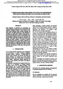
Figure 1. A Golgi-stained section of mouse cortex prepared by students in an undergraduate sophomore neuroscience class.
Golgi-Cox Procedure for Use in Undergraduate Courses
A86
Golgi-Cox solution in advance. When handling the stain solutions and treated brain tissue, two pairs of nitrile gloves should be worn in addition to lab coats and protective eye wear. Be sure to appropriately dispose of all chemical and dry waste in accordance with institutional regulations. We found that the use of a Vibratome allowed more consistent thin sections; however, to accommodate a large number of students either a handheld single-edge razor blade or two razorblades taped together is a sufficient way to cut and obtain sections of tissue.
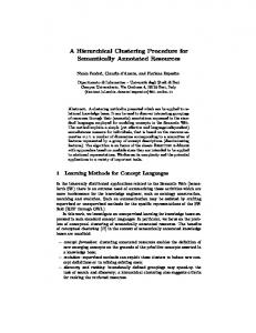
RESULTS We developed this protocol as a modification of the processing suggested in prior protocols (Paul et al., 1997), which suggested dehydration and clearing steps. By reducing the number of processing steps and using an aqueous mounting medium that eliminated the need for dehydrating the tissue, we condensed the processing time from approximately one hour to circa 15-20 minutes following cutting. The use of aqueous mounting media allowed the section to be viewed immediately after coverslipping. Our results (Figure 1, 2) were comparable to those we obtained using the longer processing method. The advantage is that students can cut and process tissue in a shorter period of time, and may have time remaining in the lab period to observe the sections. Alternatively, the instructor could fit this procedure into one extended lecture period. Slides that are intended for use in future years should be dehydrated and coverslipped according to the previously published procedure (Paul et al., 1997) for optimal results, but the aqueous mount maintains tissue quality for at least several months. We observed loss of tissue quality when slides were stored in aqueous mount for one year, perhaps due to difficulties coverslipping the slices of uneven thickness.
DISCUSSION We have described a modification of a Golgi-Cox procedure used in a teaching lab that shortens the processing time necessary following tissue sectioning. Our adapted Golgi method allows students to view stained brain sections and observe different types of neurons and
Figure 2. Golgi-stained pyramidal neuron at 40X magnification.
The Journal of Undergraduate Neuroscience Education (JUNE), Fall 2011, 10(1):A85-A87
glia in different brain areas. An undergraduate student reported “the stain itself was very clear and helpful in that we could see the complete neuron and its shape compared to other stains we have been taught. The Golgi staining process was very quick and we were able to cut our tissue and look at our slides all in one lab period” (Jennii Tran, Smith College Class of 2013). More advanced students could apply this technique to experimental questions, for example, asking if cells in different brain areas or across animals treated differently show quantifiable differences. One student doing a project on cell differentiation of hippocampal neurons paired the analysis of Golgi-stained sections with the use of ImageJ software (http://rsbweb.nih.gov/ij/) to allow quantification of images and stated that the proposed Golgi Staining method was “great because it was more focused on the structure of individual cells than other stains we have used. The procedure was much simpler than I thought it was going to be” (Grace McKay-Corkum, Smith College Class of 2014). The brain areas most popular for students have been hippocampus, cerebral cortex, striatum and cerebellum. These areas have interesting cytoarchitecture and easily accessible information in textbooks, encyclopedias and websites to help students begin to recognize the different regions and cell types. This lab procedure is useful when teaching basic brain anatomy, histological techniques and microscopy and has been used in several classes at Smith College with no difficulties. The toxic nature of the stain makes this procedure less suitable for use in high school or outreach settings, but undergraduate students have presented their slides and images to younger students interested in neuroscience in outreach activities.
REFERENCES Pannese E (2007) The contribution of Camillo Golgi to our understanding of the structure of the nervous system. Arch Ital Biol 145: 111-115. Paul CA, Beltz B, Berger-Sweeney J (1997) Golgi Sections of Adult Rat Brains. In Discovering neurons: the experimental basis of neuroscience (Paul et al., eds) pp 382-384. Cold spring Harbor, NY: Cold Spring Harbor Laboratory Press. Shankaranarayana BS, Raju TR (2004) The Golgi techniques for staining neurons. In Brain and behavior (Raju TR et al., eds) pp 108-111. Bangalore, India: National Institute of Mental Health and Neurosciences. Zhang H, Weng SJ, Hutsler JJ (2003) Does microwaving enhance the Golgi methods? A quantitative analysis of disparate staining patterns in the cerebral cortex. J Neurosci Methods 124:145-155. Received July 25, 2011; revised September 21, 2011; accepted September 27, 2011. This work was supported by Smith College CFCD and summer science stipends to KW. The authors thank the students in Neuroscience 230 for feedback on this lab exercise. Address correspondence to: Dr. Mary Harrington, Neuroscience Program, Smith College, Northampton, MA 01063. Email:
[email protected]
Copyright © 2011 Faculty for Undergraduate Neuroscience www.funjournal.org
A87