www.nature.com/scientificreports
OPEN
received: 28 September 2016 accepted: 26 January 2017 Published: 08 March 2017
A network-based method for mechanistic investigation of Shexiang Baoxin Pill’s treatment of cardiovascular diseases Hai-Yang Fang1,*, Hua-Wu Zeng2,*, Li-Mei Lin1, Xing Chen1, Xiao-Na Shen1, Peng Fu2, Chao Lv2, Qun Liu2, Run-Hui Liu2, Wei-Dong Zhang2 & Jing Zhao1,2 Shexiang Baoxin Pill (SBP), a traditional Chinese medicine formula, is commonly used to treat cardiovascular disease (CVD) in China. However, the complexity of composition and targets has deterred our understanding of its mechanism of action. Using network pharmacology-based approaches, we established the mechanism of action for SBP to treat CVD by analyzing protein-protein interactions and pathways. The computational results were confirmed at the gene expression level in microarraybased studies. Two of the SBP’s targets were further confirmed at the protein level by Western blot. In addition, we validated the theory that SBP’s plasma absorbed compounds play major therapeutic role in treating CVD. Cardiovascular disease (CVD) is a group of disorders that affect heart and blood vessels. It mainly includes coronary heart disease1, atherosclerosis2, myocardial ischemia3, myocardial fibrosis4, cerebrovascular disease5, peripheral arterial disease6, congenital heart disease, and deep vein thrombosis7,8. In comparison to common acute diseases (e.g., influenza), complex chronic diseases such as CVD cannot be completely cured. Drugs mainly regulate disease progression and relieve the symptoms. The treatment goal for these complex chronic diseases is to regulate the pathological state, thus controlling disease manifestation and retarding progression9. Shexiang Baoxin Pill (SBP) is a treasured traditional Chinese medicine (TCM) formula for cardiovascular diseases. In China, SBP has a long history of clinical application to treat coronary heart disease1, atherosclerosis2, myocardial ischemia3 and myocardial fibrosis4, with significant curative effects. Earlier studies investigated SBP’s activities in cells and animal models10–13. Recent studies revealed that SBP exerts a range of pharmacological activities, including increase the level of NO14, enhance the expression of endothelial NO synthase11, alleviate damages to blood vessels15, decrease MMP-2 expression16, and promote myocardial ischemia caused by myocardial necrosis12. Although SBP has been extensively evaluated for clinical efficacy, its mechanism of function remains unresolved. SBP is composed of seven medicinal materials, including Moschus, total gingenoside ginseng root, Styrax, Cinnamomi Cortex, Bufonis Venenum, Bovis Calculus Artifactus and Borneolum Syntheticum. Each ingredient contains a large number of chemical compounds. Like all TCM formulae, SBP is a multi-component and multi-target agent from the molecular perspective. As a result, its mode of action is most likely to be illustrated by network pharmacology-based approaches. Based on the TCM formula’s characteristics of complex components, unclear targets, and holistic regulation, we have proposed a workflow for network-based TCM pharmacology study9. It starts with the identification of compounds present in the TCM formula and corresponding targets, and ends with discovering the signaling pathways and sub-networks regulated by the TCM formula and evaluating its effects on disease network. Several studies have been conducted following this flow diagram and revealed the modes of action for different TCM formulae17.
1
Department of Mathematics, Logistical Engineering University, Chongqing, China. 2Department of Natural Medicinal Chemistry, Second Military Medical University, Shanghai, China. *These authors contributed equally to this work. Correspondence and requests for materials should be addressed to R.-H.L. (email:
[email protected]) or W.-D.Z. (email: wdzhangy@hotmail. com) or J.Z. (email:
[email protected]) Scientific Reports | 7:43632 | DOI: 10.1038/srep43632
1
www.nature.com/scientificreports/ It is clear that the identification of compounds contained in TCM formula constitutes the basis of network-based TCM pharmacology study. Two different strategies have been used regarding compound selection. Some studies included all of the identified compounds in every single component of the TCM formula. For example, Zhang et al. studied all 451 compounds from the five Chinese herbs of the TCM formula Wu-tou decoction18. On the other hand, considering that most TCM formulae are taken orally, it is assumed that only the compounds that appear in blood are biologically active. Therefore, other studies only included compounds detected in the plasma after the administration of the TCM formula19,20. We used this strategy to analyze the anti-rheumatic effects of the TCM formula Huang-Lian-Jie-Du-Tang (HLJDT), in which only 14 active compounds from HLJDT were examined21. However, very few studies have compared the effect of plasma-absorbed compounds of a TCM formula with that of a full compound set. This study is to investigate the mechanism of action for SBP to treat CVD using network pharmacology-based methods, and to elucidate if the plasma absorbed compounds are responsible for SBP’s anti-CVD therapeutic effects. Following extensive data mining, we assembled a list of CVD-related disease genes, two sets of SBP compounds, and their corresponding targets. The algorithm of random walk with restart (RWR) was applied to calculate the regulation scores of disease genes and target proteins to human protein-protein interaction (PPI) network and pathways, respectively. The computational results were verified by microarray-based experiments and two SBP targets were validated by Western blot.
Materials and Methods
Data preparation. CVD associated genes. DisGeNet is a plugin of Cytoscope22, which has collected various classes of disease associated genes. On the settings window of “Diseasegene Network” in Cytoscope, we set “select source” as “All”, “select association type” as “Any”, and “select disease class” as “Cardiovascular Disease”. In this way, we obtained 301 CVD associated genes (see Table S1 in Supplementary Material). All compounds included in SBP and their corresponding targets. SBP contains seven medicinal materials that include Moschus, total ginsenoside ginseng root, Styrax, Cinnamomi Cortex, Bufonis Venenum, Bovis Calculus Artifactus and Borneolum Syntheticum. We used several TCM databases, including TCMDS23, HIT24, TCM Database@Taiwan25, TCMID26 and 3D-MSDT27, to collect information about all compounds in SBP. Using the name of each SBP individual medicinal material as input, we searched these databases and acquired the names and chemical structures of compounds from the corresponding medicinal ingredient as output. The outputs from these databases were combined to assemble a list of all identified SBP compounds. There are 166 chemically distinct compounds in total (see Table S2 in Supplementary Material). These are considered as full compounds included in SBP. For each compound, we searched its putative targets in the following two databases: (1) Herbal Ingredients’ Targets Database (HIT)24: HIT is a comprehensive and fully curated database that contains information on the protein targets of compounds found in Chinese herbs. Based on over 3250 papers in the literature, HIT includes 1301 protein targets affected by 586 herbal compounds found in more than 1300 Chinese herbs (http://lifecenter sgst.cn/hit/). By searching the database with the name of the chemical compound, we will be able to obtain information on its target proteins if this compound is included in the HIT. (2) Search Tool for Interactions of Chemicals (STITCH)28: STITCH is a collective database from 1133 organisms (http://stitch.embl.de/), composed of interactions existing between 300,000 small molecules and 2.6 million proteins. The biggest asset of STITCH is the huge amount of data and the structural similarity comparison tool it provides. In the event that we cannot find the targets of a compound in HIT and STITCH, we can input its structure into STITCH and identify similar chemical molecules with similarity score greater than 0.9. Targets of these chemically similar compounds are considered putative targets of the queried compound. At the end, 522 distinct protein targets of SBP were obtained after integrating HIT and STITCH search results, in which 86 were identified from HIT, 416 were got from STITCH, and 20 are included in both of the databases (see Table S3 in Supplementary Material). SBP’s plasma absorbed compounds and their targets. Our earlier serum medicinal chemistry and pharmacokinetic studies identified 34 compounds in the plasma after oral administration of SBP14,29, 26 of which were the original chemical compounds found in SBP, and 8 of which were metabolites. Among the twenty six compounds from SBP, one is from Moschus, seven are from total gingenoside ginsenoside ginseng root, eight are from Bufonis Venenum, five are from Bovis Calculus Artifactus, three are from Styrax, and two are from Borneolum Syntheticum. Mining HIT and STITCH databases, we identified 113 distinct targets for these 26 original chemical compounds. Targets of the 8 metabolites cannot be found, thus they are not pursued in this study. The detailed information is provided in Table S4 of Supplementary Material. For convenience, we named all compounds included in SBP and SBP’s plasma absorbed compounds as SBPac and SBPpc, respectively. When studying TCM pharmacology at the molecular level, all compounds identified in a TCM formula are considered to be equivalent to the formula itself. Thus in this study, we think SBPac is equivalent to SBP itself. Protein-Protein Interaction Data. Protein-protein interactions in human genome were extracted from version 9.05 of STRING30, a weighted interaction database with physical and functional interactions that are integrated from multiple data sources (such as experimental data, computation-based prediction, and literature mining).
Scientific Reports | 7:43632 | DOI: 10.1038/srep43632
2
www.nature.com/scientificreports/ STRING gives each interaction a confidence score (which were normalized to the interval of [0, 1]) by a scoring system to weigh the reliability of the interaction. In order to construct a background PPI network with high confidence edges, we first filtered the STRING with threshold 0.9. Only interactions with weight above the threshold were selected for the newly constructed background network. We also checked whether CVD disease genes and SBP’s target proteins are covered in the new network. In case of existing disease or target genes that are included in the STRING but not in the new network, we lowered the threshold for these genes and their interactions, until all these genes were included in the background network. As a result, a weighted PPI background network with 9289 nodes and 57179 edges was obtained. Data of pathway gene sets. A total of 4722 pathway gene sets were derived from the C2:CP collection of MSigDB database31, which is a comprehensive integration of certain online pathway databases such as BioCarta, Reactome, and KEGG.
Microarray experiment and significantly expressed genes. Cell lines and treatment. The cell line we
used in this study is MCF7, one of the five cell lines used in connectivity map (CMAP) database as reference cells to connect biology, chemistry, and different clinical conditions including CVD32–34. MCF7 cells were obtained from American Type Culture Collection (ATCC) and maintained in MEM/EBSS (Hyclone), supplemented with 10% fetal bovine serum, 1 mM sodium pyruvate, 0.1 mM MEM non-essential amino acids, 100 unit/mL penicillin and 100 mg/mL streptomycin in a humidified environment under 5% CO2:95% Air at 37 °C. MCF7 cells were treated with SBP extract at 1μg/mL for 12 hours. Stock solutions of SBP were prepared in DMSO. Solvent (DMSO) treated cells were used as control, same as most microarray experiments32,35.
RNA isolation. Total RNA samples were extracted from MCF7 cells using Trizol reagent (Life technologies, Carlsbad, CA, US). The integrity and purity of total RNA samples were monitored by formaldehyde agarose gel electrophoresis, and quantified by spectrophotometry (NanoDrop, Wilmington, DE, USA). Microarray analysis. RNA samples were purified from cells using the QIAGEN RNeasy Kit (GmBH, Germany). Biological replica was included for each cell line. cRNA products, generated from the fragmentation of double-stranded cDNA and biotin-labeled cRNA, were pooled to perform microarray experiments using Affymetrix chip (Human U133 A 2.0), by Shanghai Biotechnology Corporation following the Affymetrix technical manual. The SBP-related microarray experiments yielded 621 differentially expressed genes, with mean expression ratios between SBPac group and control group greater than 4 or lower than 0.25. The detailed results are shown in Table S5 of Supplementary Material.
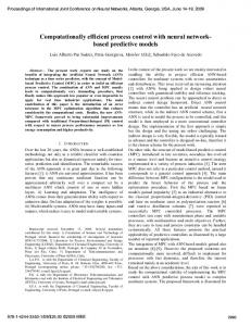
RWR-based evaluation of drug’s effect. The hypothesis for analyzing polygenic diseases and corresponding intervention in the molecule network is that genes and their products do not fulfill their biological functions independently but through prevalent interactions among different genes, and these interactions constitute a molecular network. Single gene alteration(s) incurred by diseases or drugs will affect its neighboring genes, forming a sub-network impacted by diseases or drugs. Figure 1 shows the flow diagram of determining a drug’s effect based on random walk with restart (RWR). RWR algorithm. Random walk with restart (RWR) is an improved algorithm derived from random walk algorithm36. It simulates a random walker that starts from nodes in the seed set. At any time, the walker can either randomly move to a neighboring node from current position or return to a seed node at a relatively low probability. RWR effectively measures the proximity of a node to seed set so that nodes adjacent to seed set can be prioritized. In view of this utility, RWR has been applied to predict potential disease genes by employing part of known disease genes to detect novel disease genes37–39. To be more specific, causative disease genes are considered as seed set while candidate genes in the PPI network are ranked based on their proximity to seed set scored by RWR in descending order. The genes with highest score are most likely to be disease-relevant. In this study, disease genes and drug target genes were selected as seed nodes, respectively. The extent of each gene in the network affected by the disease or drug is determined in way of scoring each gene by RWR. In general, the higher score a gene receives, the more pronounced it is affected by the disease or drug. Specifically, a node’s score at the t + 1 step is calculated as following: x t +1 = (1 − r ) Px t + rx0
(1)
P is the column-normalized adjacent matrix of the PPI network that represents the connectivity between nodes, x0 is the initial probability vector indicating a preference to seed set, and r is the restart probability that generally has a low value. For a non-bipartite and undirected graph, the equation is deemed to converge. Thus, node scores can be obtained after performing the iteration in equation (1). It is obvious that the closer the nodes connected to the seed set, the higher the scores, and vice versa. The r in the equation (1) is a parameter that varies with practical prioritization needs. In order to choose the optimal r, Erten et al. altered r values to determine how RWR affects candidates’ rank39. It was found that an r value of 0.3 outperforms others. Thus, r denoted by 0.3 is generally adopted in the ranking of sound disease genes. We set r as 0.3 in this research. Scoring disease’s effect on human PPI network. Scoring a disease’s effect on human PPI network is a process that estimates the proximity between each gene and disease genes by RWR algorithm. Disease-associated genes are viewed as the seed set. If there are m diseases genes in total, the corresponding initial component of a disease Scientific Reports | 7:43632 | DOI: 10.1038/srep43632
3
www.nature.com/scientificreports/
Figure 1. Flow diagram to evaluate the effect of a drug based on RWR.
gene in equation (1) is x0(i) = 1 /m, otherwise, x0(i) = 0. By iterating equation (1), the steady solution xdisease that represents the disease’s effect on human PPI network is obtained. Scoring drug’s effect on human PPI Network. To obtain the scores measuring the effect(s) of a drug on the background network, drug target proteins are used as seed set. If gene i is the target gene of active ingredients in the TCM formula, its corresponding component in the initial vector is set as x0(i) = 0.01, otherwise x0(i) = 0. The reason for the initial vector selection is that active ingredients in TCM formula are natural products with generally weaker effects on target proteins, in comparison to chemically defined drug molecules. Moreover, our previous study found that, the FDA-approved drug has around two orders of inhibition effect on target proteins ACE and CN than the natural compound Astragaloside, when applied at the same dose40. For each drug, their respective effect scores on PPI network xdrug are acquired with the equation (1). In order to evaluate TCM formulae in a statistically significant fashion, we set up 1000 random counterpart sets of target proteins, each of which includes the same number of proteins randomly selected from the network. We rerun RWR process on each of these random seed sets. Scoring effects of a drug to disease. In the network, the easier a node can be influenced by the seed set, the higher the score it will gain and vice versa. Therefore, genes with high scores and ranked above a defined threshold form a disease-affected sub-network that is vulnerable to diseases. Similar definition is used to establish a drug-affected sub-network. The extent of overlap between the two sub-networks is used to predict whether the drug can effectively exert its influence on the disease. This overlap can be quantitatively measured by an inner product of xdisease and xdrug so as to evaluate the performance of the drug in the treatment of the disease41. The equation is s = < x disease , x drug >
(2)
Scoring effects of a drug or disease on pathways. Signaling pathways are relatively independent biomolecule subsystems or interaction sub-networks that carry out important biological tasks and complex information processing between molecules. Scoring signaling pathways allows us to identify biological processes influenced by diseases or regulated by drugs. As shown in Fig. 2, we score the effects of a drug or disease on pathways by mapping network scores of genes calculated onto the pathway gene sets. The details are provided below. (1) Using RWR to score the effect(s) of a disease or drug on all genes in the PPI network; (2) Mapping gene scores generated in step (1) to the corresponding genes in pathways. For genes on the pathways Scientific Reports | 7:43632 | DOI: 10.1038/srep43632
4
www.nature.com/scientificreports/
Figure 2. Map network genes’ scores to the pathway gene set.
that are not included in the background network, their scores are assigned as zero (Fig. 2). (3) Adding up the scores in each pathway and denoting the average scores as disease’s or drug’s effect scores on the pathway. Evaluation measures. To determine if the TCM formula exerts significant effect(s) on the network, we generate a wide spectrum of random seed sets (e.g., 1000 sets). Each seed set is randomly generated, containing the same number of target proteins as that of the original seed set. For each random counterpart, we compute its effect scores on each gene of the background network by equation (1). Here Z-score is introduced to quantify the score difference existing between the original seed set and its counterparts. It is expressed as follows: Z=
S − Sr ∆S r
(3)
where S is the anti-disease effect score of the TCM formula, while Sr and ∆Sr represent mean and standard deviation of the scores stemming from random counterparts, respectively. Then, for each disease and drug, we take pathways with highest 5% scores, respectively. These are considered as pathways significantly influenced by the disease or regulated by the drug. The common pathways influenced by the disease and drug are used to measure drug efficacy from the biological perspective.
Target validation by Western blot. Drug preparation. SBP samples were ground into fine powder. Four grams of accurately weighed sample powder were transferred to a 250 mL round bottom flask, extracted with 52 mL mixed solvent (methanol: dichlormethane: water = 4: 2: 0.5, v/v) by ultrasonic extraction (30 min), filtered, and the filtrate collected. Detailed extraction method was described in the ref. 42. The filtrated extract sample was dried by rotation evaporation under 55 °C. A stock solution of 50 mg/mL was prepared by dissolving 5 mg extract sample in 100 μL DMSO. A total number of 26 SBP compounds detected in plasma were used to prepare SBPpc. Specifically, the stock solution of SBP’s plasma absorbed compounds was prepared by dissolving the mixture of all 26 compounds in DMSO at a concentration of 100 mg/mL (see Table S6 of Supplementary Material for contents of the compounds in the solution). Western blot analysis in Human Umbilical Vein Endothelial Cells. Human Umbilical Vein Endothelial Cells (HUVEC) were purchased from AllCells (Emeryville, CA, USA). The antibodies used for Western blot analysis are: rabbit anti-human intercellular adhesion molecule-1 (ICAM1, Abcam, Cambridge, MA, USA), rabbit anti-human cyclooxygenase 2 (COX-2, Cell Signaling Technology, Boston, MA, USA), and mouse anti-human GAPDH (Beyotime, Shanghai, China). Human tumor necrosis factor alpha (TNFα) was from PeproTech (Rocky Hill, NJ, USA). HUVECs were seeded into 60 mm cell culture plates with complete endothelial growth medium (AllCells). After reaching 80% confluence, cells were exposed to SBP extract and SBPpc in the presence or absence of TNFα (25 ng/ml) for 16 hours. The conditioned media were removed, HUVECs washed and lysed, and protein concentrations determined using the BCA protein assay kit (Beyotime, Shanghai, China). The lysate samples were stored in −80 °C. Scientific Reports | 7:43632 | DOI: 10.1038/srep43632
5
www.nature.com/scientificreports/ No
Gene Symbol
Gene function
1
COL1A1
Platelet-derived growth factor binding protein44
2
CYP2C9
Heme binding protein45
3
ESR2
4
ICAM1
5
LDLR
Receptor of low-density lipoprotein (plasma cholesterol apolipoprotein)48
MMP9
May play an essential role in local proteolysis of the extracellular matrix and in leukocyte migration49.
7
NOS3
Modulate angiogenesis, blood pressure, and calcium channel50
8
COX-2
Regulate vasoconstriction and blood pressure51
9
TGFB1
Regulation of vascular endothelial cell migration52
10
TLR4
11
VCAM1
6
A group of heme-thiolate monooxygenases46 Ligands for the leukocyte adhesion protein LFA-1 (integrin alpha-L/beta-2)47
Immune response and inflammatory process regulation53 Involved in calcium-mediated signaling pathways of intracellular calcium source54
Table 1. CVD disease genes targeted by SBP’s plasma absorbed compounds (SBPpc). Name
Target number
Effect score
Z-score
SBP’s all compounds (SBPac)
522
0.4452
12.1930
SBP’s plasma absorbed compounds (SBPpc)
113
0.1138
7.7493
Table 2. The effect scores of SBPac and SBPpc on CVD.
Samples containing equal amounts of proteins were separated by SDS-PAGE electrophoresis (10% SDS-polyacrylamide gel), and transferred onto polyvinyl membranes. The membranes were blocked with 5% dry milk in TRIS-buffered saline containing 0.1% Tween 20 (room temperature, 1 h), incubated with the specified primary antibodies (dilutions: COX-2 1:500, ICAM-1 1:1000, and GAPDH 1:1000) overnight at 4 °C. The membranes were visualized using Odessey (LI-COR, Lincoln, NE, USA).
Results and Discussion
Overlap of CVD disease genes with SBP’s target genes. From the pharmaceutical perspective, pro-
teins encoded by disease genes may serve as potential drug targets for treatment. Earlier network analysis of the DrugBank database revealed that certain FDA approved drugs, including those that treat cardiovascular diseases, preferentially target disease genes43. To determine if SBP acts in a similar manner, we counted the number of CVD disease genes that are also SBP target genes. Among the 522 target genes of SBPac, 46 are CVD disease genes, which are 15.3% of the total disease genes. However, SBPpc targeted only 11 CVD disease genes, ~3.7% of the total disease genes. Table 1 summarizes the 11 common genes and their functions. All 11 genes are associated with the functions of heart or blood vessels that are important in the etiology of CVD. Given the fact that there are 301 CVD associated genes, targeting 46 or 11 out of 301 genes is not sufficient to explain SBP’s efficacy in treating CVD.
Network analysis of SBP on CVD. To quantitatively display the effect of SBP on CVD treatment from a network perspective, we applied RWR algorithm to measure the impact of seed nodes on other nodes in the background network. By setting CVD disease genes, target genes of SBPac and SBPpc as seed nodes, respectively, we first used equation (1) to calculate the effect score vectors of CVD, SBPac, and SBPpc on the background network. Equation (2) was subsequently applied to calculate the inner products between affected vectors of CVD and SBPac, CVD and SBPpc. The network effect scores of SBPac and SBPpc on CVD were 0.4452 and 0.1138, respectively. To determine whether the network effect scores of the drug to disease are significant, we compared their scores with the scores obtained from random seed sets. Based on the 522 targets of SBPac and 113 targets of SBPpc, we generated 1000 random target gene sets from the background network, respectively. Each of these sets contained the same number of targets as SBPac and SBPpc, respectively. The scores from random seed sets could be obtained using equation (1) and (2). The average and standard deviation values of effect scores for the 1000 random counterparts of each group were calculated. The Z-scores of effect scores of SBPac and SBPpc to CVD were obtained using the equation (3). As shown in Table 2, the Z-score values are 12.193 and 7.749 of SBPac and SBPpc, suggesting that the sub-networks regulated by them overlap significantly with CVD impacted sub-network. This is because a Z-score value greater than 3 often indicates a statistically significant deviation between the actual value and the random ones. Therefore, we can conclude that both SBPac and SBPpc have significant effects to CVD. Pathways significantly regulated by SBP. We mapped the network scores of genes from the calculation
above to the 4722 pathways from the C2: CP collection of MSigDB database. For those genes on the pathways that cannot be mapped, their scores were assigned zero. The average value of all genes on each pathway was obtained. Using this approach, we acquired effect scores of CVD disease genes, SBPac and SBPpc’ target genes on each pathway, respectively.
Scientific Reports | 7:43632 | DOI: 10.1038/srep43632
6
www.nature.com/scientificreports/
Figure 3. The Venn diagram of coincide pathway number from CVD, SBPac and SBPpc regulated pathways.
We defined pathways significantly regulated by the disease or drug as those with scores ranked top 5% of the 4722 pathways. This classification led to 236 pathways affected by CVD, SBPac and SBPpc, respectively. Among the 236 pathways significantly impacted by CVD, 113 and 97 are also regulated by SBPac and SBPpc (Fig. 3), constituting 47.8% and 41% of the CVD pathways, respectively. The observation that nearly half of CVD affected pathways are regulated by SBP supports the therapeutic effect of SBP on CVD. There are 81 pathways that are significantly affected by CVD and regulated by SBPac and SBPpc both, constituting 71.7% of the SBPac regulated CVD affected pathways. Since SBPac is considered as equivalent to SBP itself, these results suggest that, SBP’s plasma absorbed compounds play major roles in SBP’s treatment of CVD.
Microarray experiment validation. Microarray-based studies were performed to verify the network analysis results. A total of 621 genes with mean expression ratios of SBP group to control group greater than 4 or less than 0.25, were considered differentially expressed genes. Among these 621 genes, only 31 and 9 genes are target genes of SBPac and SBPpc, respectively, identified by the bioinformatics method. Meanwhile, 29 of the 621 genes are CVD disease genes, among which, two genes ICAM1 and COX-2 are also target genes of SBPac and SBPpc. Both ICAM1 and COX-2 are important disease genes associated with CVD. The 621 significantly expressed genes were set as the seed nodes and the corresponding component of the gene in the initial vector as normalized differential expression ratio. Using approach similar to that described earlier, we calculated the differentially expressed genes’ network effect scores on CVD and on each of the 4722 pathways from the C2: CP collection of MSigDB database. This led to the identification of 236 pathways significantly regulated by these 621 genes with scores in the top 5%. Among them, 119 pathways are affected by CVD, which is 50.4% of all the 236 pathways. These gene expression data further support the therapeutic effect of SBP on CVD. We compared the 113 and 97 CVD affected pathways, which are also regulated by SBPac and SBPpc, with the 119 CVD affected pathways regulated by significantly expressed genes under SBP treatment, respectively. It was found that 80 of the 113 SBPac affected pathways and 70 of the 97 SBPpc affected pathways also appeared in these 119 pathways, corresponding to 67.2% and 58.8%, respectively. Specifically, 63 CVD affected pathways are also regulated by SBPac, SBPpc, and altered gene expression significantly upon SBP treatment. These results validate the reliability of SBP’s target genes and their regulating pathways identified by our bioinformatics methods. Figure 4 shows the number of genes in different intersection sets of the four gene sets, and the number of pathways affected by genes in these intersections. It could be seen that, although SBP directly act on only a few CVD disease genes, through network interactions, SBPac, SBPpc, and significantly expressed genes under SBP treatment regulate a greater number of common CVD-related signaling pathways. Although the 11 CVD genes targeted by SBPpc (AC in Fig. 4) are only 24% of the 46 CVD genes targeted by SBPac (AB in Fig. 4), the number of CVD associated pathways regulated by SBPpc (AC in Fig. 4, 97) is 86% of the number of the CVD associated pathways regulated by SBPac (AB in Fig. 4,113). Meanwhile, only few CVD disease genes targeted by SBPac and SBPpc were validated in the gene expression experiment, i.e., 6 and 2 genes, respectively (ABD and ACD in Fig. 4). In contrast, more CVD affecting pathways regulated by SBPac and SBPpc were verified in the gene expression study, i.e., 80 and 70 pathways, respectively (ABD and ACD in Fig. 4). That is to say, about 70% CVD associated pathways affected by SBPac and SBPpc were validated in the microarray-based studies. These results suggest that SBP regulates CVD associated network through multi-components and multi-targets to achieve its clinical efficacy. The high consistency between the pathways identified by the bioinformatics methods and the microarray confirmation studies exemplifies the reliability of our data mining and
Scientific Reports | 7:43632 | DOI: 10.1038/srep43632
7
www.nature.com/scientificreports/
Figure 4. Comparison of the gene number in different intersection sets of the four gene sets and the number of pathways affected by corresponding genes, where (A, B, C and D) denote CVD disease genes, SBPac’s target genes, SBPpc’s target genes, and significantly expressed genes upon SBP treatment, respectively. AB denotes the intersection set between A and B, and so on.
ID
Pathway Name
The association with CVD
1
AT1R_PATHWAY
Vasodilator and myocardial hypertrophy55
2
SPPA_PATHWAY
Vasoconstriction and platelet activation
3
EPO_PATHWAY
Increasing erythrocyte56
4
HIF_PATHWAY
Vascular remodeling and thrombopoietin57
5
IL4_PATHWAY
Hematopoietic association58
6
IL6_PATHWAY
Hematopoietic association59
7
CARDIACEGF_PATHWAY
Cardiac hypertrophy and angiotensin60
8
TPO_PATHWAY
Thrombopoietin61
9
ANGIOPOIETINRECEPTOR_PATHWAY
Angiopoietin62
10
S1P_S1P1_PATHWAY
Endothelial cell induction63
11
VEGFR1_PATHWAY
Vascular endothelial growth factor receptor64
12
LYMPHANGIOGENESIS_PATHWAY
Angiogenesis65
13
HEMATOPOIESIS_STAT3_TARGETS
Hematopoietic66
Table 3. List of selected CVD affecting pathways regulated by both SBP and SBP’s plasma absorbed components. analytical methods. It also reveals that SBP’s plasma absorbed components exert nearly the same function as that of SBP’s full compound set for CVD treatment. Among the 63 experimentally validated CVD pathways regulated by both SBPac and SBPpc (ABCD in Fig. 4, Table S7 of Supplementary Material), most are directly associated with CVD, further supporting the effectiveness of SBP to treat CVD. Table 3 lists some of these pathways that are associated with CVD.
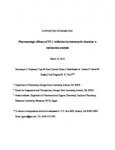
Target validation in cell-based studies. Based on above analysis, genes ICAM-1 and COX-2 are included in four different gene sets under study, i.e., CVD disease genes, SBPac’s target genes, SBPpc’s target genes, and significantly expressed genes under SBP treatment. Moreover, their encoded proteins are readily available. Thus we chose protein ICAM-1 and COX-2 for experimental validation. To validate SBP’s effects on ICAM-1 and COX-2, we conducted Western blot experiment in HUVECs. As shown in Fig. 5, SBP extract (1 μg/ml) and SBPpc (0.1 μg/ml) each induced the expression of COX-2 protein while inhibiting the expression of TNFα-induced ICAM-1 protein. Both ICAM-1 and COX-2 are expressed in endothelial cells and play important roles in the initiation and progression of cardiovascular disease. ICAM-1 facilitates the transmigration of leukocytes across vascular wall during inflammatory response. COX-2 converts arachidonic acid to various metabolites, including prostacyclin (PGI2), a major COX-2 metabolite found in endothelial cells and an effective vasodilator. These observations support the notion that SBP and its plasma absorbed compounds (SBPpc) exert their anti-CVD effects by vasodilation and anti-inflammatory action, through the up-regulation of COX-2 and down-regulation of ICAM-1.
Conclusions
This study investigated the therapeutic effect of SBP on CVD in depth using a network pharmacological approach. First, we calculated the network effect score of disease and drug on genes in the background network and further
Scientific Reports | 7:43632 | DOI: 10.1038/srep43632
8
www.nature.com/scientificreports/
Figure 5. Expression of ICAM-1 and COX-2 proteins in HUVECs treated with SBP extract and SBPpc samples for 16 hours in the presence or absence of TNFα (25 ng/ml). Specific proteins were detected by Western blot as described in the Material and Methods. Each blot is representative of three independent experiments.
analyzed the score’s significance to determine whether SBP significantly impacts CVD. Second, we mapped the scores of all genes in the background network obtained through RWR, to 4722 pathways and calculated the regulation scores of CVD, all compounds of SBP and plasma absorbed compounds of SBP to these pathways. As a result, we identified biological processes significantly regulated by SBP’s different compound sets and found most of them are associated with CVD. Finally, we validated our bioinformatics results in microarray- and Western blot-based studies. We found that, unlike FDA approved CVD drugs that mainly target specific disease genes, SBP only targets a very small fraction of CVD disease genes. However, from the perspective of network regulation, the sub-network regulated by SBP and most signaling pathways significantly regulated by SBP are overlapped with those influenced by CVD disease genes, respectively. These results suggest that SBP achieves its efficacy on CVD treatment by regulating the disease network though interactions between genes, instead of directly acting on CVD disease genes. When compared to the anti-CVD effects of all compounds included in SBP, SBP’s plasma absorbed compounds acquired significant anti-CVD effect score. Approximately 70% of CVD associated pathways affected by both SBP’s full compound set and SBP’s plasma absorbed compound set were validated in microarray-based studies, indicating that SBP’s plasma absorbed compounds play a major role in SBP’s treatment of CVD. Finally, two important CVD associated disease genes that are targeted by SBP were validated at the protein expression level by Western blot. In summary, this study applied a network pharmacology approach to determine SBP’s anti-CVD effect. It shed lights on the modern approaches to investigate TCM pharmacology and to promote the development of traditional medicine.
References
1. Wang, L., Luo, X. & Wang, Y. Evaluation on tolerability and safety of long-term administration with shexiang baoxin pill in patients with coronary heart disease of stable angina pectoris. Chinese journal of integrated traditional and Western medicine 28, 399–401 (2008). 2. Li, T.-q., Li, Y. & Fan, W.-h. Effects of She Xiang Bao Xin Pill and simvastatin on stability of atherosclerotic plaque in rabbit femoral artery. Chinese Journal of Geriatric Heart Brain and Vessel Diseases 5, 002 (2006). 3. Shen, W., Fan, W. & Shi, H. Effects of shexiang baoxin pill on angiogenesis in atherosclerosis plaque and ischemic myocardium. Chinese journal of integrated traditional and Western medicine 30, 1284–1287 (2010). 4. Wu, D., Hong, H. & Jiang, Q. Effect of shexiang baoxin pill in alleviating myocardial fibrosis in spontaneous hypertensive rats. Chinese journal of integrated traditional and Western medicine 25, 350–353 (2005). 5. Kario, K. et al. Morning surge in blood pressure as a predictor of silent and clinical cerebrovascular disease in elderly hypertensives a prospective study. Circulation 107, 1401–1406 (2003). 6. Ouriel, K. Peripheral arterial disease. The lancet 358, 1257–1264 (2001). 7. Mendis, S., Puska, P. & Norrving, B. Global atlas on cardiovascular disease prevention and control. (World Health Organization, 2011). 8. Collaborators, M. C. O. D. Global, regional, and national age-sex specific all-cause and cause-specific mortality for 240 causes of death, 1990–2013: a systematic analysis for the Global Burden of Disease Study 2013. Lancet 41, 119 (2015). 9. Zhao, J., Jiang, P. & Zhang, W. Molecular networks for the study of TCM pharmacology. Briefings in Bioinformatics 11, 417–430 (2010). 10. Jiang, P. et al. Potential biomarkers in the urine of myocardial infarction rats: a metabolomic method and its application. Molecular Biosystems 7, 824–831 (2011). 11. Xiang, L. et al. The serum metabolomic study of intervention effects of the traditional Chinese medicine Shexiang Baoxin Pill and a multi-component medicine polypill in the treatment of myocardial infarction in rats. Molecular Biosystems 8, 2434–2442 (2012). 12. Jiang, P. et al. Biomarkers in the early period of acute myocardial infarction in rat serum and protective effects of Shexiang Baoxin Pill using a metabolomic method. Journal of Ethnopharmacology 138, 530–536 (2011). 13. Xiang, L. et al. Metabolomic strategy for studying the intervention and the synergistic effects of the shexiang baoxin pill for treating myocardial infarction in rats. Evidence-based complementary and alternative medicine: eCAM 2013, 823121–823121 (2013). 14. Jiang, P. et al. Analysis of the constituents in rat plasma after oral administration of Shexiang Baoxin pill by HPLC-ESI-MS/MS. Biomedical Chromatography 23, 1333–1343 (2009). 15. Quan, Y. & Zhang, Y.-m. Efect of Shexiang Baoxin Wan on blood vessel endothelial function in patients with atrial fibrillation complicated with hypertension. Central South Pharmacy 10, 010 (2011). 16. Xuezhong, W., Ping, Z. & Yuesong, W. The Expression of MMP-2 and the Effect of Shexiangbaoxin Pill on It in DHR Heart. Journal of Emergency in Traditional Chinese Medicine 8, 060 (2006).
Scientific Reports | 7:43632 | DOI: 10.1038/srep43632
9
www.nature.com/scientificreports/ 17. Shao, L. & Zhang, B. Traditional Chinese medicine network pharmacology: theory, methodology and application. Chinese journal of natural medicines 11, 110–120 (2013). 18. Zhang, Y. et al. Uncovering pharmacological mechanisms of Wu-tou decoction acting on rheumatoid arthritis through systems approaches: drug-target prediction, network analysis and experimental validation. Scientific reports 5 (2015). 19. Wang, X. et al. Pharmacokinetics screening for multi-components absorbed in the rat plasma after oral administration traditional Chinese medicine formula Yin-Chen-Hao-Tang by ultra performance liquid chromatography-electrospray ionization/quadrupoletime-of-flight mass spectrometry combined with pattern recognition methods. Analyst 136, 5068–5076 (2011). 20. Yan, Z., Chen, Y., Li, T., Zhang, J. & Yang, X. Identification of metabolites of Si-Ni-San, a traditional Chinese medicine formula, in rat plasma and urine using liquid chromatography/diode array detection/triple–quadrupole spectrometry. Journal of Chromatography B 885, 73–82 (2012). 21. Fang, H. et al. Bioinformatics analysis for the antirheumatic effects of huang-lian-jie-du-tang from a network perspective. EvidenceBased Complementary and Alternative Medicine 2013 (2013). 22. Piñero, J. et al. DisGeNET: a discovery platform for the dynamical exploration of human diseases and their genes. Database 2015, 59P (2015). 23. Ren, T., Liu, X. & Gao, J. Introduction of “basic database system of traditional Chinese medicine”. Journal of traditional Chinese Medicine 8, 90–91 (2001). 24. Ye, H. et al. HIT: linking herbal active ingredients to targets. Nucleic acids research 39, D1055–D1059 (2011). 25. Chen, Y. C. TCM Database@Taiwan: The World’s Largest Traditional Chinese Medicine Database for Drug Screening In Silico. Plos One 6, e15939–e15939 (2011). 26. Xue, R. et al. TCMID: traditional Chinese medicine integrative database for herb molecular mechanism analysis. Nucleic Acids Research 41, D1089–D1095 (2013). 27. Qiao, X., Hou, T., Zhang, W., Guo, S. L. & Xu, X. ChemInform Abstract: A 3D Structure Database of Components from Chinese Traditional Medicinal Herbs. ChemInform 33, 481–489 (2002). 28. Kuhn, M., von Mering, C., Campillos, M., Jensen, L. J. & Bork, P. STITCH: interaction networks of chemicals and proteins. Nucleic acids research 36, D684–D688 (2008). 29. Li-ana, G. et al. Identification of volatility components of Shexiang Baoxin pill in rat plasma by GC-MS [J]. Journal of Pharmaceutical Practice 3, 014 (2012). 30. Mering, C. V. et al. STRING: a database of predicted functional associations between proteins. Nucleic Acids Research 31, 258–261 (2003). 31. Liberzon, A. et al. Molecular signatures database (MSigDB) 3.0. Bioinformatics 27, 1739–1740 (2011). 32. Lamb, J. et al. The Connectivity Map: using gene-expression signatures to connect small molecules, genes, and disease. Science 313, 1929–1935 (2006). 33. Lamb, J. The Connectivity Map: a new tool for biomedical research. Nature Reviews Cancer 7, 54–60 (2007). 34. Qu, X. A. & Rajpal, D. K. Applications of Connectivity Map in drug discovery and development. Drug Discovery Today 17, 1289–1298 (2012). 35. Zhining Wen, Z. W., Steven, Wang, Ranadheer, Ravula, Lun, Yang, Jun, Xu, Charles, Wang, Zhong, Zuo, Moses S. S., Chow, Leming, Shi & Ying, Huang. Discovery of Molecular Mechanisms of Traditional Chinese Medicinal Formula Si-Wu-Tang Using Gene Expression Microarray and Connectivity Map. Plos One 6, 1118–1136 (2011). 36. Tong, H., Faloutsos, C. & Pan, J.-Y. Random walk with restart: fast solutions and applications. Knowledge and Information Systems 14, 327–346 (2008). 37. Macropol, K., Can, T. & Singh, A. K. RRW: repeated random walks on genome-scale protein networks for local cluster discovery. BMC bioinformatics 10, 1 (2009). 38. Köhler, S., Bauer, S., Horn, D. & Robinson, P. N. Walking the interactome for prioritization of candidate disease genes. The American Journal of Human Genetics 82, 949–958 (2008). 39. Erten, M. S. Network based prioritization of disease genes. Case Western Reserve University (2010). 40. Zhao, J. et al. Therapeutic effects of astragaloside IV on myocardial injuries: multi-target identification and network analysis. PLoS One 7, e44938 (2012). 41. Colinge, J., Rix, U. & Superti-furga, G. Novel global network scores to analyze kinase inhibitor profiles. (2010). 42. Peng, J. et al. Identification of multiple constituents in the TCM-formula Shexiang Baoxin Pill by LC coupled with DAD-ESI-MSMS. Chromatographia 70, 133–142 (2009). 43. Yıldırım, M. A., Goh, K.-I., Cusick, M. E., Barabási, A.-L. & Vidal, M. Drug-target network. Nature Biotechnology 25, 1119–1126 (2007). 44. Elia, L. & Condorelli, G. RNA (Epi) genetics in cardiovascular diseases. Journal of molecular and cellular cardiology 89, 11–16 (2015). 45. Karaźniewicz-Łada, M. et al. Clinical pharmacokinetics of clopidogrel and its metabolites in patients with cardiovascular diseases. Clinical pharmacokinetics 53, 155–164 (2014). 46. Rexrode, K. M. et al. Polymorphisms and haplotypes of the estrogen receptor-βgene (ESR2) and cardiovascular disease in men and women. Clinical chemistry 53, 1749–1756 (2007). 47. Demerath, E., Towne, B., Blangero, J. & Siervogel, R. The relationship of soluble ICAM-1, VCAM-1, P-selectin and E-selectin to cardiovascular disease risk factors in healthy men and women. Annals of human biology 28, 664–678 (2001). 48. Van Gaal, L. F., Mertens, I. L. & Christophe, E. Mechanisms linking obesity with cardiovascular disease. Nature 444, 875–880 (2006). 49. Chen, K.-C. et al. OxLDL up-regulates microRNA-29b, leading to epigenetic modifications of MMP-2/MMP-9 genes: a novel mechanism for cardiovascular diseases. The FASEB Journal 25, 1718–1728 (2011). 50. Álvarez, R. et al. Association between the NOS3 (−786 T/C) and the ACE (I/D) DNA genotypes and early coronary artery disease. Nitric Oxide 5, 343–348 (2001). 51. Skarke, C. et al. Pyrosequencing of polymorphisms in the COX-2 gene (PTGS2) with reported clinical relevance. Pharmacogenomics 8, 1643–1660 (2007). 52. Azhar, M. et al. Transforming growth factor beta in cardiovascular development and function. Cytokine & growth factor reviews 14, 391–407 (2003). 53. Stoll, L. L., Denning, G. M. & Weintraub, N. L. Endotoxin, TLR4 signaling and vascular inflammation: potential therapeutic targets in cardiovascular disease. Current pharmaceutical design 12, 4229–4245 (2006). 54. Hwang, S.-J. et al. Circulating adhesion molecules VCAM-1, ICAM-1, and E-selectin in carotid atherosclerosis and incident coronary heart disease cases the Atherosclerosis Risk In Communities (ARIC) study. Circulation 96, 4219–4225 (1997). 55. Zhao, Y. et al. Angiotensin II/angiotensin II type I receptor (AT1R) signaling promotes MCF-7 breast cancer cells survival via PI3kinase/Akt pathway. Journal of cellular physiology 225, 168–173 (2010). 56. Sasaki, A. et al. CIS3/SOCS-3 suppresses erythropoietin (EPO) signaling by binding the EPO receptor and JAK2. Journal of Biological Chemistry 275, 29338–29347 (2000). 57. Ziello, J. E., Jovin, I. S. & Huang, Y. Hypoxia-Inducible Factor (HIF)-1 regulatory pathway and its potential for therapeutic intervention in malignancy and ischemia. Yale J Biol Med 80, 51–60 (2007). 58. Khaled, W. T. et al. The IL-4/IL-13/Stat6 signalling pathway promotes luminal mammary epithelial cell development. Development 134, 2739–2750 (2007). 59. Grivennikov, S. & Karin, M. Autocrine IL-6 signaling: a key event in tumorigenesis? Cancer cell 13, 7–9 (2008).
Scientific Reports | 7:43632 | DOI: 10.1038/srep43632
10
www.nature.com/scientificreports/ 60. Zhao, L., Yang, G. & Zhao, X. Rho-Associated Protein Kinases Play an Important Role in the Differentiation of Rat Adipose-Derived Stromal Cells into Cardiomyocytes In Vitro. PloS one 9, e115191 (2014). 61. Chanprasert, S., Geddis, A. E., Barroga, C., Fox, N. E. & Kaushansky, K. Thrombopoietin (TPO) induces c-myc expression through a PI3K-and MAPK-dependent pathway that is not mediated by Akt, PKC ζor mTOR in TPO-dependent cell lines and primary megakaryocytes. Cellular signalling 18, 1212–1218 (2006). 62. Fukuhara, S. et al. Angiopoietin-1/Tie2 receptor signaling in vascular quiescence and angiogenesis. Histology & Histopathology 25, 387–396 (2010). 63. Rosen, H. & Goetzl, E. J. Sphingosine 1-phosphate and its receptors: an autocrine and paracrine network. Nature Reviews Immunology 5, 560–570 (2005). 64. Roberts, D. M. et al. The vascular endothelial growth factor (VEGF) receptor Flt-1 (VEGFR-1) modulates Flk-1 (VEGFR-2) signaling during blood vessel formation. The American journal of pathology 164, 1531–1535 (2004). 65. Wissmann, C. & Detmar, M. Pathways targeting tumor lymphangiogenesis. Clinical Cancer Research 12, 6865–6868 (2006). 66. Kortylewski, M. et al. Inhibiting Stat3 signaling in the hematopoietic system elicits multicomponent antitumor immunity. Nature medicine 11, 1314–1321 (2005).
Acknowledgements
The work was supported by Professor of Chang Jiang Scholars Program, National Natural Science Foundation of China (61372194, 81260672, 8130090, 81473327, 81520108030, 81573318, 81373301, 81302658), Shanghai Engineering Research Center for the Preparation of Bioactive Natural Products (10DZ2251300), the Scientific Foundation of Shanghai China (12401900801, 13401900103, 13401900101), National Major Project of China (2011ZX09307-002-03), the National Key Technology R&D Program of China (2012BAI29B06), Chongqing Education Reform Project of Graduate (yjg152017), and the Graduate student research and innovation funding project of Chongqing China (CYB16129).
Author Contributions
J.Z., W.-D.Z. and R.-H.L. devised and guided the research project. X.C. and X.-N.S. collected the original data, H.-W.Z., P.F., Q.L and C.L. implemented biological experiments. H.-Y.F. did computation. H.-Y.F., L.-M.L. and J.Z. analyzed the results. H.-Y.F. and L.-M.L. wrote the paper.
Additional Information
Supplementary information accompanies this paper at http://www.nature.com/srep Competing Interests: The authors declare no competing financial interests. How to cite this article: Fang, H.-Y. et al. A network-based method for mechanistic investigation of Shexiang Baoxin Pill’s treatment of cardiovascular diseases. Sci. Rep. 7, 43632; doi: 10.1038/srep43632 (2017). Publisher's note: Springer Nature remains neutral with regard to jurisdictional claims in published maps and institutional affiliations. This work is licensed under a Creative Commons Attribution 4.0 International License. The images or other third party material in this article are included in the article’s Creative Commons license, unless indicated otherwise in the credit line; if the material is not included under the Creative Commons license, users will need to obtain permission from the license holder to reproduce the material. To view a copy of this license, visit http://creativecommons.org/licenses/by/4.0/ © The Author(s) 2017
Scientific Reports | 7:43632 | DOI: 10.1038/srep43632
11