of Segmental Myocardial Wall Thickening. Using Technetium-9 9m 2-Methoxy-isobutyl isonitrile ... mm) thickness would have a recovery coefficient of 99% and ... thickening from all segments were averaged to ten anatomical ... intraobserver variation of thickening measurements .... Imaging1987;1:125вâ¬â131. 8. Men R ...
A New Method for Noninvasive Quantitation of Segmental Myocardial Wall Thickening Using Technetium-9 9m 2-Methoxy-isobutyl isonitrile Scintigraphy Results in Normal Subjects Claudio Marcassa,
Paolo Marzullo,
Oberdan
Parodi, Gianmario
Sambuceti,
and Antonio
L'Abbate
CNR Institute ofClinical Physiology, Pisa, and Istituto di Patologia Medica, University ofPisa, Italy
Recently, the new 99mTc@labeled tracer 2-methoxy-iso butyl-isonitrile (MIBI) has been proposed as a “multi A quantitativeindex of regionalmyocardialwall motion obtainedfrom electrocardiogram-gated perfusionimages purpose tracer―,because first pass (7) and delayed static has been assessed.The assumptionfor the proposed acquisitions (8) provide assessment of both global left algorithmis that, accordingto the partialvolumeeffect, ventricular (LV) function and regional perfusion. Fur the recoverycountsby the instrumentationis a functionof the object size. Systo-diastolicchangesin the detected thermore, the ECG-gated acquisition of MIBI has been radioactivity would therefore reflect changes in myocardial
used to qualitatively
evaluate regional myocardial
wall
wall thickness.Ten normal volunteerswere studied in motion. control condition by @“Tc 2-methoxy-isobutyl-isonitrile Because of its kinetic characteristics, MIBI is trapped scintigraphy. Electrocardiogram (ECG)-gated images were into the myocardium (9), with a steady state concentra acquired in multiple projections. End-diastolic and end—systolic activity was measuredalong radii from the tion into the organ in the first hours following injection. centerto the edgeof the left ventride. Dataare displayed Therefore, systo-diastolic radioactivity changes re as circumferentialprofilesand the percentsystolicthick covered by the external counting depend upon varia ening determinedaccordingto the formula(end-systolic tions in wall thickness, according to the partial volume profile —and-diastolic proflle)(end-diastolic profile + back effect (10—12).Application of this intrinsic limitation ground)x 100.) The intra- and interobservervariabilities were ±5.4% and ±4.1%, respectively. Analysis of regional of nuclear devices may provide a useful tool to objec systolicthickeningshoweda heterogeneouspattern,with tively evaluate regional wall motion. The aims of this a maximalandminimumvalueof 35%and27%locatedto study were twofold: (a) to develop a method for quan the infero-apicalandto the proximalanteriorwall,respec titatingLV regional wallthickening byMIBI scintigra lively. Our valuescorrelatewell with those reportedfor phy and (b) to assess the pattern of regional LV wall normalsusingcanecomputedtomographyor nuclearmag thickening in a group of normal subjects. neticresonance. J NucIMed 1990;31:173—177
METHODS n recent years, myocardial imaging techniques have attempted to achieve the simultaneous assessment of regional perfusion and wall motion. Several imaging techniques, such as direct (1) and peripheral (2) con trast ventriculography, two-dimensional echocardiog
StudyPopulation Our study populationconsistedof 10normal volunteers(8 male, 2 female, mean age 45 ±7, range 39—51 yr) with a low probability of coronary artery disease. Preliminary clinical evaluation was accomplished in all subjects by standard chest
raphy (3,4) fast computed tomography (5) and nuclear
x-ray, two-dimensional echocardiography and bicycle maxi mal ergometric stress test. Echocardiography was performed
magnetic resonance (6) are currently available for the
in multiple views in order to evaluate all myocardial regions
regional evaluation of systolic wall thickening. How
and showed normal ventricular function in all subjects. Max
ever, none of these techniques is actually able to assess myocardial perfusion and wall motion simultaneously.
imal exercise stress test was negative in all subjects. Systemic
hypertension, valvular or other cardiac abnormalities were excluded after this evaluation in all subjects. All subjects gave
written, informed consent to participate in the study protocol. ReCeived April13,1989;revisionacceptedSept.27, 1989. For repiints contact: ClaudioMarcassa, MD,CNR Instituteof Clin@al No adverse reactions have been reported in our laboratory in more than 120 patients studied with MIBI since 1987. Physk@ogy, ViaP. Savi8, 56100 Pisa, Italy.
WallMotionQuantitationwith GatedPerfusionImages• Marcassaet al
173
Clinical Protocol All subjects were studied in the basal state after i.v. injection of 0.74 GBq (20 mCi) of MIBI (Cardiolite, Du Pont de Nemours, FRG). The tracer was injected in a fasting state; thereafter the subjects were invited to have a light meal and the images were collected 60—90mm later. Radioisotopic acquisitions were performed in anterior, 40°and 70°left anterior oblique projections. A small field, mobile gamma camera (Apex 2 l0-M, Elscint, Israel) equipped with a high resolution low energy parallel hole collimator was used, with the photopeak centered at 140 keY and a ±10% window. Sixteen frames (64 x 64 matrix) per R-R interval were ac quired in list mode, with a hardware magnification factor of 1.3, collecting at least 100,000 counts/frame. Perfusion was qualitatively assessed by two independent
observersboth on the static and gated images,accordingto previously described methods (13,14).
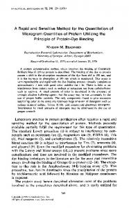
ThePrincipleof thePartialVolumeEftect The basic principle for the quantitative wall motion analysis proposed in this study is similar to that used for radionuclide
FIGURE I In vitro demonstrationof the partialvolumeeffect by the Anger
angiography,i.e. systo-diastolicchangesin detected radioac
camera used in this study. The phantom (upper left panel)
tivity. Interestingly, in ECG-gated flow images, the end-sys tolic counts obtained in a ventricular region of interest are higher than the end-diastolic ones in normal contracting seg ments. A steady state concentration of MIBI into the organ
filled with a solutionof @“Tc that simulatedmyocardialthick
has been assumed because this tracer is trapped into myocar dium and the washout rates in normal and in ischemic areas
are negligible in the first few hours following tracer injection
(9). Thecountsrecovered bytheexternal counting areinflu enced in this condition only by changes of cardiac thickness. In imaging of the heart by means of nuclear medicine devices, the anatomical size ofmyocardial wall frequently falls below the image resolution of the instrument. Under these circumstances, the partial volume related effect ( 10) induces an apparent underestimation ofthe true tracer concentration. The recovery coefficient, i.e. the ratio of apparent to true
nessina broadrangeof values(from3 to 16.5mm)is shown together with the relative image (upper right panel). An appar
entlossofcountsconcomitant withthereductionoftheobject size is demonstratedby the angular profile generatedclock wise from the top of the image (lower left panel).The lower right panel representsthe recovery coefficient(RC%)versus
the object size normalizedto the imageresolutionof our Angercamera(8 mm)(MM/FWHM);the reductionof detected
radioactivityas a functionof thicknessis particularlyevident for thicknesslower than 10 mm.
gated R-wave. The frame with the highest activity in the LV region of interest was considered as end-systolic; this was also checked by visual inspection of the LV cavity size in the
isotope concentrations, has been calculated for the Anger camera used in the present study using a phantom formed by two eccentric cylinders of plexiglas, simulating myocardial wall with variable thickness. The volume between the two
were manually drawn and the center ofgravity ofthe LV was then automatically determined. Thereafter, 60 radii (one every
cylinders was filled with a homogeneous solution of @mTc, to obtain a counting rate ofabout 1000 cps. This phantom allows the evaluation of object size in the range from 3 to 16.5 mm.
LV edge, the first radius being set in opposition to the apex. The distribution of MIBI activity was determined by calculat
In Figure 1, the demonstration of the partial volume effect in this phantom is shown. Plot ofthe recovery coefficient versus the object size, in units of the FWHM of our Anger Camera (8 mm), shows that normal systolic (15 mm) and diastolic (10 mm) thickness would have a recovery coefficient of 99% and 70%, respectively. This in vitro experiment demonstrates that in the range of physiologic thickening, the partial volume
effect induces a 29% count loss from end-systole to end diastole.
6 degrees) were automatically
drawn from the centroid to the
ing the average activity per pixel along each consecutive radius (15), in a clockwise fashion. The LV outflow region (5 radii
correspondingto 30°) wasnot used in the analysis.Data were then displayed as “circumferential profile―curves by plotting
MIBI activity against angular position and the end-diastolic and end-systolic profiles were realigned at the apex, set at 180°.Thus, the only operator-dependent
procedure was the
definition of the external edge of the LV.
The systolicwallthickeningwasthen calculatedaccording to the formula:
Calculation of RegionalWallThickening Before analysis, gated images were processed by nine-point spatial and temporal smoothing. No background subtraction was performed. The end-diastolic and end-systolic frames of the two oblique projections were analyzed. The anterior pro jection, because of the frequent superimposition of extra car diac structures, was not used in the analysis. The end-diastolic frame was defined as the image acquired at the peak of the
174
cinematic display. The LV end-diastolic and end-systolic edges
(end-systolic profile —end-diastolic profile)/(end-diastolic profile + background) x 100.
The count-based thickening profile quantitates the segmen tal wall motion as an angular function referenced from the
center of gravityof the left ventricle.The values of percent thickening from all segments were averaged to ten anatomical regions (proximal and distal anterior, septal, postero-lateral
TheJournalof NudearMedicine• Vol.31 • No.2 • February1990
and inferior walls, infero-apical wall and the apex), each one
tion of the apex and the inferior wall, the values of
consisting of 11 radii. The processing time averaged 10 mm
thickening obtainedfortheapexin theLAO 70° are
per patient.
likely to be the most reliable relative to this segment.
Statistical Analysis Correlations were determined by linear regression analysis.
Individual variation among a total of 100 LV regions analyzed ranged from 19% to 41%. The pattern of
All data are given as mean ±1 standard deviation (s.d.). A probability value (p) < 0.05 was considered significant.
subject is reported in Figure 2. The values of systolic
RESULTS
thickening in the different shown in Figure 3.
Reproducibility of Radioisotopic Thickening The values ofsystolic thickening were recalculated in all subjects by the same observer in two occasions at least one wk apart and also by a second observer. The intraobserver variation of thickening measurements was ±5.4%(y = l.lx —0.1, r = 0.86, p < 0.001). The interobserver variation was ±4.1% (y = 0.8x + 0.2, r = 0.93, p < 0.001).
Radioisotopic Thickening IntheNormalSubjects All subjects showed homogeneous distribution of the tracer within the myocardial walls. The left ventricle was always well separated from the adjacent structure so that in all cases it was possible to draw the end diastolic and end-systolic regions ofinterest. An average of 30,000 counts was collected in the LV region of interest in the end-diastolic
frame; the average counts!
pixel along the sampling radius ranged from 36 to 49 counts (42 ±5). Analysis ofthe regional systolic thick
ening showeda heterogeneouspattern, with a maximal value of 35% located to the infero-apical region and a minimum
of 27% to the proximal
anterior
segment
and with a gradual increase from the base to the apex. Because of the superimposition
*1/1,
in the LAO 40°projec
systolic thickening
obtained
in LAO 40°in a normal myocardial
regions
are
DISCUSSION In the present study, a new algorithm for quantitative analysis ofregional myocardial wall motion from ECG gated acquisition ofMIBI scintigraphy is described. The proposed method is based on a physical limitation of most nuclear medicine devices, i.e. the partial volume effect, which occurs when the object size falls below the image resolution of the instrumentation. The first observation on the relationship between the size of the myocardium and the appearance of myocar dial images has been reported by Gewirtz and co workers
using
201fl myocardial
scintigraphy
(16).
In
that study, changes of left ventricular volume induced by partial aortic or coronary occlusion caused the ap pearance of perfusion defects without any change in tracer distribution, thus demonstrating the direct rela tionship between regional wall thickness and detected radioactivity. However, in that study, no correlation between reduction
of wall thickening
and the appear
ance of perfusion defects was reported. The effect of object size on apparent regional myo cardial tracer concentration has been intensively inves
@MMT$
6@
••:@ ‘
\@
@.,
.‘ .@, @
@.-
,*—@.
g
ES
FIGURE2
Thesystolicthickeningprofile(SD ob
@pr
Wall Motion Quantitation with Gated Perfusion Images • Marcassa et al
18
tamedin LAO 40°projection in a nor mal subjects with the relative end-dia stoic and the end-systoliccount distil bution angularprofile are shown in the right and left upper panels, respec tively. In the lower panelsthe end-dia stollc(ED)andend-systolic(ES)frames are reported.
175
reported by Lanzer et al. (5) using cine computed tomography. Comparable values for systolic thickening were also obtained by Sechtem et al. (6) by nuclear magnetic resonance imaging, showing the lowest values of thickening in the posterior segments and the greatest
CD z z
in thelateralandapicalregions. Discrepancies occur in comparison with contrast
w 0 I
ventriculography.
F-
Using this technique, wall thickening
in normalsrangesfrom 30% to 150%in controlcon ditions and exceeds that measured directly by implanted
radiopaque markers (17). This discrepancy might be
SEGMENTS
FIGURE 3 Regionaldifferencesin radioisotopicsystolicthickening(% THICKENING)obtainedin 10 normalsubjects. Dataare given as mean ±1 s.d. of the mean.A heterogeneousdistribution of regional thickening is observed with a maximum value
due to the possible inclusion of trabeculations and papillary muscles in end-systolic measurements and in uncertainties in the identification of endocardial bor ders. These findings suggest that contrast ventriculo graphy should not be considered as the “gold standard― for the assessment of regional systolic thickening. Technical Considerations
Some limitations of our method should be men encountered intheinfero-apical wallanda minimumoccunng tioned. First of all, results may be influenced by back in the proximal anterior region. (ANT = anterior; S = septal;
P/L = posterolateral;I = inferior; hAP = infero-apical;AP = apical;P = proximal;D = distal).
tigated by Hoffman et al. (10) using positron emission tomography (PET). These authors showed that, when the object size falls below the image resolution
of the
ground activity. In particular, according to the formula
used for calculation, an increase in the background activity would induce a reduction in the measured wall thickening. However, in the range of heart to back ground ratio from 2: 1 to 3: 1 the calculated wall thick
ening would change no more than 10%. The effect of a marked perfusion defect must also be
instrument, the object only partially fills the sensitive
taken into
volume ofthe detectors viewing that dimension. Recon
ground would have a greater influence than for a nor mal region, because of a lower target-to-background
struction
ofthese
images resulted
in an underestimation
of the true isotope concentration in the structure (the partial volume effect). Wisenberg
et al. (12) showed
that artifactual reductions in tissue tracer concentra tions, due to the partial volume effect, can be mini mized by ECG-gated image acquisition. Parodi et al. (1 1) demonstrated that myocardial wall motion abnor malities cause underestimation of regional ‘3NH3con centration; regional fractional shortening closely corre
lated with counts recovered by the PET system (FWHM 17 mm). Although
the new generation
of gamma-cam
eras provides a better spatial resolution, minimizing the partial volume effect, an underestimation of tissue tracer concentration in cardiac images still occurs. This
account.
In this circumstance,
the back
ratio. Ad
hoc
experiments
are
needed
in order
to better
clarify the effect of these factors on count-based
thick
ening calculation.
ClinicalImplications The results of the present study should be of interest to laboratories
performing
nuclear
cardiac
imaging,
because they demonstrate the possibility of obtaining simultaneous information on regional perfusion and wall motion. An important feature ofthis radioisotopic analysis is that our method,
based on systo-diastolic
radioactivity changes, represents a non-geometrical ap proach for the assessment ofwall thickening. Moreover, is clearly shown in our phantom study, where a signif it operates on a regional basis and can characterize icantunderestimation in trueisotopeconcentration was patients with ischemic heart disease, in whom the func
observed relative to object sizes lower than
10 mm
(Fig. 2). Comparisonwith OtherTechniques Our data on the pattern of LV regional wall thick ening in normal subjects are similar to the echocardi ographic resultsreported by Haendchenet al. (3) and Zoghbi et al. (4) both in normals and in the conscious dog. These studies showed heterogeneous wall thicken ing in normal segmentswith a gradual enhancement from the base to the apex. Similar values have been
176
tional impairment
is typically regional, better than the
evaluation of global left ventricular function by the geometrical analysis of gated flow images (18). Our method overcomes the limitation ofa qualitative analy sis ofECG-gated
MIBI acquisitions
(8) for the detection
of wall motion abnormalities.
The method of analysis is simple enough for wide spread use without the need for complex computer algorithms, requires minimal operator interventions
and can be easily applied on an outpatient basis. Ap
TheJournalof NuclearMedicine• Vol.31 • No.2 • February1990
pliedto patientswithcoronaryarterydisease andre gional wall motion abnormalities, it can provide useful information about the relationship between site and
6.
extension of perfusion defects and myocardial dysfunc
tion. Results from the present study support the utilization of the proposed method to evaluate both regional myo
7.
cardialperfusion andwallmotion.However,further evaluations
in normal
subjects
and in patients
are re
quired in order to fully understand the clinical validity of this approach. Quantitative analysis of MIBI images could represent a new tool for monitoring of myocardial
function in patients with ischemic heart disease during physical and pharmacological
interventions.
This study was supported in part by a research grant from the Ministero Della Pubblica Istruzione, Italy, and by National
Research
Council
REFERENCES
(Cardiorespiratory
9.
kineticsof @“Tc-hexakis-2-methoxy-methylpropyl-isonitrile. Circu/ation 1988; 2:491—498. 10. Hoffman El, Huang SC, Phelps ME. Quantitation in positron
emission computed tomography: I. Effect of object size. J ComputAssist Tomogr 1979;5:391—400. 11. Parodi 0, Schelbert HR, SchwaigerM, Hansen H, 5dm C,
ACKNOWLEDGMENTS
87.00446.04.
8.
macologic changes in wall thickness. JAm Coil Cardio/ 1986; 8:682—692. Sechtem U, Sommerhof BA, Markiewicz W, White RD. Cheitlin MD, Higgins CB. Regional left ventricular wall thick ening by magnetic resonance imaging: evaluation in normal persons and patients with global and regional dysfunction. AmiCardio/ 1987;59:145—151. Sic SI, Holman BL. Dynamic myocardial imaging in is chemic heart disease: use of @“Tc-isonitriles. Am J Cardiac Imaging1987;1:125—131. Men R, Maddahi J, Roy L, Berman DS. Gated RP-30 perfusion study after stress predicts myocardial viability [Ab stract]. JAm Coil Cardio/ 1987; 9:27A. Okada RD. Glover D, Gaffney T, Williams S. Myocardial
group)
Grant
Hoffman EJ. Cardiac emission tomography: Underestimation of regional tracer concentrations due to wall motion abnor malities. J Comput Assist Tomogr 1984; 8:1083—1092. 12. Wisenberg G, Schelbert HR. Hoffman El et al. In vivo quantitation of regional myocardial blood flow by positron emission computed tomography. Circu/ation 1981; 63:1248— 1258. 13. Parodi 0, Marzullo P. Bencivelli W, Galli M, L'Abbate A.
Microspheresin the assessmentof both myocardial contrac tility and perfusion. In: C. Raynaud ed. Nuc/ear Medicine 1. Gelberg HJ, Brundage BH, Glantz 5, Parmley WW. Quanti tative left ventricular wall motion analysis: a comparison of and Biology. Paris:PergamonPress;1982:3053—3056. area, chord and radial methods. Circu/ation 1979; 59:99 1— 14. Carpeggiani C, L'Abbate A, Marzullo P et al. Multiparametric 1000. approach to diagnosis of non Q wave acute myocardial in 2. Mancini GB, Norris SL, Peterson KL et al. Quantitative farction.Am J Cardiol 1989:in press. assessment of segmental wall motion abnormalities at rest 15. Burow RD, Pond M, Schafer AW, Becker L. Circumferential profiles:a new method for computer analysis of @°‘Tl myo and after atrial pacing using digital intravenous ventriculo cardial perfusion images. JNuc/Med 1979; 20:771—777. graphy. JAm Coil Cardio/ 1983; 2:70—76. 3. Haendchen RV, Wyatt HL, Maurer G et al. Quantification 16. Gewirtz H, Grotte GJ, Strauss HW et al. The influence of left ventricular volume and wall motion on myocardial images. of regional myocardial function by two-dimensional echocar diography. I. Patterns of contraction in the normal left yen Circu/ation 1979;6:1172—1177. tricle. Circu/alion 1983; 67:1234—1245. 17. Mitchell JH, Wildenthal K, Mullins CB. Geometrical studies of the left ventricle utilizing biplane cinefluorography. Fed 4. Zoghbi WA, Charlat ML, Bolli R et al. End-systolic radius to Proc1969;28:1334—1343. thickness ratio: an echocardiographic index of regional per 18. Najim YC, Ellam SV, Timmis AD, Maisey MN, Sowton E. formance during reversible myocardial ischemia in the con sciousdog.JAmCoilCardio/1987; 10:1113—1121. Simultaneous evaluation of left ventricular function and per 5. Lanzer P, Garrett J, Lipton MJ et al. Quantitation of regional fusion: a new computerized method [Abstract]. Eur J Nuc/ Med1988; 14:516. myocardial function by cine computed tomography. Phar
WallMotionQuantitationwith GatedPerfusionImages• Marcassaet al
177