Quattlebaum and Carner (1980) recently described a vapor fixation method using osmium tetroxide followed by air drying that gave better results with a fungus, ...
0038-91 53/83/5806-0347$02.00/0 STAINTECHNOLOGY Copyrigh: 0 1983 by The Williams k Wilkins Co.
Vol. 58, No. 6 Printed in U.S.A.
A NEW METHOD USING HEXAMETHYLDISILAZANE FOR PREPARATION OF SOFT INSECT TISSUES FOR SCANNING ELECTRON MICROSCOPY
Biotech Histochem Downloaded from informahealthcare.com by University of Connecticut on 10/26/12 For personal use only.
JAMES
L. NATION,Department of Entomology and Nematology, University of Florida, Gainesville, Florida 3261 1
ABSTRACT. A new rapid procedure for preparing soft internal tissues from insects that allows air drying was found to compare favorably with tissues prepared by critical point drying. In the new procedure, tissues were fixed in 1% glutaraldehyde,dehydrated through a graded ethanol series, immersed in hexamethyldisilazane (HMDS) for 5 minutes, and air dried. Tissues prepared by both the HMDS treatment and by critical point drying were coated with gold for scanning electron microscopy.Tissues prepared by the HMDS treatment did not shrink or distort upon air drying and excellent surface detail was preserved. The HMDS treatment required about 5 minutes, whereas the critical point drying procedure required about 1.5 hours.
Scanning electron microscopy (SEM) has become an invaluable tool in biological research. Hard structures such as the exoskeleton of many insects usually present no problems and can be air dried prior to coating with metal for SEM. Soft tissues, such as internal organs, often must be chemically fixed or hardened, carefully dehydrated and dried. Simple air drying, even of chemically hardened tissues, usually results in some collapse, shrinking and wrinkling. Several alternatives to air drying have been developed. The most commonly used procedure is the critical point drying technique (Hayat 1978, Cohen 1974, Anderson 1951, 1966). Some soft tissues have also given good results when frozen or freeze dried (Nei 1974, Boyde and Echlin 1973, Echlin and Morton 1973, Boyde and Wood 1969). In some cases air drying has been applied successfully after treating the tissues with fluids of low surface tension, for example, with acetone or propylene oxide (Bessis and Weed 1972, Boyde 1972, Boyde and Wood 1969). Quattlebaum and Carner (1980) recently described a vapor fixation method using osmium tetroxide followed by air drying that gave better results with a fungus, Beauvaria spp., than either critical point drying or freeze drying. During SEM studies on the internal structure of insects, I found that soft tissues such as gut, Malpighian tubules and various glands could be prepared for SEM without critical point drying if the tissues were dehydrated through a graded ethanol series, immersed in hexamethyldisilazane (HMDS) and air dried. This paper compares the results of the critical point drying procedure with the results of the HMDS treatment followed by air drying.
MATERIALSAND METHODS Tissues were dissected from decapitated insects immersed in a physiological saline, and immediately transferred for 5 min to 1% glutaraldehyde in 0.1 M Florida Agricultural Experimental Station Journal Series No. 4 17 1 347
Biotech Histochem Downloaded from informahealthcare.com by University of Connecticut on 10/26/12 For personal use only.
348
STAIN TECHNOLOGY
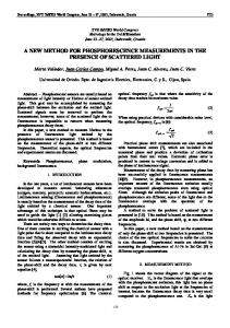
FIG. 1. Comparison of the HMDS treatment with critical point drying of soft tissues from insects. A, Malpighian tubule from a cricket, C'yllus nssi?nih, critical point dried. X 600. B, Malpighian tubule from G. ctssiinilis, HMDS treatment. Arrow points to spiral muscle; some hemocytes are adhering. X 600. C, Hemocytes adhering to tissue from G. assirnilis, critical point dried. X 1100. D, Hemocytes adhering to caecal glands from a mole cricket, Scapteriscus nrletus, HMDS treatment.')< 2300.
cacodylate buffer, pH 7. Some dissections were performed with the insect immersed in buffered glutaraldehyde, but no advantage was found with the tissues studied here. Pieces of tissue from 2 mm to 10 mm long and 2 mm thick were washed in distilled water (5 min) and then dehydrated through ethanol solutions of 70%, 8596, 95% and loo%, with 5 min in each. The tissues were then immersed in HMDS (Applied Sciences, Inc.) for 5 min, air
Biotech Histochem Downloaded from informahealthcare.com by University of Connecticut on 10/26/12 For personal use only.
HEXAMETHYLDISILAZANE IN SEM PREPARATIONS
349
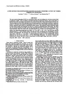
FIG. 2. Soft tissues prepared by HMDS treatment. A, Anterior hindgut from a mole cricket, S. v i r i w s , showing gut wall, musculature, and tracheal tubes. X 69. B, Trachea and musculature from gut of S. uichus. X 130. C, Trachea from S. uirinus showing taenedial windings, X 1300. D, Bacteria adhering to the lumen surface of the gut of the house cricket, Arheta domesticus. X 5000.
dried at room temperature, and mounted on stainless steel stubs with double sticky tabs. T h e tissues were coated immediately with gold in an Eiko 1B-2 ion coater, or stored in a desiccator over anhydrous Drierite until coated. After gold coating, specimens were kept in a desiccator. Tissues to be critical point dried were treated as described above until they were in 100% ethanol. They then were transferred to a small wire-basket
350
STAIN TECHNOLOGY
holder in a Balzers Critical Point Dryer, model CPD 010, and flooded with liquid C 0 2 at 5 C. After 5 min the C 0 2 was slowly vented. Flooding and venting as above were repeated 5 times to purge all ethanol. Finally, with the specimen immersed in Con, the temperature was raised over 10 min to 42 C for critical point drying and CO2 was slowly vented over about 20 min. The entire procedure from first immersion in C 0 2 to removal from the dryer took about 1.5 h and required constant monitoring.
Biotech Histochem Downloaded from informahealthcare.com by University of Connecticut on 10/26/12 For personal use only.
RESULTS T h e HMDS treatment gave results as good as those from critical point drying, with a great saving in time and without complex equipment. Tissues prepared by critical point drying (Figs. 1A and 1C) are compared with those prepared by HMDS treatment (Figs. 1B and 1D). Figs. 1A and 1B clearly show the small muscles that run in spiral fashion along the length of the Malpighian tubules of prthopteran insects. Several hemocytes adhering to the tubules can be seen in each preparation and are especially well shown in Fig. 1C (critical point dried) and in Fig. 1D (HMDS treatment). This method has been used with success on a variety of insect tissues, some of which are shown in Fig. 2, A-D. DISCUSSION Probably most laboratories engaged in SEM work have critical point drying equipment available, and it is not proposed that the HMDS treatment can eliminate all need for critical point drying. T h e HMDS process seems very satisfactory for insect internal tissues, and it may prove useful with other tissues. Its chief advantage lies in the speed with which small pieces of tissue can be prepared for gold coating as compared with the critical point drying technique (about 5 min vs 1.5 h). In addition, the method is inexpensive and does not require special equipment or skills. T h e manner in which HMDS works on tissue preparations has not been investigated. Hexamethyldisilazane is a reagent commonly used in gas chromatography to prepare silyl ethers of compounds with one or more reactive hydrogen atoms, such as sugars, amino acids, alcohols and numerous other compounds. It is not known whether HMDS reacts with some of these compounds in tissues, or simply reacts with the ethanol remaining and thereby dries the tissues as excess HMDS evaporates. It seems probable, however, that HMDS does form some silyl ethers of the more reactive compounds in tissues, and it may crosslink proteins and stiffen the tissue so that it resists collapse upon drying. Because of its possible reactivity with tissue compounds, one should avoid contact with the skin and use HMDS in a fume hood. ACKNOWLEDGMENTS
I thank Kathy Dennis for able technical assistance and Mrs. Glinda Benson for typing the manuscript. Dr. Greg Erdos provided assistance in learning SEM instrumentation. SEM facilities and critical point drying equipment were
HEXAMETHYLDISILAZANE I N SEM PREPARATIONS
351
provided by the University of Florida, IFAS SEM Laboratory. Critical review of the manuscript and helpful suggestions were given by Dr. Ellis Matheny, Entomology and Nematology Dept., and by Dr. Henry Aldrich, Ultrastructure Laboratory, University of Florida.
Biotech Histochem Downloaded from informahealthcare.com by University of Connecticut on 10/26/12 For personal use only.
REFERENCES Anderson, T. F. 1951. Techniques for the preservation of three-dimensional structure in preparing specimens for the electron microscope. Trans. N. Y. Acad. Sci. 13: 130-134. Anderson, T. F. 1966. Electron microscopy of microorganisms. In: Physical Techniques in Biologml Researrh. Pollister, A. W., ed., 2nd ed., Vol. IIIA, Academic Press, New York. pp. 319-387. Bessis, M. and Weed, R. I. 1972. Preparation of red blood cells (RBC) for SEM: a survey of various artifacts. In: Scanning Electron Mirroscopy, Proc. 5th Ann. Scan. E. M. Symp., I. I. T. Research Institute, Chicago. pp. 289-296. Boyde, A. 1972. Biological specimen preparation for the scanning electron microscope: an overview. In: Sranning Elerlron Mirroscopy, Proc. 5th Ann. SEM Symp., I. I. T. Research. Institute, Chicago. pp. 257-264. Boyde, A. and Echlin, P. 1973. Freezing and freeze-drying-a preparative technique for scanning electron microscopy. In: Sranning Electron Microscopy, Proc. 6th Ann. SEM Symp., 1. I. T. Research Institute, Chicago. pp. 759-766. Boyde, A. and Wood, C. 1969. Preparation of animal tissue for surface scanning electron microscopy. J. Microsc. 90: 221-249. Cohen, A. L. 1974. Critical point drying. In: Principles and Techniques ofScunning Electron Microscopy, Vol. I , Biological Applications. Hayat, M. A., ed., Van Nostrand Reinhold, New York. pp. 44112. Echlin, P. and Moreton, R. 1973. The preparation, coating, and examination of frozen biological materials in the scanning electron microscope. In: Scanning Electron Microscopy, Proc. 6th Ann. SEM Symp., I. I. T. Research Institute, Chicago. pp. 325-332. Hayat, M. A. 1978. Introdurtion to Biological Scanning Electron Microscopy. Univ. Park Press, Baltimore. pp. 131-166. Nei, T. 1974. Cryotechniques. In: Principles and Techniques of Scanning Electron Microscopy, Vol. 1, Biologrrol Applira/ions. Hayat, M. A., ed., Van Nostrand Reinhold, New York. pp. 113-124. Quattlebaum, E. C. and Carner, G. R. 1980. A technique for preparing Beauveria spp. for scanning electron microscopy. Can. J. Bot. 58: 1700-1703.