conserved DNA sequence motifs of animal retroviruses and its ... cell lines (Hodgkin's lymphoma, Burkitt's lymphoma) have thus far been investigated using ...
Journal of General Virology (2001), 82, 2205–2213. Printed in Great Britain ...................................................................................................................................................................................................................................................................................
A PCR primer system for detecting oncoretroviruses based on conserved DNA sequence motifs of animal retroviruses and its application to human leukaemias and lymphomas Thomas Burmeister, Stefan Schwartz and Eckhard Thiel Freie Universita$ t Berlin, Medizinische Klinik III, Hindenburgdamm 30, 12200 Berlin, Germany
Many C- and D-type retroviruses are known to cause a broad spectrum of malignant diseases in animals. Certain genome regions of these animal retroviruses are highly conserved between different animal species. It should be possible to detect new members of the retrovirus family with consensus PCR primers derived from these conserved sequence motifs. The consensus PCR primers developed in this study are generic enough to detect nearly all known oncogenic mammalian and avian exogenous C- and D-type retroviruses but do not amplify human endogenous retroviral sequences. In contrast to previous investigations, the present study involved highly stringent PCR conditions and truly generic PCR primers. Forty-four samples from patients with various immunophenotyped malignant diseases (acute and chronic T-/B-cell lymphocytic leukaemias, acute myeloid leukaemias, T-/B-cell lymphomas, chronic myeloproliferative disorders) and three cell lines (Hodgkin’s lymphoma, Burkitt’s lymphoma) have thus far been investigated using these PCR primers. The fact that no retroviruses have been found argues against an involvement of known animal oncoretroviruses or related hitherto undetected human retroviruses in the aetiopathogenesis of these diseases. The retrovirus detection system developed here may be used to confirm suspected retroviral involvement in other (malignant or nonmalignant) human diseases as well as to identify new animal retroviruses.
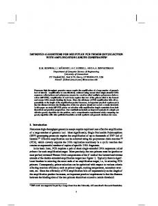
Introduction Retroviruses are known to cause various malignant and non-malignant diseases in animals over a wide range of species. In contrast, only four genuine human retroviruses – human immunodeficiency virus (HIV) types 1 and 2 and human Tlymphotropic virus (HTLV) types 1 and 2 – have been isolated thus far. HIV-1\-2 cause acquired immunodeficiency syndrome, and HTLV-1 has been identified as the key aetiological agent in adult T-cell leukaemia as well as in tropical spastic paraparesis\HTLV-associated myelopathy, a non-malignant neurological disorder. The association of HTLV-2 with human diseases is not well documented. Little is known about its seroprevalence, particularly since many tests used for serological detection of HTLV infection do not discriminate between HTLV-1 and -2. Retrovirus-induced diseases in animals have been described since the beginning of the last century. There are six known genera of exogenous retroviruses infecting mammals or birds (Hunter et al., 2000 ; Fig. 1). Viruses Author for correspondence : Thomas Burmeister Fax j49 30 8445 4468. e-mail tbu!gmx.net
0001-7616 # 2001 SGM
of the first four genera cause mainly (but not exclusively) malignant diseases, while the lentiviruses lead to chronic nonmalignant degenerative diseases. Spumaviruses are not known to cause any disease at all. What makes it attractive to search for exogenous retroviruses in human diseases ? First, there is a striking discrepancy between the wide variety of known animal retroviruses and the small number of their known human counterparts. The assumption that humans and their phylogenetic ancestors have indeed been the target of multiple retroviral infections in the past is substantiated by the fact that an estimated 1–2 % of the human genome consists of sequences of retroviral origin (e.g. endogenous retroviruses), i.e. relicts of prehistoric retrovirus infections of germline cells (Urnovitz & Murphy, 1996). Secondly, the pathogenesis and aetiology of many malignant human diseases are not yet well understood despite the increasing amount of pertinent experimental data, which leaves open the possibility of a causative viral agent, as is the case in many animal species. Thirdly, epidemiological data suggest that strong exogenous factors (such as infectious agents, e.g. viruses) are involved in the pathogenesis of certain human malignant diseases and there have been some hitherto CCAF
T. Burmeister, S. Schwartz and E. Thiel
Fig. 1. Exogenous retroviruses in mammals and birds.
unconfirmed reports on the isolation of novel putatively exogenous retroviral sequences from human tumours. The objective of this study was to construct a set of primers based on conserved nucleotide sequence motifs in animal viruses that should be able to detect human homologues. The basic assumption was that viral genome regions conserved between different animal species may reflect some essential feature of the viral genome that may also be present in human viral homologues, if any exist. The main problem here is that, as mentioned above, the human genome contains numerous endogenous retroviral sequences with a potential for false positive PCR results when using highly degenerate primers under low-stringency PCR conditions. This problem was mastered by selecting PCR primers very carefully and using specific PCR techniques (‘ long PCR ’ with proofreading polymerases). The newly developed PCR system was applied to a variety of DNA samples obtained from patients with various malignant haematological diseases.
Methods
Patient material. Bone marrow or peripheral blood mononuclear cell samples from patients with various malignant haematological diseases (Table 1) were obtained from residual material archived in liquid nitrogen. CCAG
Nearly all samples had been taken at the time of diagnosis without previous treatment. DNA from the two cell lines L428 and KM-H2 (Marafioti et al., 2000), both derived from Hodgkin cells was kindly supplied by H. Stein (Berlin, Germany). DNA from cell line Raji was obtained from Clontech.
Immunophenotyping and classification of diseases. Immunophenotypic classification of diseases followed the principles outlined in Ludwig et al. (1994) and Bene et al. (1995). Lymphomas were classified according to the REAL classification scheme (Harris, 1997), acute myeloid leukaemias according to the FAB scheme (Bennett et al., 1985).
DNA preparation. In all samples mononuclear cells had been isolated by either Ficoll gradient centrifugation or red blood cell lysis. DNA was prepared from the cells using an alkaline lysis-based method with subsequent isopropanol precipitation (Puregene, Biozym Diagnostik). DNA was dissolved in Tris–EDTA buffer at a photometrically determined concentration (GeneQuant II DNA\RNA calculator, Pharmacia) of approximately 100 ng\µl.
PCR. PCR was performed in a Perkin Elmer 9600 thermocycler using the Expand 20kB PLUS system (Boehringer Mannheim) with a 25 µl reaction mix containing 400 nM primers and 500 µM deoxyribonucleotides as well as polymerase mix (containing a mixture of proofreading Pwo polymerase and Taq polymerase). Buffer conditions and mineral oil overlay followed the recommendations of the supplier. The reaction mix was always prepared as a minimally 5-fold master mix to compensate for pipette inaccuracies and was used immediately after
PCR primers for detecting enteroviruses
Table 1. Patient samples investigated No. of samples investigated
Disease/subentity Acute lymphocytic leukaemia (ALL) Cortical T-ALL Pre-T-ALL Common ALL B-ALL Acute myeloid leukaemia (AML) AML-M1 AML-M2 AML-M4 B-lineage non-Hodgkin’s lymphoma Chronic lymphocytic leukaemia Immunocytoma Plasmacytoma\plasma cell leukaemia Prolymphocytic leukaemia Follicular lymphoma Mantle cell lymphoma Hairy cell leukaemia Splenic lymphoma with villous lymphocytes Burkitt’s lymphoma cell line Raji T-lineage non-Hodgkin’s lymphoma Chronic lymphocytic leukaemia Prolymphocytic leukaemia Se! zary syndrome Chronic myeloproliferative disorders Chronic myeloid leukaemia in chronic phase or blast crisis Polycythaemia vera Osteomyelofibrosis Hodgkin’s lymphoma Hodgkin cell-derived cell lines L428, KM-H2
1 1 1 3 1 2 2 7 2 5 1 2 1 1 1 1 3 1 1 4 3 1 2
included in every PCR. Cycling conditions were as follows : modified ‘ hot-start ’ technique (Chou et al., 1992) (putting the vials in the cycler at � 80 mC), denaturation at 92 mC for 2 min, 15 cycles for 10 s at 92 mC, for 30 s annealing at the PCR-specific temperature (Table 2), and for a product-specific extension time (Table 2) at 68 mC, 20 additional cycles as described but with a 10 s increment per cycle in extension time, a 7 min final extension at 68 mC, and cooling to 4 mC. Aliquots of the PCR mixture were analysed on a 1 % agarose gel and visualized under UV illumination after ethidium bromide staining.
Primers. Primer sequences are given in Table 2. PCR primers were obtained from Metabion Inc. (Planegg-Martinsried, Germany) and HPLCpurified. All primers were synthesized with two phosphorothioate nucleotides at their 3h end to prevent degradation by the 3h–5h exonuclease activity of Pwo polymerase and subsequent mispriming of truncated primers (Skerra, 1992). BLAST 2.0 (Altschul et al., 1997) was used to check whether PCR primer sequences exhibited a high degree of homology to known human genomic sequences, since this could cause unwanted PCR artefacts.
Sequence alignments. Sequence alignments were done using Clustal X 1.6 (Thompson et al., 1997) on an Apple Macintosh PPC computer. All nucleotide sequences were obtained from the EMBL\ GenBank\DDBJ database. The accession numbers are given in Table 3.
Virus-infected genomic DNA. Virus-infected genomic animal DNA or virus-infected cell lines were supplied by M. Spiegel, Tu$ bingen, Germany [packaging cell line PA317 (Miller & Buttimore, 1986) harbouring a partially truncated amphotropic murine leukaemia virus (MuLV)], K. Venugopal, Newbury, UK (avian DNA infected with ALV), H. Ellerbrok, Robert-Koch-Institut Berlin, Germany [cell line MT-2 (Miyoshi et al., 1981) infected with HTLV-1] and M. Sharp, Edinburgh, UK (sheep kidney DNA infected with JSRV). A plasmid containing the entire proviral genome of BLV (Sagata et al., 1985) was provided by P. Blankenstein, Berlin, Germany. Plasmid pAMS (Miller & Buttimore, 1986), containing a hybrid amphotropic\ecotropic MuLV provirus, was obtained from the ATCC (F45167), EMBL\GenBank\DDBJ accession no. AF010170).
Results preparation. Two-hundred ng of genomic sample DNA was added to each tube. A negative control (leukocyte DNA from two healthy individuals) and a positive control (virus-infected DNA, see below) were
The primary aim of this study was to identify highly conserved genome regions in animal retroviruses suitable for constructing generic PCR primers. Thus the EMBL\GenBank\
Table 2. PCR primers with annealing temperatures (Ta ), elongation times and PCR product sizes Elongation time
Product length (kb)
Primer pair
Sequences 5h–3h
Ta (mC)
P-tRNA POL-Cm W-tRNA POL-Ca P-tRNA POL-3 K12-tRNA GAG-D HβG forward HβG reverse
CADKTGGGGGCTCGTCCGGGAT TTCATTCTTTCTACCTGACCTGARCTYTGGG TCATTTGGTGACCCCGACGTGAT ARKGGCCAYTGRTYAABCCABACAGG CADKTGGGGGCTCGTCCGGGAT GGCCTGGAGGCGYTCHRGTTTAAMGG CANBTGGCGCCCAACGTGGGGC CAWTKTTCAAAAAAYTCAGATTTCCA CACAAGGGCTACTGGTTGCCGATT AGCTTCCCAACGTGATCGCCTTTCTCCCAT
67
4 min
5n0
61
1 min 45 s
2n5
65
1 min 30 s
1n8
61
45 s
62
18 min
1n2 28n8
CCAH
T. Burmeister, S. Schwartz and E. Thiel
Table 3. EMBL/GenBank/DDBJ accession numbers of viral sequences used in the nucleotide alignments Virus HTLV-1 HTLV-2 STLV-1 STLV-2 STLV-L BLV MuLV
FeLV GaLV MPMV SRV-1 SRV-2 ENTV JSRV ALV MMTV
EMBL/GenBank/DDBJ accession number J02029 (Japanese ATL isolate), AF033817, L03561, D13784 (Caribbean isolate), L02534 (Melanesian isolate), U19949 (isolate from an ATL patient), AF042071 (isolate from Germany), L36905 (from a patient with posttransfusional spastic paraparesis) M10060, L11456 (Guyami Indian isolate), Y14365 (Congolese Bambute Efe Pygmy isolate), X89270 (Italian isolate), L20734, Y13051 (African isolate, subtype b) Z46900 (from Celebes macaques), AF074966 (isolate Tan90 from Central African Republic) Y14570 (STLV-PP from Pan paniscus), U90557 (from Pan paniscus) Y07616 (STLV-PH969 from a Hamadryas baboon) K02120 (Japanese isolate), AF033818 J02255 (Moloney MuLV), AF033811 (Moloney MuLV), Z11128 (Friend MuLV FB29), D88386 (variant of Friend MuLV), M93134 (variant of Friend MuLV strain PVC-211), Y13893 (strain PVC-441), U94692 (Rauscher MuLV), X57540 (strain CAS-BR-E), J01998 (strain AKV), K03363 [strain RadLV\VL3(T+L+)], U13766 (strain MCF1233), U63133 (from BL6 melanoma cells) M18247 [subgroup A (FeLV-FAIDS)], AF052723 (strain Rickard subgroup A) M26927 (strain SEATO and SF) M12349 (strain MPMV\6A), AF033815 M11841 (strain L47.1) M16605, AF126467 (strain D2\RHE\OR) Y16627 (British isolate) M80216 (South African isolate), AF105220 (British isolate) Z46390 (ALV HPRS-103 subgroup J), M37980 (ALV-RSA) M15122, D16249 (from JYG Chinese wild mice), AF033807
DDBJ database was searched for nucleotide sequences of animal retroviruses. The search was limited to viruses belonging to genera 2–4 depicted in Fig. 1, since they are the only ones known to be involved in the induction of malignancies. Though clearly associated with malignant diseases, mammalian type B viruses were excluded from this study for two reasons. First of all, the fact that mouse mammary tumour virus (MMTV) is the only known member makes this group unsuitable for pursuing our central objective of characterizing highly conserved genome regions by alignment of sequences from different animal species. The second reason relates to the fact that the human genome harbours several copies of endogenous retroviruses of the HERV-K family that are remarkably intact and display very high nucleotide sequence homology to MMTV (Mayer et al., 1999 ; To$ njes et al., 1999). Putative highly conserved genome regions of type B retroviruses are probably conserved in members of the closely related HERV-K family, which makes it very difficult to construct consensus primers for exogenous viruses alone. Only complete viral genomic sequences (containing the entire viral sequence between the two LTRs plus at least one of the U3, R and U5 regions) were taken into consideration, since incomplete sequence fragments and partial clones could originate from viruses that are somehow defective or truncated and therefore replication-incompetent. Endogenous retroviruses (ERVs) were not included, even if complete in the sense of possessing gag, pol and env regions with flanking LTRs, CCAI
because ERVs usually have non-functional genes due to premature stop codons, frame-shift mutations and defective splice sites. Thus, for effective replication, they may rely on help obtained from endogenous or exogenous retroviruses via trans-complementation. The inclusion of such defective or truncated sequences would have distorted the final alignment and complicated the characterization of conserved regions. The accession numbers of all sequences thus obtained are listed in Table 3. The following sections discuss each group of viruses separately and then summarize the results obtained with the constructed primers. Alpharetroviruses
Two complete isolates of avian leukosis virus (ALV) could be retrieved from the database (Table 3). The alignment showed very high overall nucleotide sequence similarity (� 95 %) with only slight differences, especially in the regions encoding the env proteins. Thus no single highly conserved genome region for primer design was readily identifiable. One primer (W-tRNA) was designed to be complementary to the tRNA binding site of ALV (coding for tryptophan-tRNA), since the nucleotide sequence of tryptophan-tRNA in humans is nearly identical to that of those avian species under consideration. The second primer was constructed by additional alignments of the two avian isolates with proviral nucleotide sequences of the most closely related animal retroviruses from other groups, i.e. type B (MMTV) and simian
PCR primers for detecting enteroviruses
type D viruses. Alignment with mammalian type C viruses discussed above yielded no genome regions of significant homology. The mixed ALV–MMTV–simian type D alignment disclosed one sufficiently conserved pol region that was used to construct the second primer (primer POL-Ca) (Fig. 2 a). The PCR was tested and optimized with serial dilutions of ALVinfected avian DNA as a positive control. Type D retroviruses (subgroup of Betaretroviruses)
Retroviruses of this subgroup exhibit a characteristic morphology of the virion particle and have therefore been classified as a separate group within the family Retroviridae (Coffin, 1992). The group includes simian [Mason–Pfizer monkey virus (MPMV), simian retrovirus (SRV)] and ovine [jaagsiekte retrovirus (JSRV), enzootic nasal tumour virus (ENTV)] members. Altogether eight complete isolates were available in the EMBL\GenBank\DDBJ database (Table 2). The simian type D retroviruses are not known to cause malignant diseases, although the first isolate was obtained from a breast carcinoma in a rhesus monkey (Chopra & Mason, 1970). There are no known human members in this group, although a number of established human cell lines producing type D retroviruses have been described (reviewed in Bohannon et al., 1991). These isolates probably arose from laboratory contamination with simian viruses or may in some cases have been obtained from severely immunocompromised (AIDS) patients who were accidentally infected through close contact with animals (Bohannon et al., 1991). JSRV (Palmarini et al., 1997) and the closely related ENTV (Cousens et al., 1999) are known to cause tumours of the lung or upper respiratory tract in infected sheep. The construction of generic PCR primers for this group was complicated by the fact that type D viruses display high nucleotide sequence similarity to type B viruses, which in turn are closely related to the HERV-K family, as mentioned above. One primer was chosen to be complementary to the lysine-1,2-tRNA binding site (primer K12tRNA) ; the other was constructed from a region in the central part of the gag gene. The PCR conditions were optimized by using serial dilutions of JSRV-infected sheep DNA as a positive control. Gammaretroviruses
Type C retroviruses have been reported in a variety of mammals and birds. Three well-characterized exogenous retroviruses whose complete viral genomes have been published were included in this study : murine leukaemia virus (MuLV), feline leukaemia virus (FeLV) and gibbon ape leukaemia virus (GaLV). Reticuloendotheliosis viruses (REVs) cause infected birds to develop lymphoproliferative disorders in rare cases. REVs were not included in this study, because no complete isolate was available in the EMBL\GenBank\DDBJ database. Altogether twelve complete isolates of MuLV, two
of FeLV and one of GaLV were retrieved from the EMBL\ GenBank\DDBJ database (Table 3). Global alignment of the sequences showed several homologous regions in the gag and pol genes. Two regions turned out to be suitable for primer design : the binding site for proline-tRNA (primer P-tRNA) and a region from the 3h part of the pol gene (primer POL-Cm) (Fig. 2 c). The proline-tRNA binding site is universal and characteristic for all viruses of this group. Its integrity and conservation is necessary for efficient virus replication, since proline-tRNA is used as a primer for the synthesis of the complementary DNA strand during the virus replication cycle. The latter region harbours the catalytic centre of the integrase, and its high degree of conservation is thus understandable. PCR conditions were optimized using PA317-DNA and serial dilutions of pAMS as positive controls. Deltaretroviruses
This is the only group of viruses under investigation here that includes members pathogenic to humans. HTLV-1 infection is endemic in parts of Japan, the Caribbean, South America and Central Africa. With 95 % nucleotide sequence identity, STLV is the simian counterpart that causes T-cell neoplasms in a variety of Old World monkeys or apes (Gessain & de The! , 1996). Bovine leukaemia virus (BLV) has a genetic organization similar to that of HTLV\STLV and causes lymphoproliferative disorders in cattle. Twenty-one nucleotide sequences of proviruses available in the EMBL\GenBank\ DDBJ database were collected (Table 3), and the entire sequences were aligned. The 14 human isolates included isolates from different parts of the world and different ethnic groups (see Table 3). Five simian isolates and two complete isolates of BLV were included. The alignment showed several gag, pol and even env regions of moderate homology, but only two regions proved to be sufficiently conserved to allow the creation of highly stringent primer sequences with low degeneracy (Fig. 2). One region is the primer binding site for proline-tRNA (Primer P-tRNA). The other highly conserved region is located in the 3h region of the viral protease gene (primer POL-3). This sequence TYYCCKTTAAACYDGARCGCCTCCAGGCCY corresponds to the site in the HTLV\ STLV\BLV protease (prt) where ribosomal frameshifting (‘ ribosomal slippage ’) can occur. This ribosomal frameshifting takes place in an estimated 5 % of the cases and leads to translation of the complete viral gag\prt\pol precursor polyprotein. If no frameshifting occurs, the translation is terminated some nucleotides downstream at stop codons, resulting in a gag\prt precursor polyprotein. For correct synthesis of viral proteins, it is very important for ribosomal frameshifting to always occur at the same position, since too early an occurrence could prevent correct synthesis of the protease and polymerase. The high conservation level of this sequence motif is thus understandable. The PCR was tested and optimized with DNA from an HTLV-1-infected human cell line (MT-2) and CCAJ
T. Burmeister, S. Schwartz and E. Thiel
Fig. 2. Construction of consensus primers. The genome regions from which consensus primers were derived are displayed for each of the four retrovirus groups under investigation. Each virus isolate is characterized by its EMBL/GenBank/DDBJ accession number (see Table 3). Arrows indicate primer locations. (a) Alpharetroviruses : One primer is derived from the tryptophan-tRNA binding site common to the ALV group ; the other was constructed by alignment of the two available avian isolates with type D virus genomes and is located in the viral protease gene. (b) Type D viruses : One primer is derived from the lysine-1,2-tRNA binding site ; the other is located in the 3h part of the gag gene. The deviations of M11841 from the consensus lysine-1,2-tRNA motif at position 20 and 22 in K12-tRNA were disregarded, since they are not found in other SRV-1 isolates (Heidecker et al., 1987) and probably result from cloning or sequencing artefacts causing truncation of the primer binding site (Power et al., 1986). (c) Gammaretroviruses : One primer is derived from the proline-tRNA binding site, the other from a part of the retroviral integrase. The deviation of J02255 from the consensus motif at position 14 in POL-Cm was disregarded. (d ) Deltaretroviruses :
CCBA
PCR primers for detecting enteroviruses
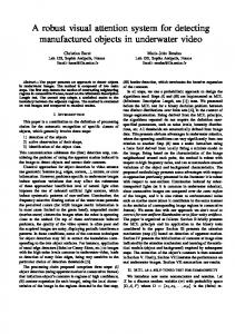
least 2000 malignant cells per PCR). DNA integrity was ensured by subjecting each sample to a control PCR (primers HβG forward and HβG reverse, Table 1) yielding a 28n8 kb amplification product (Fig. 3). Samples that did not meet this standard were excluded from the investigation. As has been stated each PCR included a negative control (200 ng leukocyte DNA) that yielded no PCR product. Thus altogether 44 samples were analysed applying the primers and PCR conditions described above and in Table 2. Their characteristics are listed in Table 1. No retroviruses were detected in any of the samples investigated.
Discussion Fig. 3. Agarose gel of the PCR products using virus-infected genomic DNA as positive control (10 ng DNA in each case). Lanes 1 and 7, λ/HindIII standard (Gibco BRL) ; lane 2, gammaretrovirus PCR (PA317 DNA) ; lane 3, alpharetrovirus PCR (ALV-infected avian DNA) ; lane 4, deltaretrovirus PCR (MT-2 DNA) ; lane 5, type D PCR (JSRV-infected sheep DNA) (the type D PCR yielded three additional bands at approx 2n5 kb when using JSRV-infected sheep DNA ; these bands were not visible when testing human DNA and probably resulted from sheep genomic sequences and were thus not considered further) ; lane 6, HβG control PCR (200 ng human DNA).
with a plasmid containing the entire BLV provirus as a positive control (Fig. 3).
Testing of patient samples
The final conditions in each PCR allowed the detection of less than 500 pg of retrovirus-infected genomic DNA, which is estimated to be equivalent to less than 50 infected cells per reaction mix (assuming 1 cell l 10 pg DNA). However, the type D PCR may have lower sensitivity because the sheep genome harbours an estimated 15–20 copies of endogenous type D viruses closely related to JSRV (Palmarini et al., 2000), which may be partially coamplified when using JSRV-infected sheep DNA as a positive control in the PCR. Since nearly all of the oncogenic animal retroviruses under consideration here are known to cause malignant haematological diseases in animals (with the exception of JSRV and ENTV), the PCR primers were applied to DNA samples obtained from humans with various malignant haematological diseases (Table 1). Diagnoses were confirmed by cytomorphology and\or immunophenotyping and in each sample case at least 10 % of the total cell volume (in most cases more that 50 %) was confirmed to belong to the malignant cell population. This ensured that all samples contained a sufficiently high percentage of malignant cells (at
This study has established a PCR system for detecting putative human (onco-) retroviruses. Highly conserved genome regions of animal retroviruses were characterized, and consensus primers were derived from these regions. The fact that these regions are conserved between widely differing animal species is indicative of their vital importance for the replication capacity of the respective group of viruses. They may be expected to be conserved in genetically highly related human counterparts as well. The primers thus obtained should then be capable of amplifying such putative human retroviruses, if any exist. Different primers were developed for each subgroup of animal retroviruses. The use of carefully optimized primer sequences, PCR conditions and specific PCR techniques (long PCR with proofreading polymerases) made it possible to construct consensus primers capable of specifically amplifying exogenous animal retroviruses without amplifying human endogenous retroviral elements. Sensitivity turned out to be high enough for this purpose, since virus-infected genomes could be detected at a dilution of less than 0n1 %. The PCR detection system was applied to a wide variety of DNA samples obtained from humans with malignant haematological diseases. No retroviruses were found in any of the samples investigated. This suggests that none of the diseases under investigation here is caused by a retrovirus related to known type C or D retroviruses. Of course it does not exclude the possibility that hitherto unknown retroviruses may play a role in human malignancies, since the PCR system used here was designed to detect retroviruses genetically related to already known ones. Retroviruses with tRNA binding sites differing from those investigated (proline, lysine-1,2, tryptophan) could not be detected by the PCR primers used here. Some previous investigators also used PCR-based methods in their search for retroviruses but the primers they used were either not generic enough to also include distantly related virus strains or had many mismatches in the primer binding regions
One primer is derived from the proline-tRNA binding site ; the other is located at the beginning of the pol gene. The additional G in U90557 at position 21 was disregarded, since it deviates from the known sequences of human, simian, murine and feline proline-tRNA and may be the result of a sequencing/cloning artefact.
CCBB
T. Burmeister, S. Schwartz and E. Thiel
or did not discriminate between sequences of endogenous and exogenous retroviral origin (Shih et al., 1989 ; Donehower et al., 1990 ; Medstrand & Blomberg, 1993 ; Li et al., 1996 ; Dube et al., 1997). For example, the ‘ universal ’ primers for detecting retroviruses developed by Donehower et al. could not distinguish between sequences of exogenous and endogenous origin and relied on concentrated and well-purified retrovirus preparations. In addition, the stringency of the PCR conditions had to be quite low (annealing temperature of 37 mC for the first 10 cycles) because of the many possible nucleotide mismatches in the primer regions. The detection method was based on DNA analysis. It could be argued that RNA-based analysis would circumvent some of the difficulties caused by endogenous retroviral elements, since most HERVs are not expressed on the RNA level. However, a large number of HERV mRNA transcripts have been observed in human cells, and especially those with close nucleotide sequence similarity to known exogenous viruses are most likely to be expressed, as exemplified by HERV-K (To$ njes et al., 1999). It must also be pointed out that the expression of retroviral proteins or RNA is not absolutely necessary for malignant transformation. Type B and type C viruses are known to act oncogenically by transactivation of genes near their integration site through promoters located in the viral LTRs. The U3 region of the type C LTR contains sequences for control and regulation of viral transcription with binding sites for several factors influencing tissue-specific expression and regulation of expression of both virus-encoded and cellular proteins (Fan, 1997 ; Barat & Rassart, 1998). Even in those cases where viral proteins are known to act as oncogenic transactivators, e.g. the HTLV-1 Tax protein, their expression may vary considerably during a cellular lifetime and can reach very low levels that are difficult to detect. Thus, DNA-based analysis appears to be more reliable and suitable for our purpose. There has been speculation regarding a direct involvement of animal retroviruses in the causation of human malignant diseases (Johnson, 1994 ; Spiegelman et al., 1974). Currently, there is little epidemiological or experimental evidence pointing to zoonotic viral causes of human malignancies but few investigations have been published on this topic. It should be noted that many animal retroviruses are capable of infecting human cells at least in vitro (Sommerfelt, 1999). GaLV and FeLV subtype B use the same cell surface receptor (Takeuchi et al., 1992), and this receptor is present on at least a subset of human bone marrow cells (Morgan et al., 1993). The recently identified receptor for xenotropic MuLVs is present on human cells derived from various tissues (Levy, 1999). JSRV also seems to be capable of infecting human cells (Rai et al., 2000). The results of this study, however, rule out an involvement of known exogenous oncogenic animal retroviruses or related endogenous counterparts in the aetiopathology of the diseases investigated here. HTLV-1 is a special case because it is the only known oncogenic human retrovirus. Since its first CCBC
description, many investigations have focussed on the possible role of this virus in other malignant T-cell disorders. This study confirms previous reports that showed no association of HTLV-1\-2 with any disease under investigation here. In summary, the results of this study suggest that none of the human diseases investigated here are caused by a known oncogenic animal retrovirus or a related but hitherto undiscovered human retroviral counterpart. The PCR system developed here has proven useful and reliable in searching for human oncoretroviruses related to known animal ones, since it is both generic, i.e. based on conserved consensus sequence motifs, and specific, i.e. capable of discriminating between exogenous and human endogenous retroviruses. Though applied to human haematological diseases in this study, the system is in no way limited to that pathological spectrum and may be applied to any human disease suspected of retroviral involvement. Though not developed and optimized for this purpose, it could even be useful in the search for unknown exogenous or endogenous animal retroviruses. This work was in part supported by a grant from the German Federal Ministry of Health (Bundesministerium fu$ r Gesundheit). We thank Drs M. Spiegel, K. Venugopal, P. Ha$ rko$ nen, M. Sharp, P. Blankenstein, H. Stein and H. Ellerbrok for providing us with virus-infected DNA samples or clones (mentioned above). We are indebted to Dr M. Otto (Go$ ttingen, Germany) for assistance with the software. We also wish to thank Ms B. Komischke, Ms R. Lippold and Ms A. Sindram for skillful technical assistance and Dr J. Weirowski for critically reading the manuscript.
References Altschul, S. F., Madden, T. L., Schaffer, A. A., Zhang, J., Zhang, Z., Miller, W. & Lipman, D. J. (1997). Gapped BLAST and PSI-BLAST : a
new generation of protein database search programs. Nucleic Acids Research 25, 3389–3402. Barat, C. & Rassart, E. (1998). Nuclear factors that bind to the U3 region of two murine myeloid leukemia-inducing retroviruses, Cas-Br-E and Graffi. Virology 252, 82–95. Bene, M. C., Castoldi, G., Knapp, W., Ludwig, W. D., Matutes, E., Orfao, A. & van ’t Veer, M. B. (1995). Proposals for the immunological
classification of acute leukemias. European Group for the Immunological Characterization of Leukemias (EGIL). Leukemia 9, 1783–1786. Bennett, J. M., Catovsky, D., Daniel, M. T., Flandrin, G., Galton, D. A., Gralnick, H. R. & Sultan, C. (1985). Proposed revised criteria for the
classification of acute myeloid leukemia. A report of the French– American–British Cooperative Group. Annals of Internal Medicine 103, 620–625. Bohannon, R. C., Donehower, L. A. & Ford, R. J. (1991). Isolation of a type D retrovirus from B-cell lymphomas of a patient with AIDS. Journal of Virology 65, 5663–5672. Chopra, H. C. & Mason, M. M. (1970). A new virus in a spontaneous mammary tumor of a rhesus monkey. Cancer Research 30, 2081–2086. Chou, Q., Russell, M., Birch, D. E., Raymond, J. & Bloch, W. (1992).
Prevention of pre-PCR mis-priming and primer dimerization improves low-copy-number amplifications. Nucleic Acids Research 20, 1717–1723. Coffin, J. M. (1992). Structure and classification of retroviruses. In The Retroviridae, vol. 1, pp. 19–50. Edited by J. A. Levy. New York : Plenum Press.
PCR primers for detecting enteroviruses Cousens, C., Minguijon, E., Dalziel, R. G., Ortin, A., Garcia, M., Park, J., Gonzalez, L., Sharp, J. M. & de las Heras, M. (1999). Complete
sequence of enzootic nasal tumor virus, a retrovirus associated with transmissible intranasal tumors of sheep. Journal of Virology 73, 3986–3993. Donehower, L. A., Bohannon, R. C., Ford, R. J. & Gibbs, R. A. (1990).
The use of primers from highly conserved pol regions to identify uncharacterized retroviruses by the polymerase chain reaction. Journal of Virological Methods 28, 33–46. Dube, S., Bachman, S., Spicer, T., Love, J., Choi, D., Esteban, E., Ferrer, J. F. & Poiesz, B. J. (1997). Degenerate and specific PCR assays
for the detection of bovine leukaemia virus and primate T cell leukaemia\lymphoma virus pol DNA and RNA : phylogenetic comparisons of amplified sequences from cattle and primates from around the world. Journal of General Virology 78, 1389–1398. Fan, H. (1997). Leukemogenesis by Moloney murine leukemia virus : a multistep process. Trends in Microbiology 5, 74–82. Gessain, A. & de The! , G. (1996). Geographic and molecular epidemiology of primate T lymphotropic retroviruses : HTLV-I, HTLV-II, STLV-I, STLV-PP, and PTLV-L. Advances in Virus Research 47, 377–426. Harris, N. L. (1997). Principles of the revised European–American Lymphoma Classification (from the International Lymphoma Study Group). Annals of Oncology 8(Suppl 2), 11–16. Heidecker, G., Lerche, N. W., Lowenstine, L. J., Lackner, A. A., Osborn, K. G., Gardner, M. B. & Marx, P. A. (1987). Induction of simian acquired
immune deficiency syndrome (SAIDS) with a molecular clone of a type D SAIDS retrovirus. Journal of Virology 61, 3066–3071. Hunter, E., Casey, J., Hahn, B., Hayami, M., Korber, B., Kurth, R., Neil, J., Rethwilm, A., Sonigo, P. & Stoye, J. (2000). Family Retroviridae. In
Virus Taxonomy. Seventh Report of the International Committee on Taxonomy of Viruses, pp. 369–387. Edited by M. H. V. van Regenmortel, C. M. Fauquet, D. H. L. Bishop, E. B. Carstens, M. K. Estes, S. M. Lemon, J. Maniloff, M. A. Mayo, D. J. McGeoch, C. R. Pringle & R. B. Wickner. San Diego : Academic Press. Johnson, E. S. (1994). Poultry oncogenic retroviruses and humans. Cancer Detection and Prevention 18, 9–30. Levy, J. A. (1999). Xenotropism : the elusive viral receptor finally uncovered. Proceedings of the National Academy of Sciences, USA 96, 802–804. Li, M. D., Lemke, T. D., Bronson, D. L. & Faras, A. J. (1996). Synthesis and analysis of a 640-bp pol region of novel human endogenous retroviral sequences and their evolutionary relationships. Virology 217, 1–10. Ludwig, W. D., Raghavachar, A. & Thiel, E. (1994). Immunophenotypic classification of acute lymphoblastic leukaemia. Baillieres Clinical Haematology 7, 235–262. Marafioti, T., Hummel, M., Foss, H.-D., Laumen, H., Korbjuhn, P., Anagnostopoulos, I., Lammert, H., Demel, G., Theil, J., Wirth, T. & Stein, H. (2000). Hodgkin and Reed–Sternberg cells represent an
expansion of a single clone originating from a germinal center B-cell with functional immunoglobulin gene rearrangements but defective immunoglobulin transcription. Blood 95, 1443–1450. Mayer, J., Sauter, M., Racz, A., Scherer, D., Mueller-Lantzsch, N. & Meese, E. (1999). An almost-intact human endogenous retrovirus K on
human chromosome 7. Nature Genetics 21, 257–258. Medstrand, P. & Blomberg, J. (1993). Characterization of novel reverse transcriptase encoding human endogenous retroviral sequences similar to type A and type B retroviruses : differential transcription in normal human tissues. Journal of Virology 67, 6778–6787.
Miller, A. D. & Buttimore, C. (1986). Redesign of retrovirus packaging
cell lines to avoid recombination leading to helper virus production. Molecular and Cellular Biology 6, 2895–2902. Miyoshi, I., Kubonishi, I., Yoshimoto, S. & Shiraishi, Y. (1981). A T-cell line derived from normal human cord leukocytes by co-culturing with human leukemic T-cells. Japanese Journal of Cancer Research (GANN) 72, 978–981. Morgan, R. A., Dornsife, R. E., Anderson, W. F. & Hoover, E. A. (1993).
In vitro infection of human bone marrow by feline leukemia viruses. Virology 193, 439–442. Palmarini, M., Fan, H. & Sharp, J. M. (1997). Sheep pulmonary adenomatosis : a unique model of retrovirus-associated lung cancer. Trends in Microbiology 5, 478–483. Palmarini, M., Hallwirth, C., York, D., Murgia, C., de Oliveira, T., Spencer, T. & Fan, H. (2000). Molecular cloning and functional analysis
of three type D endogenous retroviruses of sheep reveal a different cell tropism from that of the highly related exogenous Jaagsiekte retrovirus. Journal of Virology 74, 8065–8076. Power, M. D., Marx, P. A., Bryant, M. L., Gardner, M. B., Barr, P. J. & Luciw, P. A. (1986). Nucleotide sequence of SRV-1, a type D simian
acquired immune deficiency syndrome retrovirus. Science 231, 1567–1572. Rai, S. K., DeMartini, J. C. & Miller, A. D. (2000). Retrovirus vectors bearing jaagsiekte sheep retrovirus Env transduce human cells by using a new receptor localized to chromosome 3p21. 3. Journal of Virology 74, 4698–4704. Sagata, N., Yasunaga, T., Tsuzuku-Kawamura, J., Ohishi, K., Ogawa, Y. & Ikawa, Y. (1985). Complete nucleotide sequence of the genome of
bovine leukemia virus : its evolutionary relationship to other retroviruses. Proceedings of the National Academy of Sciences, USA 82, 677–681. Shih, A., Misra, R. & Rush, M. G. (1989). Detection of multiple, novel reverse transcriptase coding sequences in human nucleic acids : relation to primate retroviruses. Journal of Virology 63, 64–75. Skerra, A. (1992). Phosphorothioate primers improve the amplification of DNA sequences by DNA polymerases with proofreading activity. Nucleic Acids Research 20, 3551–3554. Sommerfelt, M. A. (1999). Retrovirus receptors. Journal of General Virology 80, 3049–3064. Spiegelman, S., Axel, R., Baxt, W., Kufe, D. & Schlom, J. (1974). Human cancer and animal viral oncology. Cancer 34(suppl.), 1406–1420. Takeuchi, Y., Vile, R. G., Simpson, G., O’Hara, B., Collins, M. K. & Weiss, R. A. (1992). Feline leukemia virus subgroup B uses the same cell
surface receptor as gibbon ape leukemia virus. Journal of Virology 66, 1219–1222. Thompson, J. D., Gibson, T. J., Plewniak, F., Jeanmougin, F. & Higgins, D. G. (1997). The CLUSTAL X windows interface : flexible strategies for
multiple sequence alignment aided by quality analysis tools. Nucleic Acids Research 25, 4876–4882. To$ njes, R. R., Czauderna, F. & Kurth, R. (1999). Genome-wide screening, cloning, chromosomal assignment, and expression of fulllength human endogenous retrovirus type K. Journal of Virology 73, 9187–9195. Urnovitz, H. B. & Murphy, W. H. (1996). Human endogenous retroviruses : nature, occurrence, and clinical implications in human disease. Clinical Microbiology Reviews 9, 72–99.
Received 2 January 2001 ; Accepted 13 April 2001
CCBD