Bioinformatics Advance Access published October 20, 2011
VizPrimer: a web server for visualised PCR primer design based on known gene structure Yang Zhou1,2†, Wubin Qu1†, Yiming Lu1, Yanchun Zhang1, Xiaolei Wang3, Dongsheng Zhao3, Yi Yang2* and Chenggang Zhang1,4* 1
Beijing Institute of Radiation Medicine, State Key Laboratory of Proteomics, Cognitive and Mental Health Research Center, Beijing 100850; 2College of Life Science, Sichuan University, Key Laboratory of Bio-Resources and Eco-Environment of MOE, Chengdu 610064; 3Beijing Institute of Health Service and Medical Information, Beijing 100850, China and 4School of Life Science, Anhui Medical University, Anhui, 230032.
Associate Editor: Prof. John Quackenbush
ABSTRACT Summary: The visualisation of gene structure plays an important role in PCR primer design, especially for eukaryotic genes with a number of splice variants that users need to distinguish between via PCR. Here, we describe a visualised web server for primer design named VizPrimer. It utilises the new IT (Information Technology) tools, HTML5 to display gene structure and JavaScript to interact with the users. In VizPrimer, the users can focus their attention on the gene structure and primer design strategy, without wasting time calculating the exon positions of splice variants or manually configuring complicated parameters. In addition, VizPrimer is also suitable for the design of PCR primers for amplifying open reading frames and detecting single nucleotide polymorphisms (SNPs). Availability: VizPrimer is freely available at http://biocompute.bmi.ac.cn/CZlab/VizPrimer/. (Supported browsers: Chrome [•5.0], Firefox [•3.0], Safari [•4.0], Opera [•10.0]). Contacts: CZ (
[email protected]); YY (
[email protected])
INTRODUCTION Alternative splicing is widely found for eukaryotic genes, and it plays important roles in biological processes, such as increasing protein diversity. The variant-specific PCR technique requires an unique region of a transcript variant for the primer design to distinguish between the specific variant from other isoforms. Existing programs, such as Primer3 (Rozen and Skaletsky, 2000), PerlPrimer (Marshall, 2004) and Primer Premier (Singh, et al., 1998), usually require users to enter or paste the DNA template sequence and to specify the unique region manually. Another program, PRIMEGENS-v2, added a new algorithm, HSPD (Srivastava, et al., 2011), to design homolog-specific PCR primers without any prior information of the splice variants, but the algorithm requires much longer search times to generate homologspecific primers. Importantly, none of these programs supports the graphic display of the splice variants of eukaryotic genes. However, the visualisation of gene structure makes a more direct impression
*
To whom correspondence should be addressed. authors wish it to be known that, in their opinion, these authors contributed equally to this work. †The
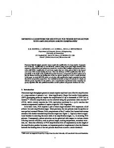
and provides more information on the spatial relationships to users than the currently used, sequence-based information. In fact, with the rapid development of IT (Information Technology) tools, especially HTML5 and JavaScript, the user experience of PCR primer design can be largely improved. The configuration of complicated parameters and options can be avoided by some mouse clicks and simple dragging operations. Therefore, we developed VizPrimer, a web server for visualised PCR primer design based on known gene structure. There are several features that distinguish VizPrimer from other traditional PCR primer design programs: 1) Instead of pasting the template DNA sequence, VizPrimer accepts official gene symbols, NCBI GeneIDs, accession numbers, official full gene names and “fuzzy” words as key words to fetch the gene records from the server; 2) Instead of directly displaying the sequence on the screen, VizPrimer graphically displays the gene structure (for all of the splice variants) with the genome location, as other genome browsers do (Fig. 1), to help users find the unique region of a splice variant for PCR primer design; 3) Rather than asking users to configure or input the complicated parameters, VizPrimer provides an intuitive and easy-control platform to help users to specify the primer-picking region and other operations; 4) Designing PCR primers in VizPrimer is flexible and, depending on the primer design strategy and positions of primer icons, VizPrimer can design PCR primers for the amplification of specific exons and open reading frames and the identification of transcript variants and single nucleotide polymorphisms (SNPs) and 5) In combination with our previous work, MFEprimer (Qu, et al., 2009), VizPrimer acts as a single package, which includes gene structure visualisation, a primer design and a specificity-checking service to help users design high-quality PCR primers.
IMPLEMENTATION There are five modules in VizPrimer: 1) User-input query module; 2) gene structure display module; 3) user-interactive module; 4) primer design module and 5) primer specificity-check module. As mentioned above, the user-input query module in VizPrimer accepts the official gene symbols, NCBI GeneIDs, accession numbers, official full gene names and fuzzy words as key words to fetch gene records from the server. In addition, VizPrimer allows
© The Author (2011). Published by Oxford University Press. All rights reserved. For Permissions, please email:
[email protected]
1
Downloaded from http://bioinformatics.oxfordjournals.org/ by guest on August 25, 2015
*
the users to upload their own cDNA and genomic DNA sequence for visualising the gene structure. GMAP (Wu and Watanabe, 2005) was used as a backend program for the gene structure prediction. There are two layers of gene structure that are displayed. First, a global view of the gene structure is presented, including all of the splice variants; the resolution is dynamic and can be easily controlled. Second, the detailed gene structure can be shown at a resolution of even one base pair. The Genome Mapping data (ftp://ftp.ncbi.nih.gov/genomes/MapView) was used to display the splice variant images at the gene level. The JavaScript and HTML5 element “canvas” was implemented in the gene structure visualisation. A JavaScript-based graphics demonstration has been widely used in bioinformatic and cheminformatic visualisations due to its low cost on the server-side and free plug-in feature on the browser-side.
Unlike the currently and widely used programs, the PCR primer design work in VizPrimer is similar to playing video games. Without the need to calculate the position of an exon, the user merely puts/drags the primer icons (right and left arrowheads) on the selected exon, and the amplicon size and primer position will be simultaneously displayed when moving the forward/reverse primer icons (Fig. 1). All of these operations are controlled by JavaScript on the browser-side which is fast and does not need to communicate with the server. There are four options available in VizPrimer to select the optimal PCR primers for special demands: “Exon locked” (for splice variant analysis), “3’-terminus free” (for gene expression analysis), “5’-terminus locked” (for ORF analysis) and “3’terminus locked” (for SNP analysis). The “Exon locked” option indicates that VizPrimer will search the optimal primers from the full region of the selected exon, which is useful for the identification of the expression of the splice variant containing this exon. The “3’-terminus free” option means that VizPrimer will search the optimal primers globally from the full-length cDNA sequence. The “5’-terminus locked” option means that the 5’terminus of the primer is locked on the position indicated by the primer icons, as denoted by the right and left arrowheads; this option is suitable for ORF primer design to amplify the region from the start codon (“ATG”) to or before the stop codon (“TAA/TAG/TGA”). Of course, the users can also specify their own cDNA region to be amplified by using this option. In addition, the “3’-terminus locked” option will be useful for SNP PCR primer design. The kernel primer design program is a Primer3 (version 2.2.3) command-line program. The result page will return five PCR primer pairs by default and the graphic gene structure with the designed primers located on it. Moreover, a link to the MFEprimer-2.0 (http://biocompute.bmi.ac.cn/CZlab/MFEprimer2.0/) specificity evaluation results is provided for each primer pair.
2
DISCUSSION The design of PCR primers for eukaryotic genes in the identification of splice variants is a complex and tedious task. The currently available primer design programs do not have a function of graphically displaying the gene structure to help users locate the unique regions among the splice variants. Using HTML5 and JavaScript for adding interactivity to a web page is a new paradigm of web computing nowadays. This combination is also encouraged by scientific applications due to the fast progress in the performance of JavaScript engines and HTML5-compatible browsers. There are several scientific applications relying on the JavaScript and “canvas”element already that are currently available, including Anno-J (Lister, et al., 2008) and ChemDoodle (http://web.chemdoodle.com/). Therefore, by integrating these new technologies, the widely used primer design program, Primer3, and the specificity-checking program, MFEprimer, VizPrimer will be a beneficial visual PCR primer design tool. In the future, we will add the function of visualised multiple PCR primer design to VizPrimer.
ACKNOWLEDGEMENTS Funding: We are grateful to the National Basic Research Project (973 program) (2012CB518200), the General Program (81170558, 30900862) of National Natural Science Foundation of China, the State Key Laboratory of Proteomics of China (SKLP-Y200906, SKLP-K201004, SKLP-O201002) for support.
REFERENCES Lister, R., O'Malley, R.C., Tonti-Filippini, J., Gregory, B.D., Berry, C.C., Millar, A.H. and Ecker, J.R. (2008) Highly integrated single-base resolution maps of the epigenome in Arabidopsis, Cell, 133, 133 523-536. Marshall, O.J. (2004) PerlPrimer: cross-platform, graphical primer design for standard, bisulphite and real-time PCR, Bioinformatics, 20, 20 2471-2472. Qu, W., Shen, Z., Zhao, D., Yang, Y. and Zhang, C. (2009) MFEprimer: multiple factor evaluation of the specificity of PCR primers, Bioinformatics, 25, 25 276-278. Rozen, S. and Skaletsky, H. (2000) Primer3 on the WWW for general users and for biologist programmers, Methods Mol Biol, 132, 132 365-386. Singh, V.K., Mangalam, A.K., Dwivedi, S. and Naik, S. (1998) Primer premier: program for design of degenerate primers from a protein sequence, Biotechniques, 24, 24 318-319. Srivastava, G.P., Hanumappa, M., Kushwaha, G., Nguyen, H.T. and Xu, D. (2011) Homolog-specific PCR primer design for profiling splice variants, Nucl. Acids Res., 39, 39 e69. WU, T.D. AND WATANABE, WATANAB E, C.K. (2005) GMAP: A GENOMIC MAPPING AND ALIGNMENT ALIGNMENT PROGRAM FOR MRNA AND EST SEQUENCES BIOINFORMATICS 21, 18591859- 1875.
Downloaded from http://bioinformatics.oxfordjournals.org/ by guest on August 25, 2015
Fig. 1. Visualisation of the mouse neuronatin (Nnat) gene structure using VizPrimer. The right and left arrowheads represent the forward and reverse PCR primers, respectively.
The overall computing time for both the PCR primer design and primer specificity evaluation were approximately 1 second on average, without considering the network communication time. VizPrimer requires browsers supporting HTML5, such as Chrome (≥5.0) and Firefox (≥3.0).