Keywordsâsleep apnea hypopnea; portable system; respiratory effort detection; automatic .... The respiration status determined by sleep experts based on the.
A Portable Monitoring System with Automatic Event Detection for Sleep Apnea Level-IV Evaluation Jhao-Cheng Wu1, Chia-Wei Wang1, Yuan-Hao Huang1,2, Hau-Tieng Wu3, Po-Chiun Huang1, Yu-Lun Lo4, 5 1
Department of Electrical engineering, National Tsing-Hua University, Taiwan. Institute of Communications Engineering, National Tsing-Hua University, Taiwan 3 Department of Mathematics and Department of Statistical Science, Duke University, Durham, NC, USA 4 Department of Thoracic Medicine, Healthcare Center, Chang Gung Memorial Hospital, Taiwan 5 Chang Gung University, School of Medicine, Taipei, Taiwan 2
Abstract— To meet the demands on a comfortable screening, or even diagnostic, equipment without interfering with the sleep, this study develops a level IV portable system, equipped with two tri-axial accelerometers (TAA) measuring the thoracic and abdominal respiratory efforts, and one oximeter measuring the oxygen saturation (SpO2), to identify obstructive sleep apnea (OSA), central sleep apnea (CSA), and hypopnea (HYP) events. The prototype integrates all the hardware and software for physiological information extraction. In addition, an automatic event detection algorithm is proposed to reduce the laborintensive work on scoring the events. Based on 63 subjects, with 80% data for training and 20% for validation, the classification accuracy of the apnea hypopnea-index (AHI) is 84.13%. The results indicate that the proposed algorithm has great potential to classify the severity of patients in clinical examinations for both the screening and the homecare purposes. Keywords—sleep apnea hypopnea; portable system; respiratory effort detection; automatic apnea/hypopnea event detection.
I.
INTRODUCTION
Sleep apnea syndrome (SAS) is a common sleep disorder that affects among 14% adult men and 5% adult women [1]. There are two main kinds: obstructive sleep apnea (OSA) and central sleep apnea (CSA). Subjects with SAS usually have morning headache, heartburn, loud snoring, and irritability. It has been well known that SAS is intimately associated with several diseases, like cardiovascular diseases [2] and stroke [3]. Although SAS has received a great amount of attention, most patients with SAS (approximately 80-90% of all patients) are not aware of it and untreated [4]. Therefore, a convenient screening tool is urgently needed. Conventional diagnosis of SAS depends mainly on overnight polysomnography (PSG) examination, which provides comprehensive sleep-related information, in the sleep laboratories of hospitals. However, PSG is a labor-intensive, uncomfortable, and high-cost study. Moreover, the “first night effect” is inevitable so the test result might be deteriorated. The need for an easy-to-install, cheap, and accurate method to screen SAS at home is clear. Many studies tried to simplify SAS diagnosis by employing several physiological signals [5]. Some researchers identified the OSA epoch using ECG. Others used nasal airflow to detect the OSA features. But the multi-lead ECG setup is annoying, and the airflow instruments usually cause discomfort of the patients during sleep. Since the thoracic and abdominal
movements are directly related to the respiratory effort [6], this research aims to develop an algorithm to identify the OSA, CSA, and HYP events based on the behaviors of the thoracic and abdominal signals measured at home. Thereby, doctors can preliminarily estimate the severity of the SAS patients and reduce the cost of the PSG examination. The rest of this paper is organized as follows. Section II shows the system diagram and describes the requirements for the portable monitoring system. Section III presents the frontend prototype, including hardware and signal processing techniques, for respiratory effort detection. Section IV introduces the proposed automatic event detection algorithm. Section V demonstrates the clinical test results. Finally, section VI concludes this paper. II.
SYSTEM REQUIREMENT AND DESIGN
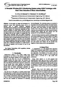
Airflow and respiration effort detections are essential for sleep apnea monitoring. During the OSA event, the airflow is low. The thoracic and abdominal movements are small in amplitude and paradoxical in phase. During the CSA event, the thorax and abdomen are silent, so the amplitudes are almost like zero and no oscillation could be observed. Hypopnea events, on the other hand, cannot be distinguished by respiratory effort, since the changes in the movement are not significant during the hypopnea events. According to the 2012 AASM guidelines, any event with 3% drop in blood oxygen level is counted in the sleep apnea and hypopnea event number. In this study, we extract the features from SpO2 to identify the hypopnea events. In a full-featured PSG clinical test, the oro-nasal cannulas to sense the airflow are bothersome for sleep. Improper belt strain makes the respiration effort detection with the piezoelectric gauge or respiratory inductive plethysmography less sensitive. For easy setup, minimal sleep influence and reliable performance under different body posture, we propose a screening system for level IV portable evaluation [7] usage. Fig. 1 shows the proposed system architecture. This system contains two kinds of sensing devices: a pulse oximeter for monitoring oxygen desaturation, and inertial sensing [8][9] for respiratory effort detection. All the recorded data is transmitted wirelessly to the mobile device. The mobile device is an interface to display the real-time waveform for calibration and system information to user. At back end, the data server
executes an automatic event detection algorithm. The medical doctors then receive the whole night report for evaluation.
information from tri-axis acceleration waveforms. Considering the calculation complexity, phase ambiguity for small breath signal, and the most important, the possible time shift due to transformation and multipath data fusion [11], this work employs a simple while effective method called dominant axis selection (DAS). We select the axis signal with reasonable pattern (frequency range of 0.2 to 0.3 Hz and stable amplitude) to represent as the respiratory waveform. Fig. 3(c) compares the output using DAS method with the data sensed by the piezo belt in the reference PSG. The key features are kept and the phase shift is small.
Fig. 1. Block diagram of the proposed sleep apnea monitoring system. There are front-end circuits for physiological signal detection. The mobile device serves as the user interface. An automatic event detection algorithm is executed on the server. The system then reports whole night SAS results to doctors.
III.
RESPIRATORY EFFORT DETECTION FRONT END
Fig. 2 shows the hardware prototype [10]. The system contains a pulse oximeter for SpO2 detection and two tri-axial accelerometer (TAA) sensors for respiratory monitoring. A micro-controller AT328P (Atmel) executes the data acquisition flow then transfer the data to a server wirelessly by a Bluetooth module (HC05). Using the embedded ADC in micro-controller, the sample rate for TAA sensors is 226Hz with 8-bit resolution. The respiratory pattern then is extracted from the thoracic and abdominal movement signals, denoted as TAA-THO and TAA-ABD respectively.
Fig. 3. The 3D trajectory angle (degree) of the respiratory movement by putting the accelerometer (a) on the thorax and (b) on the abdomen. The normalized respiratory patterns detected by a piezo belt and by an accelerometer with the dominant axis selection (DAS).
IV.
AUTOMATIC EVENT DETECTION ALGORITHM
Fig. 4 shows the signal processing flow chart of the proposed sleep apnea and hypopnea detection algorithm. The THO and ABD signals are from two accelerometers and the SpO2 signal is from an oximeter. We first extract apnea-related features from the THO and ABD signals. These features are fed into support vector machine (SVM) to generate the classifiers. Then, the OSA, CSA and HYP events are classified by the state machine. The SVM state results are compared with the desaturation state determined by oxygen desaturation to generate the final results. The number of the detected events are accumulated to obtain the numbers of overnight apnea and hypopnea events, thereby determining the apnea hypopneaindex (AHI).
Fig. 2. The prototype system and configuration for sleep apnea monitoring. In clinical tests, a full-featured PSG system is attached to be the ground truth. Two TAAs are attached on the backside of piezo belts. The probe of oximeter is applied to the forefinger of right hand, that is not shown in this photo.
In sensor node, TAA (ADXL335, Analog Device) has three analog outputs. Each output signal is connected to an amplifier (x4) and a low-pass filter (1Hz). There are wires to connect a sensor node to a data hub. In data hub, the thoracic and abdominal movement signals are post amplified by band-pass variable-gain amplifiers (1x to 10x) and notch filters (60Hz). These analog waveforms are converted to the digital domain by an ADC with time-sharing. Fig. 3(a) and 3(b) depicts the trajectory curves of the thoracic and abdominal respiratory efforts recorded by accelerometers. There are several methods, like principal component analysis (PCA) [8], angular motion extraction [9], and piecewise linear approximation, proposed to extract the respiratory movement
Fig. 4. The flow diagram of the proposed automatic apnea and hypopnea event detection algorithm based on accelerometer and oximeter signals.
In the following, the key functions in the proposed automatic event detection algorithm are introduced.
support vector machine (SVM) is considered based on the standard radial based function. The respiration status determined by sleep experts based on the 2007 guidelines of American Academy of Sleep Medicine (AASM) was referred to as the ground truth in the training process. Four binary SVM classifiers were trained by using the 10-dimensional features in the training data, which consist of four features from respiration efforts and six features extracted from the SpO2 signal. Classifier CLF_NA distinguishes normal (N) and apnea (A); CLF_NH distinguishes normal (N) and hypopnea (H); CLF_AH distinguishes apnea (A) and hypopnea (H); CLF_OC distinguishes OSA (O) and CSA (C). C. State Machine With the physiological nature, only one of four states, NOR, CSA, OSA, HYP, will be presented at one time. Therefore, the finite state machine is a proper technique to be the flow controller. There are six rules guiding the state transition, depending on the L previous states of SVM, and the covariance value Cov. In this design, L is the tape number of state machine and set as 12 after careful system estimations. For the n-th current window, the state transits based on the following rules: transition (1)
Fig. 5. Each operation of the proposed automatic apnea and hypopnea event detection algorithm.
(2) (3)
A. Feature Extraction The filtered two-channel TAA signals were down-sampled from 226Hz to 4Hz and segmented by a 10-second window, and were slid every 0.5 seconds. The sampling rate of the oxygen saturation (SpO2) is 1Hz. This signal was segmented by a 20-second window and was slid every 0.5 seconds. For respiratory effort extraction, based on the physiological phenomenon, two features, amplitude ratio (AR) and frequency ratio (FR) of two TAA signals, were used to identify OSA and CSA events [12]. The covariance (Cov) between two TAA signals is also used as an auxiliary index to quantify the paradoxical movement since it is a significant respiratory characteristic of OSA events.
(4) (5)
For oxygen desaturation, six features including minimum, maximum, median, mean, variance of the first derivative, and the difference between median and minimum of SpO2 are used. B. Support Vector Machine To learn the relationship between the apnea events and the extracted respiratory features for prediction purpose, the kernel
(6)
condition
The state machine thus provides a prediction of sleep apnea events every 0.5 seconds, based on which the location of each apnea event, as well as the total number of events are outputted. D. SpO2 Desaturation Detection For all epochs classified as normal, if there is a desaturation determined by the desaturation features, that epoch is corrected to the HYP event. After the correction, we obtain the final whole night sleep apnea annotation. V.
CLINICAL TEST RESULTS
There are 63 clinical subjects for this study. Table I tabulates their demographic details. The test protocol has been approved and executed at the sleep laboratory in Chang Gung Memorial Hospital (CGMH), Linkou, Taiwan. The TAA sensors are put on the subject chest and abdomen to record the thoracic and abdominal movements. The SpO2 was recorded at a 1-Hz sampling from the PSG data acquisition system (Philips Respironics). A smartphone collects the data from prototype system. Besides of the prototype proposed, a standard PSG was performed with at least 6 hours of sleep to confirm the presence or absence of sleep apnea and hypopnea. Table I. Demographic details of the enrolled subjects
distinguished successfully. The accuracy of the proposed algorithm, although evaluated under two different clinical databases, is also improved. VI.
CONCLUSION
This research develops a prototype system that incorporates two tri-axis accelerometers and an oximeter to analyze respiratory efforts and oxygen desaturation to detect SAS events. Three-dimensional data sensed by TAAs were first converted into one-dimensional data by the proposed DAS method. Different features of the THO/ABD movement and SpO2 signals are used to identify sleep apnea and hypopnea events by SVM classifiers. Afterwards, the SVM classifiers are trained and tested to identify OSA, CSA, and HYP events in overnight sleep recordings with intra-individual models. SpO2 desaturation detector not only detects hypopnea events but also enhances the event-by-event accuracy. The proposed prototype system has shown its capability to evaluate the severity of SAS patients for both the screening and the homecare purposes. ACKNOWLEDGMENT Hau-tieng Wu acknowledges the support of Sloan Research Fellowships, FR-2015-65363. This work was supported by the Ministry of Science and Technology (MoST), Taiwan, under grant number MOST 104-2220-E-007-019. REFERENCES [1]
Here a common technique for machine learning non-overfitting model is applied. We split the initial dataset in an 80:20 ratio. 80% patterns of each subject were used to train the model, and the rest 20% patterns were used for testing purposes. Then apply the model to the overnight data themselves to generate the final state. The detection performance can be expressed by AHI severity confusion matrix, as shown in Table II. The diagonal elements of the confusion matrix are the numbers of subjects that have the same predicted AHI severity by the proposed algorithm as the sleep expert. With the proposed algorithm of intra individual model combined with SpO2 desaturation, the detection accuracy of AHI severity is 84.13%. Table II. Confusion matrix of the proposed algorithm and PSG system labeled by experts.
Compared to our previous work that uses two piezo belts for sensing the thoracic and abdominal movement signals [12], with the help of SpO2 information, the hypopnea event can be
P. E. Peppard, et al, “Increased prevalence of sleep-disordered breathing in adults,” Am. J. Epidemiol., vol. 177, no. 9, pp. 1006–1014, Apr. 2013. [2] S. Golbidi, et al, “Cardiovascular consequences of sleep apnea,” Lung, vol. 190, no. 2, pp. 113–32, 2012. [3] H. Yaggi, et al, “Obstructive sleep apnea as a risk factor for stroke and death,” New Engl. J. Med., vol. 353, no. 19, pp. 2034–41, 2005. [4] T. Young, et al, “Burden of sleep apnea: rationale, design, and major findings of the Wisconsin Sleep Cohort study,” Wis. Med. Journal, vol. 108, pp.246-249, 2009. [5] H. Al-Angari and A. Sahakian. “Automated recognition of obstructive sleep apnea syndrome using support vector machine classifier.” IEEE Trans. Info. Tech. in Biomedicine, vol. 6, pp.363-368, May 2012. [6] P. Dehkordi, et al, “Monitoring torso acceleration for estimating the respiratory flow and efforts for sleep apnea detection,” Int. Conf. IEEE EMBS, pp. 6345–6348, 2012. [7] R. Ferber, et al, “Portable recording in the assessment of obstructive sleep apnea. ASDA standards of practice,” Sleep, vol. 17(4), pp.378392, 1994. [8] A. Bates, M. J. Ling, J. Mann and D. K. Arvind, “Respiratory rate and flow waveform estimation from tri-axial accelerometer data”, IEEE Body Sensor Networks Conf., pp.144-150, 2010. [9] A. Jin, B. Yin, G. Morren, and R. M. Aarts, “Performance evaluation of a tri-axial accelerometry-based respiration monitoring for ambient assisted living”, Int. Conf. IEEE EMBS, pp.5677-5680, 2009. [10] C.-W. Wang, A prototype of home-monitoring system for patents with sleep-disordered breathing, Master thesis, National Tsing Hua Univ, Taiwan, 2014. [11] P. Varady, S. Bongar and Z. Benyo, “Detection of airway obstructions and sleep apnea by analyzing the phase relation of respiration movement signals’’, IEEE Trans. Instrument and Measurement, vol. 52, pp.2-6, Feb. 2003. [12] Y.-Y. Lin, H.-T. Wu, C.-A. Hsu, P.-C. Huang, Y.-H. Huang, and Y.-L. Lo, “Sleep apnea detection based on thoracic and abdominal movement signals of wearable piezo-electric bands,” IEEE J. Biomedical and Health Informatics, vol. 21, pp.1533-1545, Nov. 2017.