Analytical Biochemistry 431 (2012) 1–3
Contents lists available at SciVerse ScienceDirect
Analytical Biochemistry journal homepage: www.elsevier.com/locate/yabio
Notes & Tips
A regularly spaced and self-revealing protein ladder for anti-tag Western blot analysis Chien-Han Kao a,1, Chiu-Min Cheng b,1, Kuo-Hsiang Chuang c, Chih-Hung Chuang d, Shey-Cherng Tzou e, Ta-Chun Cheng a,1, Yuan-Chin Hsieh a, Kuang-Wen Liao e, Yun-Ming Wang e, Long-Sen Chang f, Steve R. Roffler g, Fang Ming Chen h,⇑, Tian-Lu Cheng f,i,⇑ a
Graduate Institute of Medicine, Kaohsiung Medical University, Kaohsiung, Taiwan Department of Aquaculture, National Kaohsiung Marine University, Kaohsiung, Taiwan c Graduate Institute of Pharmacognosy, Taipei Medical University, Taipei, Taiwan d Institute of Basic Medical Sciences, National Cheng Kung University, Tainan, Taiwan e Department of Biological Science and Technology, National Chiao Tung University, Hsin-Chu, Taiwan f Institute of Biomedical Sciences, National Sun Yat-Sen University, Kaohsiung, Taiwan g Institute of Biomedical Sciences, Academia Sinica, Taipei, Taiwan h Department of Surgery, Faculty of Medicine, College of Medicine, Kaohsiung Medical University, Kaohsiung, Taiwan i Department of Biomedical Science and Environmental Biology, Kaohsiung Medical University, Kaohsiung, Taiwan b
a r t i c l e
i n f o
Article history: Received 5 June 2012 Received in revised form 28 July 2012 Accepted 1 August 2012 Available online 9 August 2012 Keywords: 14-Epitope tag protein ladder Epitope tags Anti-epitope tag antibodies Protein molecular estimation Sodium dodecyl sulfate–polyacrylamide gel electrophoresis Western blot analysis
a b s t r a c t We designed a protein ladder (hereafter referred to as ‘‘Mega-tag’’) that contains 14 of the most commonly used epitope tags fused to molecular weight markers. The Mega-tag ladder can be simultaneously visualized when anti-tag antibodies are used to detect epitope-tagged recombinant proteins in Western blots. The logarithm of molecular weights and relative mobility of the Mega-tag protein ladder are highly correlated (R2 = 0.997 ± 0.00232), indicating that the dye-free Mega-tag protein ladder is accurate. It can also serve as a positive control for anti-epitope tag immunoblots. The Mega-tag protein ladder should provide a convenient and precise tool for Western blot analysis. Ó 2012 Elsevier Inc. All rights reserved.
Accurate determination of protein molecular weights is important for Western blot analysis [1], in which investigators often rely on prestained protein ladders [2,3]. However, covalent linkage of dyes to proteins alters their electrophoretic mobility [4]. In addition, prestained protein ladders generally cannot be detected on X-ray films, and manual alignment is required to mark the position of molecular weight ladders on the film. Bennett and coworkers developed a biotinylated protein ladder that could be visualized in Western blots using streptavidin-conjugated horseradish peroxidase [5]. However, covalent attachment of biotin groups alters the electrophoretic mobility of the standard proteins in the ladder [6]. Other solutions to directly visualize the protein ladder on Western ⇑ Corresponding authors. Address: Department of Biomedical Science and Environmental Biology, Kaohsiung Medical University, Kaohsiung, Taiwan (T.-L. Cheng). Fax: +886 7 3114679 (F.M. Chen), +886 7 3227508 (T.-L. Cheng). E-mail addresses:
[email protected] (F.M. Chen),
[email protected] (T.-L. Cheng). 1 These authors contributed equally to this work. 0003-2697/$ - see front matter Ó 2012 Elsevier Inc. All rights reserved. http://dx.doi.org/10.1016/j.ab.2012.08.001
blots include development of a dye-free GFP protein ladder [7], protein A fusion calibration proteins [8], and the EasySee Western marker (Spark Biologicals Technology) that consists of fusion proteins incorporating the immunoglobulin G (IgG)2-binding domains of both protein A and protein G [9]. These protein ladders can be directly visualized on immmunoblots, but the native protein conformation must be maintained for these ladders to function properly. Thus, these protein ladders cannot be completely denatured, which may affect their electromobility in sodium dodecyl sulfate–polyacrylamide gel electrophoresis (SDS–PAGE). Hexahistidine-tagged [10] and S-tagged [11] protein ladders (Perfect Protein Western Markers, Novagen, Cat. No. 69149-3) are useful for Western blot analysis of His-tagged or S-tagged fusion proteins with anti-hexahistidine tag or S tag antibodies. However, only hexahistidine-tagged
2 Abbreviations used: IgG, immunoglobulin G; SDS–PAGE, sodium dodecyl sulfate– polyacrylamide gel electrophoresis; PCR, polymerase chain reaction; TRX, thioredoxin; MBP, maltose-binding protein; GST, glutathione S-transferase.
2
Notes & Tips/Anal. Biochem. 431 (2012) 1–3
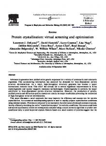
Fig.1. Western blot analysis of the Mega-tag protein ladder by anti-epitope tag antibodies. (A) The Mega-tag fusion protein, consisting of hexahistidine, HA, T7, E, VSV-G, V5, AU5, S, HSV, FLAG, Lumio, Glu-Glu, cMYC, and AU1 epitope tags, was generated by assembly PCR and then fused with a 5-kDa spacer (gray box), TRX, MBP, or GST. Bacteria expressing the Mega-tag marker proteins were separated by SDS–PAGE and then were transferred to nitrocellulose membranes. The Mega-tag protein ladder was directly visualized on Western blots using anti-epitope tag antibodies. (B–E) Individual (B) or mixed (C–E) bacteria expressing the Mega-tag protein ladder were stained with anti-S tag antibody (B), anti-HA antibody (C), anti-cMYC antibody (D), or anti-FLAG antibody (E), followed by horseradish peroxidase (HRP)-conjugated anti-goat IgG (B) or HRPconjugated anti-mouse IgG (C–E). The molecular weights of a commercial prestained marker (kDa) are indicated on the left.
and S-tagged recombinant proteins, respectively, can be detected in conjunction with these protein ladders. Multi-Tag Marker (Roche, Cat. No. 1828649) contains GST-, HA-, cMYC-, and His-tags but is still limited to fusion proteins incorporating these tags. In addition, the size increment of the component markers is not regular, rendering it less convenient to interpret. In this article, we describe a regularly spaced and self-revealing protein ladder for accurate determination of the molecular weights of epitope-tagged proteins in both SDS–PAGE and Western blots. We selected the 14 most widely used epitope tags to create the Mega-tags protein ladder. The epitope tags (hexahistidine, HA, T7, E, VSV-G, V5, AU5, S, HSV, FLAG, Lumio, Glu-Glu, cMYC, and AU1) [12] were joined together using assembly polymerase chain reaction (PCR) [13,14] and then subcloned into pRSET-B to form the pRSET-14 plasmid. The 14-tag gene was then fused with defined numbers of thioredoxin (TRX), maltose-binding protein (MBP), or glutathione S-transferase (GST) [12] genes to construct plasmids encoding the epitope-tagged protein ladder (Fig. 1A). Ideally, each of the protein markers should be recognized by all of the corresponding anti-epitope tag antibodies. Therefore we analyzed the recombinant protein markers produced in bacteria by Western blot analysis using anti-tag antibodies. All of the component protein markers were detected by anti-S tag antibody (Fig. 1B). A clear molecular weight ladder was visible in Western blots when bacteria expressing the individual markers were pooled and analyzed by Western blot using anti-HA tag antibody (Fig. 1C), anti-cMYC tag antibody (Fig. 1D), and anti-FLAG tag antibody (Fig. 1E). Similar results were obtained using other anti-tag antibodies in Western blots (see Table S1 in supplementary material). These results demonstrate that the Mega-tag protein ladder can be visualized for
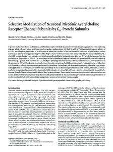
concurrent detection of epitope-tagged recombinant proteins in Western blots using the same anti-tag antibodies. No additional protocols or reagents are required for simultaneous detection of the epitope-tagged proteins and the protein ladder. Relative mobility of a protein in SDS–PAGE, defined as the ratio of distance a protein migrated to the distance of the running front, is inversely proportional to the logarithm of the molecular weight (kDa) of that protein. Linear regression analysis demonstrated the relative mobilities and the logarithmic molecular weights Mr of the component protein markers. Then we calculated the R2 values from the immunoblotting results of 6 anti-epitope tag antibodies. The anti-S tag Ab (Fig. 1B), anti-HA Ab (Fig. 1C), anti-cMYC Ab (Fig. 1D), anti-FLAG Ab (Fig. 1E), anti-V5 Ab (Fig. S1a in supplementary material), and anti-His Ab (Fig. S1b) showed R2 values of 0.9993 (Fig. 2), 0.9970, 0.9945, 0.9930, 0.9977, and 0.9976, respectively. The average R2 value is 0.997 ± 0.00232, indicating that the Mega-tag protein ladder is accurate. The correlation coefficient of the corresponding linear regression analysis for a commercial prestained marker (Fermentas, Cat. No. SM0671) was appreciably lower (R2 = 0.966 ± 0.0119), possibly owing to altered electrophoretic mobilities caused by dye molecules on the marker proteins. Collectively, our data show that the Mega-tag protein ladder is highly accurate. In addition to accurate determination of protein molecular weights, the Mega-tag protein ladder can also act as a positive control, providing a convenient tool for Western blot analysis. Inclusion of positive controls in Western blots is important to verify antibody activity and allow correct interpretation of results. Thus, a 52-kDa multiple tag fusion protein (GeneScript, Cat. No. M0101) and 12-Epitope Tag Protein Marker Lysate (Rockland Immunochemicals,
3
Notes & Tips/Anal. Biochem. 431 (2012) 1–3
Fig.2. Relationship between protein molecular weight and electrophoretic mobility. The relative mobilities of the Mega-tag protein ladder (d) or prestained markers (s) (Fermentas, Cat. No. SM0671) were plotted against the logarithm of the molecular weight of each protein (y axis). Simple linear regression was used to determine the best-fit lines.
Cat. No. MB-301-100) were previously developed as positive controls for Western blots. However, the multiple tag fusion protein is a single protein and does not allow determination of the molecular weights of target proteins. By contrast, our Mega-tag protein ladder can act as a positive control for 14 different types of anti-tag antibodies in Western blot studies. The power of the Mega-tag protein ladder is based on several factors. First, the 14 most commonly used epitope tags are included in the Mega-tag protein ladder, allowing direct visualization of both markers and tagged recombinant proteins in Western blots. Second, only the standard procedure of the Western blot technique is required to visualize the ladder. Third, the ladder can also act as a positive control for anti-epitope tag antibodies in Western blots. Fourth, the protein ladder can be boiled and processed using standard sample preparation procedures for accurate migration in SDS–PAGE. Fifth, the Mega-tag protein ladder can also be used as SDS–PAGE markers after Coomassie blue or silver staining. A possible improvement of the Mega-tag protein ladder is to add dye-modified low-, medium-, or high-molecular-weight proteins, allowing direct visualization during SDS–PAGE and after protein transfer to nitrocellulose membranes. Based on these advantages, the Mega-tag protein ladder appears to possess great potential for direct molecular weight estimation on SDS–PAGE and Western blots. Acknowledgments This work was supported by grants from the National Research Program for Biopharmaceuticals, National Science Council, Taipei, Taiwan (NSC 100-2325-B-037-001 and 100-2321-B-037-006), the Department of Health, Executive Yuan, Taiwan (DOH100-TD-N111-010 and DOH100-TD-C-111-002), and the Grant of Biosignature in Colorectal Cancers, Academia Sinica, Taiwan, and Kaohsiung Medical University Research Foundation (KMUER014 and KMUER012).
Appendix A. Supplementary data Supplementary data associated with this article can be found, in the online version, at http://dx.doi.org/10.1016/j.ab.2012.08.001. References [1] H. Towbin, T. Staehelin, J. Gordon, Electrophoretic transfer of proteins from polyacrylamide gels to nitrocellulose sheets: procedure and some applications, Proc. Natl. Acad. Sci. USA 76 (1979) 4350–4354. [2] V.C. Tsang, K. Hancock, A.R. Simons, Calibration of prestained protein molecular weight standards for use in the ‘‘Western’’ or enzyme-linked immunoelectrotransfer blot techniques, Anal. Biochem. 143 (1984) 304–307. [3] D. Parkinson, J.D. Redshaw, Visible labeling of proteins for polyacrylamide gel electrophoresis with dabsyl chloride, Anal. Biochem. 141 (1984) 121–126. [4] M.M. Compton, S.A. Lapp, R. Pedemonte, Generation of multicolored, prestained molecular weight markers for gel electrophoresis, Electrophoresis 23 (2002) 3262–3265. [5] D. Della-Penna, R.E. Christoffersen, A.B. Bennett, Biotinylated proteins as molecular weight standards on Western blots, Anal. Biochem. 152 (1986) 329–332. [6] M. Neumaier, U. Fenger, C. Wagener, Transblot studies with biotin-labeled proteins: electrophoretic mobilities and detection limits, Anal. Biochem. 156 (1986) 76–80. [7] M. Chang, H.Y. Hsu, H.J. Lee, Dye-free protein molecular weight markers, Electrophoresis 26 (2005) 3062–3068. [8] C. Lindbladh, K. Mosbach, L. Bulow, Standard calibration proteins for Western blotting obtained by genetically prepared protein A conjugates, Anal. Biochem. 197 (1991) 187–190. [9] Y.M. Zhang, L. Geng, D.W. Huang, Generate Western blot protein marker from a single construct, Anal. Biochem. 390 (2009) 206–208. [10] R.R. Burgess, T.M. Arthur, B.C. Pietz, Mapping protein–protein interaction domains using ordered fragment ladder far-Western analysis of hexahistidinetagged fusion proteins, Methods Enzymol. 328 (2000) 141–157. [11] K. Daskalow, P. Boisguerin, B. Jandrig, R. Volkmer, B. Micheel, J.A. Schenk, Epitope mapping of antibodies against S-tagged fusion proteins and molecular weight markers, Biosci. Biotechnol. Biochem. 72 (2008) 346–351. [12] K. Terpe, Overview of tag protein fusions: from molecular and biochemical fundamentals to commercial systems, Appl. Microbiol. Biotechnol. 60 (2003) 523–533. [13] R. Rydzanicz, X.S. Zhao, P.E. Johnson, Assembly PCR oligo maker: a tool for designing oligodeoxynucleotides for constructing long DNA molecules for RNA production, Nucleic Acids Res. 33 (2005) W521–W525. [14] K. Nakajima, Y. Yaoita, Construction of multiple-epitope tag sequence by PCR for sensitive Western blot analysis, Nucleic Acids Res. 25 (1997) 2231–2232.