Aug 11, 1995 - chromatin assembly factor 1 anda 56-kDa histone-binding protein. (nucleosome assembly). MICHAEL BULGER, TAKASHI ITO, ROHINTON T.
Proc. Natl. Acad. Sci. USA Vol. 92, pp. 11726-11730, December 1995 Biochemistry
Assembly of regularly spaced nucleosome arrays by Drosophila chromatin assembly factor 1 and a 56-kDa histone-binding protein (nucleosome assembly)
MICHAEL BULGER, TAKASHI ITO, ROHINTON T. KAMAKAKA, AND JAMES T. KADONAGA* Department of Biology, 0347, and Center for Molecular Genetics, Pacific Hall, Room 2216, University of California, San Diego, La Jolla, CA 92093-0347
Communicated by Bruno H. Zimm, University of California at San Diego, La Jolla, CA, August 11, 1995
thought of as histone chaperones, are involved in the assembly of chromatin (6-8, 24, 25). Neither CAF-1 nor NAP-I was identified as activities that were required for nucleosome assembly with the fractionated Xenopus extracts. Yet chromatin that is reconstituted with purified nucleoplasmin and/or N1/N2 does not possess regularly spaced nucleosomal arrays, and it has been postulated that there may be additional factors that are required for the generation of the periodic spacing of nucleosomes (24-28). Thus, to date, a variety of histonebinding factors that randomly deposit nucleosomes have been characterized, but the set of factors, whether in purified or partially purified fractions, that is sufficient for the assembly of regularly spaced nucleosome arrays has not yet been fractionated and identified. In this work, we have sought to analyze the mechanism of assembly of regularly spaced nucleosomal arrays by fractionation and purification of factors from a crude extract, termed the S-190, that is derived from predominantly postblastoderm Drosophila embryos (22, 23). These studies have led to the identification of two activities, termed Drosophila CAFs 1 and 4 (dCAF-1 and dCAF-4), that are required in addition to core histones, ATP, and DNA for the efficient assembly of periodic nucleosomal arrays.
To ascertain the mechanism by which nuABSTRACT cleosomes are assembled by factors derived from Drosophila embryos, two proteins termed Drosophila chromatin assembly factors (CAFs) 1 and 4 (dCAF-1 and dCAF-4) were fractionated and purified from a Drosophila embryo extract. The assembly of chromatin by dCAF-1, dCAF-4, purified histones, ATP, and DNA is a process that generates regularly spaced nucleosomal arrays with a repeat length that resembles that of bulk native Drosophila chromatin and is not obligatorily coupled to DNA replication. The assembly of chromatin by dCAF-1 and dCAF-4 is nearly complete within 10 min. The dCAF-1 activity copurified with the Drosophila version of chromatin assembly factor-i (CAF-1), a factor that has been found to be required for the assembly of chromatin during large tumor (T) antigen-mediated, simian virus 40 (SV40) origin-dependent DNA replication. The dCAF-4 activity copurified with a 56-kDa core-histone-binding protein that was purified to >90% homogeneity. DNA (and hence, chromatin) is a central component of many cellular processes; thus, the mechanism by which chromatin is assembled is relevant to a broad range of biological phenomena (for reviews, see refs. 1-5). The analysis of nucleosome assembly in vivo has revealed that assembly commences immediately after DNA replication but may not be obligatorily coupled to DNA replication. On the other hand, biochemical studies of chromatin assembly have led to the identification of core-histone-binding factors, which include nucleoplasmin, N1/N2, nucleosome assembly protein 1 (NAP-1), and RNA, that can mediate the DNA-replication- and ATP-independent deposition of nucleosomes that do not exhibit regular nucleosomal spacing (see, for example, refs. 6-11). In addition, a factor termed chromatin assembly factor 1 (CAF-1) has been found to be required for nucleosome assembly during large tumor (T) antigen-mediated, simian virus 40 (SV40) origindependent DNA replication (12-17). CAF-1 has been characterized by using a biochemical complementation assay with a CAF-1-deficient DNA replication extract, and purified human CAF-1 appears to exhibit a preference for the assembly of nucleosomes onto newly synthesized DNA relative to bulk nonreplicated DNA (refs. 12-14; but also see refs. 18 and 19). Up to the present, however, the factors that function along with CAF-1 to assemble chromatin have not been identified. It also has been demonstrated that factors present in crude extracts of either Xenopus or Drosophila can mediate the DNA-replication-independent assembly of nucleosomes that are approximately regular in spacing (which we will henceforth refer to as "regularly spaced nucleosomes") in the presence of ATP (see, for example, refs. 20-23). Biochemical characterization of Xenopus extracts (derived from either oocytes or unfertilized eggs) has revealed that the core-histone-binding factors, nucleoplasmin and N1/N2, which may perhaps be
MATERIALS AND METHODS Chromatin Assembly. Chromatin assembly reactions were performed as described (22, 23) with slight modification. Circular plasmid DNA was relaxed by incubation with purified recombinant Drosophila topoisomerase I for 30 min under conditions identical to those described for the native enzyme (29, 30). A standard chromatin assembly reaction (75-,u final volume) was carried out as follows: relaxed circular DNA (600 ng; with topoisomerase I) was combined with dCAF-1 [24 ,tg of protein from the 0.25-0.5 M potassium phosphate fraction eluted from the hydroxylapatite column in Fig. 1A (except as indicated in Figs. 3B and 4B)], dCAF-4 [40 ,Ag of protein from the 0.3-0.4 M NaCl fraction eluted from the Q-Sepharose column in Fig. 1A (except as indicated in Fig. 4B)], purified Drosophila core histones (500 ng), 3 mM ATP (and ATP regeneration system comprising 30 mM phosphocreatine and 1 ,tg of creatine phosphokinase per ml), and bovine serum albumin as a carrier protein (150 ng). Where indicated, purified Drosophila histone Hi was also included. All reactions, except as indicated in Fig. 1E, were incubated at 27°C for 4 hr. In reactions containing 3 mM ATP, MgCl2 was added to a final concentration of 5 mM, while in reactions that did not contain ATP, MgCl2 was added to final concentration of 2 mM. It is also pertinent to note that chromatin assembly with the purified factors was not sensitive to the Mg2+ concentration, as the extent of chromatin assembly was not affected by Abbreviations: CAF-1, chromatin assembly factor I; NAP-I, nucleosome assembly protein I; T antigen, large tumor antigen; dCAF-1 and dCAF-4, Drosophila CAFs 1 and 4; SV40, simian virus 40. *To whom reprint requests should be addressed.
The publication costs of this article were defrayed in part by page charge payment. This article must therefore be hereby marked "advertisement" in accordance with 18 U.S.C. §1734 solely to indicate this fact.
11726
Proc. Natl. Acad. Sci. USA 92 (1995)
Biochemistry: Bulger et al. A
B Drosophila embryo extract (S-190)
dCAF-1 dCAF-4 ATP +
DEAE-Sepharose
-25-
10.1
Io
1 0.5 M NaCI
dCAF-1, dCAF-4
+ +
++
++
++
+.
+.
+
SP-Sepharose lo010-25 dCAF-4
0.5 dCAF-1
0.3 l 0.4 dCAF-4
-N
R
1I1.0 M NaCI
.-s
Hydroxylapatite
Q-Sepharose 0
11727
1.0 M NaCI 0
0.01
0.5 M KP, T0.25 dCAF-1
1
23
45
67
C dCAF-1 dCAF-4 ATP
+
+9
+
+
+
+
+
_
+
+
D
z o
_
X-
2
o
_-
m CO,
3
4
x -s 4 CX) e 't _ CMJ
=
_
-
4 5 6 7 8 9J 23 1 M
M
M
M
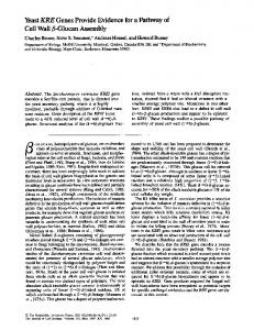
FIG. 1. Assembly of chromatin by partially purified dCAF-1 and dCAF-4 yields regularly spaced nucleosomal arrays. (A) Scheme for the fractionation of the Drosophila S-190 extract. (B) DNA supercoiling analysis. Chromatin assembly reactions were carried out with relaxed circular DNA (3.25-kbp plasmid), and dCAF-1, dCAF-4, and ATP were included, as indicated. After 4 hr, the samples were deproteinized. The resulting DNA was subjected to 1% agarose gel electrophoresis and then visualized by staining with ethidium bromide. The positions of relaxed (R), supercoiled (S), and nicked circular (N) forms of DNA are denoted. (C) Micrococcal nuclease digestion analysis. For each reaction condition, the different lanes represent increasing concentrations (from left to right) of micrococcal nuclease used to digest the chromatin as described in text. Chromatin assembly reactions were carried out in either the presence or the absence of the indicated factors. The samples were partially digested with various concentrations of micrococcal nuclease and then deproteinized. The resulting DNA fragments were subjected to agarose gel electrophoresis and visualized by staining with ethidium bromide. The molecular mass marker (lanes M) is the 123-bp ladder (GIBCO/BRL). (D) Electron microscopy. Chromatin was assembled and then purified by 30-50% sucrose gradient sedimentation, desalted with Sepharose CL-4B (Pharmacia Biotech), and analyzed by electron microscopy. (E) Time course of assembly by dCAF-1 and dCAF-4, as measured by the DNA supercoiling assay. Lanes: 1, relaxed circular DNA used in the assembly reaction; 2-9, aliquots from a chromatin assembly reaction that were removed and deproteinized at the indicated times.
variation of the Mg2+ concentration from 1.3-7 mM (unpublished data). Each lane in the DNA supercoiling and micrococcal nuclease assays represents the DNA derived from one unit reaction. Assays. In the micrococcal nuclease assays, four 75-,ul aliquots were taken from a single larger scale reaction mixture. To each aliquot, 0.1 M CaCl2 was added to 3 mM, and then micrococcal nuclease was added to 0.11, 0.23, 0.45, or 0.90 unit/ml. Digestion was allowed to proceed for 10 min, and the reaction was stopped by addition of EDTA (pH 8.0) to 10 mM and of RNase A to 20 ng/,il. In the DNA supercoiling assays, the reaction products sometimes appear as a single band at the bottom of the gel. This band represents a distribution of highly supercoiled DNAs that are not resolved under the electrophoretic conditions. For each of the reactions shown, we have also resolved these supercoiled species by electrophoresis in the presence of chloroquine (data not shown), and those results confirmed our interpretation of the agarose gels shown in the figures. The T antigen- and SV40 origin-dependent SV40 DNA replication reactions for replicative CAF-1 activity were carried out as described (12, 13). Reactions under all conditions, whether shown or indicated as unpublished data,
were performed a minimum of two times (but usually three or more times) to ensure reproducibility of the data. Supplementary material that provides a detailed description of the
fractionation and purification of the assembly factors is available upon request.
RESULTS AND DISCUSSION Assembly of Chromatin by Partially Purified dCAF-l and dCAF-4. The initial fractionation and partial purification of chromatin assembly factors dCAF-1 and dCAF-4 are depicted in Fig. 1A. To monitor the proper assembly of chromatin and to minimize potential artifacts that could have arisen during the fractionation, we employed three assays-DNA supercoiling (31, 32), micrococcal nuclease digestion (33), and electron microscopy (34). At this early stage of the fractionation, the dCAF-1 and dCAF-4 activities were not yet completely resolved, and there was some contaminating RNA in the dCAF-1 fraction (data not shown), which had contributed to ATPindependent nucleosome deposition (11). Nevertheless, the assembly of chromatin was found to be most efficient in the presence of dCAF-1, dCAF-4, and ATP (Fig. 1 B and C).
Biochemistry: Bulger et al. Electron microscopy of the chromatin revealed that the templates were fully assembled (Fig. ID). Furthermore, quantitative analysis of the DNA supercoiling by two-dimensional agarose gel electrophoresis revealed an average of 21 nucleo11728
3.25-kbp template in the absence of histone HI, which corresponds to 1 nucleosome per 155 bp of DNA (data not shown). Further purification of the factors, as shown below, subsequently clarified the requirement for dCAF-1, dCAF-4, and ATP for efficient chromatin assembly. Rate of Chromatin Assembly by dCAF-1 and dCAF-4. We have also investigated the rate of chromatin assembly by dCAF-1 and dCAF-4. These experiments revealed that chromatin assembly was nearly complete after 10 min, as measured by DNA supercoiling analysis (Fig. IE) and by the micrococcal nuclease digestion assay (unpublished data). By comparison, with a Xenopus S-150 oocyte extract (35), 1-2 hr of reaction time is required to achieve the same extent of assembly that is observed in 10 min with dCAF-I and dCAF-4. Under these conditions, assembly is nearly complete within 10 min, which is comparable to the rate of chromatin assembly that has been observed in vivo (36-39). Nucleosome Repeat Length of Chromatin Assembled with dCAF-1 and dCAF-4. We then examined factors that affect the nucleosome repeat length. In the absence of histone Hi, the repeat length varied with the monovalent salt concentration in the reaction medium as follows: 155 bp (21 nucleosomes per 3.25-kbp plasmid) at 7 mM KCl; 162 bp (20 nucleosomes per plasmid) at 57 mM KCl; and 180 bp (18 nucleosomes per plasmid) at 107 mM KCl (Fig. 2A). It has been shown previously that the incorporation of histone Hi into chromatin during assembly in vitro increases the nucleosome repeat length (see, for example, refs. 21, 22, and 40). Moreover, the apparent absence of Hi in the yeast Saccharomyces cerevisiae corresponds with the short (165 bp) nucleosome repeat length in that organism. We have therefore examined the effect of histone HI upon nucleosomal spacing upon assembly by dCAF-1 and dCAF-4 and found that the repeat length of the HI-containing chromatin (190 bp; 17 nucleosomes per 3.25kbp plasmid) resembled that of native Hi-containingDrosophila chromatin (Fig. 2B) and was not significantly affected by variation of the KCl concentration in the reaction medium from 7 to 107 mM (unpublished data). These results indicate that the assembly of histone Hicontaining chromatin by dCAF-1 and dCAF-4 yields a nucleosomal array with a repeat length that resembles that of bulk native (HI-containing) chromatin from Drosophila embryos. In the absence of HI, the repeat length increases with the concentration of monovalent salt in the reaction medium. It is possible that both the salt and the histone HI, which has lysine-rich N- and C-terminal tails, act similarly as charged species to affect the repeat length, which may be a function of histone-histone and/or histone-DNA interactions. The dCAF-1 Fraction Contains the Drosophila Version of Human CAF-1. Next, we sought to characterize the active components of dCAF-1 and dCAF-4, and to this end we have found that the dCAF-I fraction contains a functional homologue of human CAF-1. The chromatographic column fractions shown in Fig. lA were tested for CAF-1 activity by carrying out T antigen- and SV40 origin-dependent DNA replication reactions with a CAF-1-deficient replication extract (12, 13) (Fig. 3A). This experiment revealed that the nonreplicative dCAF-1 activity (as shown in Fig. 1A) copurifies with the replicative CAF-1 activity through three chromatographic purification steps. In separate studies, we have also purified the Drosophila version of CAF-1 to -30% homogeneity by using the SV40 replication assay (R.T.K., M.B., P. D. Kaufman, B. Stillman, and J.T.K., unpublished data), and this highly purified Drosophila CAF-1 substitutes for the dCAF-I fraction in the nonreplicative assembly reaction (Fig. 3B). It is therefore likely that an active component in the somes
per
Proc. Natl. Acad. Sci. USA 92 (1995)
B Reconstituted Chromatin
Hi
+
A
107
57
m--i
M
M
7 mM KCI
m
*> _I
Ca co
z
m
>
_' c
2 r--iz
--
M
M
FIG. 2. The nucleosomal repeat length is affected by monovalent salt concentration and histone Hi. For each reaction condition, the different lanes represent increasing concentrations (from left to right) of micrococcal nuclease used to digest the chromatin as described in text. (A) Modulation of nucleosomal spacing by monovalent salt concentration in the absence of histone HI. Assembly reactions, which contain 7 mM KCl in the reaction medium, were performed with the indicated concentrations of additional KCI, and the resulting chromatin was then subjected to micrococcal nuclease digestion analysis. (B) Chromatin assembled with histone HI possesses a repeat length that resembles that of native Drosophila chromatin. The reaction was performed in the presence of 1.5 molecules of histone HI per 200 bp of DNA, and the chromatin was subjected to micrococcal nuclease digestion analysis. The same results were obtained with 1.0-2.5 molecules of HI per 200 bp of DNA in the assembly reaction (unpublished data). The native chromatin standard was prepared by micrococcal nuclease treatment of nuclei from Drosophila embryos. The molecular mass marker (lanes M) is the 123-bp ladder (GIBCO/
BRL).
dCAF-I fraction is the Drosophila homologue of human CAF-1. Moreover, with the highly purified Drosophila CAF-1 preparation, it is evident that the efficient and rapid assembly of chromatin requires dCAF-1, dCAF-4, and ATP. Yet, because the dCAF-1 has not been purified to homogeneity, it remains possible that there are other chromatin assembly factors in the dCAF-1 fraction. dCAF-4 Is a 56-kDa Core-Histone-Binding Protein. We then examined whether proteins in the dCAF-4 fraction contained core-histone-binding activity by glycerol-gradient sedimentation analysis. Examination of 12 independent sedimentation analyses (with five different dCAF-4 preparations), 1 of which is depicted in Fig. 4A, consistently revealed that a predominant protein in dCAF-4 with an apparent molecular mass of 56 kDa, termed p56, sediments in the upper-middle region of the gradient in the absence of core histones (Fig. 4A Upper, fractions 6 and 7) but comigrates with all of the four core histones to a lower region of the gradient when dCAF-4 was combined with the histones (Fig. 4A Lower, fractions 8 and 9). For comparison, the purified core histones remain at the top of the gradient (fractions 1 to 3) in the absence of dCAF-4 (unpublished data). In addition, at least one minor component of dCAF-4, such as the polypeptide with an apparent molecular mass of 120 kDa, also appeared to comigrate with the core histones in fractions 8 and 9, but only a fraction of this protein shifted with the core histones, which is in contrast to the quantitative shift observed with p56.
Proc. Natl. Acad. Sci. USA 92 (1995)
Biochemistry: Bulger et al.
11729
A 0
0
o
.
o 0
0
XA 4raL2 i)
........ AwIL,.;,, .....
dCAF-1 - dCAF-4 + + ATP +-
++
+ +
- -
++
+-
+-
]N R
..-..
-s
-S 56
34
1 2
carried out a series of reactions with purified dCAF-1, ATP, and each of the glycerol-gradient fractions shown in Fig. 4A Lower. This experiment, which is presented in Fig. 4B, revealed
To determine whether the fast-sedimenting core histonedCAF-4 complexes obtained by glycerol-gradient sedimentation were functionally active for chromatin assembly, we
B 01C
e0
A w- N0flI W0 N.
Owv-NC 0 y-w--
205 l116 /97 4 66 S..~ ..''',.... ..
8 ...
v
......
Purified dCAF-1 Glycerol Gradient Fraction
e kDa
Z
iLm _
Is I~~~~
CO)f.43
R%
_
~r-CMM V &
t
P
OrC'JW.N
0
4
7-
v
-: N R
45
.. ,
-36
.
-S
29
24
Bottom
Top
C 205
497.4
''
'iU
116
-66
45
p56-
-36 -29 24
Core [ Histones
D+
kDa 205- _
ATP
116--
974-: _
466
66- -
-p56 45-
.
Nicked Circular DNA Linear DNA :. Relaxed Circular DNA - Supercoiled DNA
-I
\20 14.2
Top
Bottom
FIG. 4. dCAF-4 is a 56-kDa core-histone-binding protein. (A) A predominant 56-kDa protein in the dCAF-4 fraction comigrates with the four core histones during glycerol gradient sedimentation. The dCAF-4 fraction was sedimented through glycerol gradients in the absence (Upper) or the presence (Lower) of purified core histones. The fractions were then subjected to electrophoresis in 4-25% (wt/vol) polyacrylamide gradient gels containing SDS, and the proteins were visualized by staining with Coomassie brilliant blue R-250. The positions of molecular mass markers, core histones, and p56 are indicated. (B) The fast-sedimenting dCAF-4-core histone complexes are active for chromatin assembly, as determined by DNA supercoiling analysis. Assembly reactions were performed with relaxed circular DNA, ATP, purified dCAF-1, and the indicated glycerolgradient fractions from the experiment shown inA Lower. In the leftmost lane, core histones were included at a histone/DNA ratio of 0.83:1 (wt/wt). (C) Analysis of purified dCAF-4 by 10% (wt/vol) polyacrylamide/SDS gel electrophoresis. Protein was visualized by staining with Coomassie
brilliant blue R-250. (D) Assembly of chromatin by dCAF-1 and purified p56/dCAF-4 as determined by DNA supercoiling analysis. Chromatin assembly reactions were carried out for 4 hr with dCAF-1 (-30% purity), the preparation of purified p56/dCAF-4 that is shown in C, relaxed circular DNA, and purified Drosophila core histones in either the presence or the absence of ATP as indicated.
11730
Biochemistry: Bulger et al.
that the glycerol-gradient fractions containing the fastsedimenting core histone-p56 complexes were able to substitute for both dCAF-4 and core histones in the assembly reaction. We therefore purified p56 to >90% homogeneity by ammonium sulfate precipitation and chromatography on butylSepharose (Pharmacia Biotech) (Fig. 4C). The activity of the purified p56 was indistinguishable from that of the dCAF-4 fraction, as determined by the DNA supercoiling assay (Fig. 4D) and the micrococcal nuclease digestion assay (unpublished data). Hence, p56/dCAF-4 is a Drosophila core-histonebinding protein, and it may be related to histone-binding factors, such as nucleoplasmin, N1/N2, or NAP-I, which have been characterized in Xenopus, yeast, and humans. Summary and Perspectives. In this study, we have been able to assemble chromatin in vitro efficiently when dCAF-1, p56/ dCAF-4, histones, DNA, and ATP are all present, and the results suggest that the assembly of chromatin by dCAF-1 and dCAF-4 is an ATP-dependent process that yields regularly spaced nucleosomal arrays with high efficiency. It is our hope that these studies will lead to a better understanding of the function of assembly factors in the context of DNA replication and cell growth and in the diverse biological processes that are dependent upon both the integrity and the plasticity of chromatin structure. We thank B. Zimm, E. P. Geiduschek, J. Gottesfeld, E. Blackwood, T. Burke, M. Pazin, J. Tyler, and S. Paranjape for critical reading of the manuscript. We are grateful to P. Kaufman and B. Stillman for advice regarding CAF-1 and for the gift of SV40 large T antigen and T-antigen expression vectors; T. Hsieh for the recombinant Drosophila topoisomerase expression plasmid; E. Blackwood for help in the production of T antigen antibodies; and L. Washington for assistance with the electron micrographs. J.T.K. is a Presidential Faculty Fellow. This work was supported by grants to J.T.K. from the National Institutes of Health (GM 46995), National Science Foundation, and Council for Tobacco Research. 1. van Holde, K. E. (1989) Chromatin (Springer, New York). 2. Wolffe, A. P. (1992) Chromatin: Structure and Function (Academic, San Diego). 3. Gruss, C. & Sogo, J. M. (1992) BioEssays 14, 1-8. 4. Svaren, J. & Chalkley, R. (1990) Trends Genet. 6, 52-56. 5. Paranjape, S. M., Kamakaka, R. T. & Kadonaga, J. T. (1994) Annu. Rev. Biochem. 63, 265-297. 6. Laskey, R. A., Honda, B. M., Mills, A. D. & Finch, J. T. (1978) Nature (London) 275, 416-420. 7. Sealy, L., Cotten, M. & Chalkley, R. (1986) Biochemistry 25, 3064-3072.
Proc. Natl. Acad. Sci. USA 92 (1995) 8. Kleinschmidt, J. A., Seiter, A. & Zentgraf, H. (1990) EMBO J. 9, 1309-1318. 9. Ishimi, Y., Hirosumi, J., Sato, W., Sugasawa, K., Yokota, S., Hanaoka, F. & Yamada, M. (1984) Eur. J. Biochem. 142,431-439. 10. Ishimi, Y. & Kikuchi, A. (1991) J. Biol. Chem. 266, 7025-7029. 11. Nelson, T., Wiegand, R. & Brutlag, D. (1981) Biochemistry 20, 2594-2601. 12. Stillman, B. (1986) Cell 45, 555-565. 13. Smith, S. & Stillman, B. (1989) Cell 58, 15-25. 14. Smith, S. & Stillman, B. (1991) EMBO J. 10, 971-980. 15. Smith, S. & Stillman, B. (1991)J. Biol. Chem. 266, 12041-12047. 16. Kamakaka, R. T., Kaufman, P. D., Stillman, B. W., Mitsis, P. G. & Kadonaga, J. T. (1994) MoL Cell. Bio. 14, 5114-5122. 17. Kaufman, P. D., Kobayashi, R., Kessler, N. & Stillman, B. (1995) Cell 81, 1105-1114. 18. Gruss, C., Gutierrez, C., Burhans, W. C., DePamphilis, M. L., Koller, T. & Sogo, J. M. (1990) EMBO J. 9, 2911-2922. 19. Lassle, M., Richter, A. & Knippers, R. (1992) Biochim. Biophys. Acta 1132, 1-10. 20. Glikin, G. C., Ruberti, I. & Worcel, A. (1984) Cell 37, 33-41. 21. Becker, P. B. & Wu, C. (1992) Mol. Cell. Bio. 12, 2241-2249. 22. Kamakaka, R. T., Bulger, M. & Kadonaga, J. T. (1993) Genes Dev. 7, 1779-1795. 23. Bulger, M. & Kadonaga, J. T. (1994) Methods Mol. Genet. 5, 241-262. 24. Sapp, M. & Worcel, A. (1990) J. Biol. Chem. 265, 9357-9365. 25. Zucker, K. & Worcel, A. (1990) J. Biol. Chem. 265, 14487-14496. 26. Tremethick, D. J. & Frommer, M. (1992) J. Biol. Chem. 267, 15041-15048. 27. Tremethick, D. J. & Drew, H. R. (1993) J. Biol. Chem. 268, 11389-11393. 28. Drew, H. R. (1993) J. Mol. Bio. 230, 824-836. 29. Hsieh, T., Brown, S. D., Huang, P. & Fostel, J. (1992) Nucleic Acids Res. 20, 6177-6182. 30. Javaherian, K., Tse, Y.-C. & Vega, J. (1982) Nucleic Acids Res. 10, 6945-6955. 31. Germond, J. E., Hirt, B., Oudet, P., Gross-Bellard, M. & Chambon, P. (1975) Proc. Natl. Acad. Sci. USA 72, 1843-1847. 32. Simpson, R. T., Thoma, F. & Brubaker, J. M. (1985) Cell 42, 799-808. 33. Noll, M. & Kornberg, R. D. (1977) J. Mol. Biol. 109, 393-404. 34. Thoma, F., Koller, T. & Klug, A. (1979) J. Cell Biol. 83, 403-427. 35. Ruberti, I. & Worcel, A. (1986) J. Mol. Biol. 189, 457-476. 36. Worcel, A., Han, S. & Wong, M. L. (1978) Cell 15, 969-977. 37. Ryoji, M. & Worcel, A. (1984) Cell 37, 21-32. 38. Cusick, M. E., Lee, K.-S., DePamphilis, M. L. & Wassarman, P. M. (1983) Biochemistry 22, 3873-3884. 39. Smith, P. A., Jackson, V. & Chalkley, R. (1984) Biochemistry 23, 1576-1581. 40. Rodriguez-Campos, A., Shimamura, A. & Worcel, A. (1989) J. Mol. Biol. 209, 135-150.