A robotics platform for automated batch fabrication of high density, microfluidics-based DNA microarrays, with applications to single cell, multiplex assays of secreted proteins Habib Ahmad, Alex Sutherland, Young Shik Shin, Kiwook Hwang, Lidong Qin et al. Citation: Rev. Sci. Instrum. 82, 094301 (2011); doi: 10.1063/1.3636077 View online: http://dx.doi.org/10.1063/1.3636077 View Table of Contents: http://rsi.aip.org/resource/1/RSINAK/v82/i9 Published by the American Institute of Physics.
Related Articles Electrical detection of DNA immobilization and hybridization by streaming current measurements in microchannels Appl. Phys. Lett. 99, 183702 (2011) Microscale pH regulation by splitting water Biomicrofluidics 5, 046502 (2011) Membrane-integrated microfluidic device for high-resolution live cell imaging Biomicrofluidics 5, 046501 (2011) Microfluidic droplet encapsulation of highly motile single zoospores for phenotypic screening of an antioomycete chemical Biomicrofluidics 5, 044103 (2011) Controlled transport of superparamagnetic beads with spin-valves Appl. Phys. Lett. 99, 143703 (2011)
Additional information on Rev. Sci. Instrum. Journal Homepage: http://rsi.aip.org Journal Information: http://rsi.aip.org/about/about_the_journal Top downloads: http://rsi.aip.org/features/most_downloaded Information for Authors: http://rsi.aip.org/authors
Downloaded 04 Nov 2011 to 131.215.220.186. Redistribution subject to AIP license or copyright; see http://rsi.aip.org/about/rights_and_permissions
REVIEW OF SCIENTIFIC INSTRUMENTS 82, 094301 (2011)
A robotics platform for automated batch fabrication of high density, microfluidics-based DNA microarrays, with applications to single cell, multiplex assays of secreted proteins Habib Ahmad, Alex Sutherland, Young Shik Shin, Kiwook Hwang, Lidong Qin,a) Russell-John Krom, and James R. Heathb) NanoSystems Biology Cancer Center and Division of Chemistry and Chemical Engineering, California Institute of Technology, MC 127–72, 1200 East California Boulevard, Pasadena, California 91125, USA
(Received 11 June 2011; accepted 15 August 2011; published online 16 September 2011) Microfluidics flow-patterning has been utilized for the construction of chip-scale miniaturized DNA and protein barcode arrays. Such arrays have been used for specific clinical and fundamental investigations in which many proteins are assayed from single cells or other small sample sizes. However, flow-patterned arrays are hand-prepared, and so are impractical for broad applications. We describe an integrated robotics/microfluidics platform for the automated preparation of such arrays, and we apply it to the batch fabrication of up to eighteen chips of flow-patterned DNA barcodes. The resulting substrates are comparable in quality with hand-made arrays and exhibit excellent substrate-to-substrate consistency. We demonstrate the utility and reproducibility of robotics-patterned barcodes by utilizing two flow-patterned chips for highly parallel assays of a panel of secreted proteins from single macrophage cells. © 2011 American Institute of Physics. [doi:10.1063/1.3636077] I. INTRODUCTION
We describe an integrated robotics/microfluidics platform for automating the molecular printing technique of microfluidics flow-patterning,1–5 which can be harnessed to produce miniaturized DNA or antibody microarrays. There are multiple patterning techniques that can produce microscopic molecular patterns.6–10 However, any such technique has tradeoffs that must be balanced against the desired application. These include the degree of multiplexing, the achievable feature shape, size, and pitch, the coverage and spotto-spot variability of the molecular features, the surface area that can be patterned, and the throughput of the patterning approach. For example, traditional spotted DNA arrays are typically characterized by 150 μm spot sizes, patterned at ∼300 μm pitch, and can readily permit a degree of multiplexing of a few hundred. In addition, chip-scale spotted arrays can be produced in moderate throughput, and can yield a high molecular coverage, at high purity, on a given spot. In terms of reproducibility, typical spot-to-spot coverage variation can be from 5%–10% across individual substrates, and 10%–30% between substrates.11, 12 These factors make spotted arrays useful for a host of biological assays; they provide a standard against which other molecular patterning techniques can be compared. The microfluidics flow-patterning approach discussed here is utilized to form barcode-structured antibody arrays. We have demonstrated that those barcode arrays enable unique clinical applications in which a large number of proteins are assayed from very small sample sizes. For example, we utilized a microchip platform called the single-cell a) Present address: The Methodist Hospital Research Institute; 6670 Bertner
ST, Mail Stop R7-121, Houston TX 77030.
b) Author to whom correspondence should be addressed. Electronic mail:
[email protected]. 0034-6748/2011/82(9)/094301/8/$30.00
barcode chip (SCBC) to perform a comprehensive functional analysis of rare cells from clinical specimens.13 This chip was composed of ∼1000 separate microchambers into which single cells or small, defined cell colonies were isolated. Each microchamber contained two duplicate copies of an antibody array. We used the SCBC to profile quantitatively the levels of 12 secreted (functional) proteins from single tumorantigen-specific T cells collected from a melanoma cancer patient responding to adoptive T cell immunotherapy.14 A full functional analysis of those T-cells required co-analysis of several hundred such single cell assays, and the interpretation of the results required direct comparisons between cancer patient samples and those collected from healthy donors. This application highlights the unique combination of requirements that were fulfilled with the microfluidics flowpatterning technique. As examples, we needed a reasonably high multiplexing capacity and a 10-fold higher array density than is achievable with conventional spotted arrays. The high density is critical since each single-cell assay required 26 separate biomolecular assays to be executed within each of the 2–3 nl volume microchambers. High assay sensitivity for single cell profiling is required, implying the need for a high surface coverage of the patterned biomolecules. In addition, it was necessary to integrate several hundred measurements into a single analysis, and to directly compare data sets between patients and healthy donors. These demands required patterning uniformity over a ∼6 cm2 chip surface area, and low chipto-chip variability. Finally, algorithms for digitizing the raw fluorescence data from the individual barcode stripes required a high level of pattern structure uniformity across the chip surface to aid in automated data capture and digitization. These requirements were met by using barcodes that were patterned with an optimized chemistry,15 but were hand prepared by skilled researchers. This last point ultimately limits broader applications. Here we describe an integrated microfluidics/
82, 094301-1
© 2011 American Institute of Physics
Downloaded 04 Nov 2011 to 131.215.220.186. Redistribution subject to AIP license or copyright; see http://rsi.aip.org/about/rights_and_permissions
094301-2
Ahmad et al.
robotics tool for automating the flow patterning technique. The tool produces barcode patterned substrates at a rate that is comparable with array spotter tools, while retaining the characteristics needed for a demanding, quantitative clinical application. The challenges associated with automating the production of flow patterned microarrays are twofold. First, there is the problem of scale. The microarray features are initially patterned as ∼0.8 m long, 20 μm wide stripes of ssDNA that meander over the surface of an aminated or poly-L-lysinecoated glass microscope slide. Most applications yield improved performance as the DNA loading within a given stripe is increased,16, 17 and so it is important to optimize for both high and uniform loading across the entire length of these channels. The aspect ratio of each stripe (105 –106 ), coupled with the loading requirements, places severe demands on the flow patterning chemistry. In addition, a full microarray pattern is composed of between 10 and 50 stripes, each of which represents a distinct ssDNA sequence. Thus, a robotics system must self-align a large number of injectors with a given elastomeric mold at an alignment precision of order 100 μm, and it must do so many times across a ∼1 m2 tray, in order to sequentially and automatically address many chips. The second challenge relates to the mechanical characteristics of the elastomeric flow patterning mold. This mold is only weakly bonded to the glass surface; it is removed once the patterning process is complete. In addition, the individual stripes within a barcode are separated from one another by as little as 20 μm, which is the wall thickness of microchannels in the flow patterning template. Thus, the machine’s injector head must mate and then disengage each elastomeric chip very gently, and the intermediate DNA injection process must be executed at low pressures to prevent both wholesale elastomer delamination and localized channel-to-channel delamination, both of which lead to chip failure. We first give a brief overview of the robotics-driven sequential production of up to 18 flow-patterned glass slides, followed by a statistical evaluation of their quality – both in terms of feature variability on a given slide, and across different slides produced in the same run. We then use these substrates for multiplexed assays of secreted proteins from single cells, and we statistically compare data sets of single cell assays between hand-patterned and robotics-patterned slides, and between two different robotics-patterned slides.
II. EXPERIMENTAL SECTION A. Robotics design
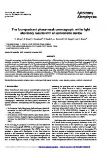
The robotics-driven flow patterning apparatus is shown in Figure 1(a). Major components of the robotics are numerically labeled in the figure, including the chip support tray (1), the injector module (2), the DNA solution reservoirs (4), and the translation motors (5). A detailed scheme of the injector module is presented in Fig. 1(b); the injector employs a standard microfluidics interfacing scheme wherein stainless steel pins are inserted into punched access holes that bridge the top surface of the PDMS, flow patterning molds with the microchannel/glass surface interfaces below.18 The stainless
Rev. Sci. Instrum. 82, 094301 (2011)
FIG. 1. (Color) The robotics platform for automated microfluidics flowpatterning of glass slides. (a) An overview of the instrument as implemented. Substrates are arrayed on the slide stage (1) and thereafter are addressed sequentially by a mobile injector head (2). A camera system (3) images the substrate access holes to guide alignment as the injector interfaces each substrate, and reagents are supplied from a set of adjacent microvials (4). Mechanical motion in the x, y, z, and θ axes is effected by a combination of linear stages and stepper drives (5). (b) Drawing showing detail of the injector assembly. This drawing illustrates the pin interface used to engage each substrate and the pneumatic pressure plate which prevents delamination when disengaging. (c) An image from the camera system during substrate alignment. The field of view encompasses just one corner of the substrate; green circles (enhanced for clarity) indicate access holes in the PDMS that have been recognized by the software pattern recognition algorithm and are used for fine adjustment of the injector head prior to interfacing. Scale bar: 2 mm.
steel pins are embedded within a laser-drilled acrylic “injector plate,” and are arranged according to a pre-determined pattern that matches the substrate access holes (Fig. 1(c)). This scheme allows for a high density of fluidic inputs, and it reduces substrate filling to a parallel process. However, the scheme also introduces a challenge related to the alignment of the pins to the access holes: the pins are 650 μm in diameter while the access holes are only 500 μm. The dimensional mismatch forces the elastomer to expand upon interfacing and thereby form a leak-proof seal around the pins. However, the soft nature of the elastomer also means that misalignment of the two components can lead to unwanted deformation or unintended puncturing of the PDMS, instead of smooth mating of pin and hole. The problem is compounded by the fact that all the pins must be aligned simultaneously, leaving very small tolerances for angular misalignment of the injector plate. As such, substrate-injector alignment is done in two phases. A pre-alignment is achieved by virtue of plastic cutouts on the substrate tray, which loosely define the locations of the (up to) 18 PDMS flow patterning chips. Finer alignment is provided by a Cognex IS5400 camera system
Downloaded 04 Nov 2011 to 131.215.220.186. Redistribution subject to AIP license or copyright; see http://rsi.aip.org/about/rights_and_permissions
094301-3
Ahmad et al.
mounted to the side of the injector head. Prior to engaging each substrate, the camera is positioned directly over the chip and images the access hole pattern, comparing it to a pretrained image using built-in pattern recognition algorithms (Figure 1(c)); x, y, and θ deviations are reported to the control software which adjusts the appropriate translation stages and re-images the substrate iteratively until a null deviation reading is achieved. The injector head is then shifted a precalibrated distance to align with the substrate and is slowly lowered into place until the pins sink 1 mm into their corresponding access holes. Once engaged, DNA solutions are supplied to the injector head from a set of adjacent, disposable microvials via short lengths of Tygon tubing. The delivery of precisely metered, microliter scale aliquots is typically accomplished by external syringe pumps, but here we offload metering responsibility to the PDMS chips themselves. Specifically, the microfluidic channels are fabricated with a set of input access holes, but no output holes, thereby creating a closed system upon substrate engagement. Because PDMS is air permeable, a pressurized solution injected into the input ports can displace air within the microchannel until it reaches the end.18 In this way, a very precise volume, defined by the input access hole and microchannel dimensions, is metered into each channel. The onchip metering allows for a relatively simple implementation of the pressure system used to drive solutions, as depicted in Figure 2. Briefly, the solution-containing microvials are connected to a pair of three-way solenoid valves (Gems Sensors M-series) that can be configured to connect one of three inputs to the vials. The inputs establish either positive pressure, a vent, or a closed system within the vials. The latter two states are achieved by virtue of an open and a sealed input on the solenoids. The positive pressure input derives from a compressed air source which is regulated to the pressure required to drive solutions through their microfluidic channels. Typical pressures range from 2–5 psi, and are set with inverse proportion to the pattern density of the chip in order to prevent cross-contamination of solutions from adjacent channels. An electronically controlled proportional valve is integrated in the pressure line and opens gradually to introduce the pressure via a gentle ramp, thereby avoiding splashing of the solutions in their microvials. After engaging a substrate, the microvials are pressurized to drive their solutions into the microchannels. Once filling is complete, the solenoids reconfigure to vent the
Rev. Sci. Instrum. 82, 094301 (2011)
microvials, and then reconfigure again to create a closed system prior to disengaging. This final state helps to balance hydrostatic pressures and prevent leaking from the injector pins in the disengaged state. Disengaging the injector head from a substrate requires additional engineered components; because the injector pins form a tight seal with their corresponding substrate access holes, care must be exercised to prevent the PDMS mold and glass substrate from delaminating while extracting the pins. The injector plate is therefore fitted with four pneumatic pistons whose rods secure a second “pressure plate” to its underside. Matching through holes in the pressure plate enable the injector plate pins to protrude beneath it during substrate engagement and manipulation. When disengaging a substrate, this pressure plate is extended to brace the PDMS firmly against the slide tray while the pins are extracted. The entire injector/pressure plate assembly slides into a slot on the machined injector carriage and is reproducibly located via two shoulder screws. This modular implementation makes it easy to swap injector heads with different pin configurations from run to run, thereby allowing significant flexibility in substrate design. B. Substrate fabrication
We standardized virtually all aspects of the microfluidic flow channel chip dimensions, and streamlined their production.19, 20 To generate PDMS substrates, a deep reactive ion etched Si master is clamped between two machined aluminum plates; the upper plate contains cutouts that will define the substrate dimensions, and the master is positioned such that its features are aligned within these cutouts (Figure 3). The resulting dimensional uniformity, particularly in thickness, obviates the need for sophisticated depth control when interfacing the injector head with substrates; a precalibrated constant is sufficient. The most critical substrate features, however, are the access holes which bridge microfluidic channels with the top side of the substrate; these must be positioned very precisely and reproducibly relative to one another because the rigidly defined injector plate interfaces with them simultaneously. We therefore developed a template to mold access holes as the substrate cures. Specifically, stainless steel wires are embedded into a laser-drilled acrylic plate in the required pattern. After pouring PDMS into the alu-
FIG. 2. (Color online) Schematic representation of the instrument’s simplified pressure system for driving reagents.
Downloaded 04 Nov 2011 to 131.215.220.186. Redistribution subject to AIP license or copyright; see http://rsi.aip.org/about/rights_and_permissions
094301-4
Ahmad et al.
Rev. Sci. Instrum. 82, 094301 (2011)
to originally mold the holes. The thin membranes are retained in the outlet ports. This means that we are able to generate a dead-end filled substrate that yields extremely consistent metering volumes. The substrate also fills relatively quickly due to the high air permeability of the thin membranes at the output ports. C. Software and operation
FIG. 3. (Color) The aluminum stencil used to fabricate each PDMS substrate standardizes the overall patterned dimensions and the access hole placement and size. A silicon wafer bearing barcode microfeatures is first sandwiched between two aluminum plates; PDMS precursor is poured into the cutouts and acrylic plates for molding access holes are affixed on top.
minum/silicon mold assembly (Fig. 3), this plate is secured to the top side such that the wires extend into the PDMS below. Upon curing, the plate is removed, leaving behind the templated inlet and outlet ports. The wires do not extend completely to the underlying Si mold, which prevents damage to the Si mold. Thus, a very thin membrane of PDMS at the bottom of each access hole is retained. For the inlet ports, these membranes are easily punctured in a single step by pressing the substrate onto the top side of the same acrylic plate used
The mechanical components of the instrument are controlled by custom software written in National Instruments (NI) Labwindows/CVI. Stage motion is powered by a standard 6K 4-Axis Motion Controller, while a NI DAQ card (PCI-6052E) provides digital and analog outputs to regulate an array of relays, solenoids, and proportional valves. The software presents an interface that allows users to click which of the 18 microchip positions on the substrate tray are to be processed. Once a run is initiated, the software assumes active control of all components, and processes the marked substrates sequentially without further intervention. Figure 4 illustrates the instrument’s process flowchart for a typical barcoding run; from the user perspective, it simply consists of laying out the substrates in their tray, filling the microvials with DNA solutions, and loading the appropriate configuration files before pressing a button to start the run. As such, the user can pattern up to 18 barcode substrates with