REVIEW ARTICLE
CELLULAR NEUROSCIENCE
published: 18 September 2014 doi: 10.3389/fncel.2014.00290
A synaptic mechanism for network synchrony Simon T. Alford * and Michael H. Alpert Department of Biological Sciences, University of Illinois at Chicago, Chicago, IL, USA
Edited by: Sergey M. Korogod, National Academy of Sciences of Ukraine, Ukraine Reviewed by: Patrik Krieger, Ruhr University Bochum, Germany Stefano Taverna, Italian Institute of Technology, Italy *Correspondence: Simon T. Alford, Department of Biological Sciences, University of Illinois at Chicago, M/C 068, Rm 4285, 840 West Taylor Street, Chicago, IL 60607, USA e-mail:
[email protected]
Within neural networks, synchronization of activity is dependent upon the synaptic connectivity of embedded microcircuits and the intrinsic membrane properties of their constituent neurons. Synaptic integration, dendritic Ca2+ signaling, and non-linear interactions are crucial cellular attributes that dictate single neuron computation, but their roles promoting synchrony and the generation of network oscillations are not well understood, especially within the context of a defined behavior. In this regard, the lamprey spinal central pattern generator (CPG) stands out as a well-characterized, conserved vertebrate model of a neural network (Smith et al., 2013a), which produces synchronized oscillations in which neural elements from the systems to cellular level that control rhythmic locomotion have been determined. We review the current evidence for the synaptic basis of oscillation generation with a particular emphasis on the linkage between synaptic communication and its cellular coupling to membrane processes that control oscillatory behavior of neurons within the locomotor network. We seek to relate dendritic function found in many vertebrate systems to the accessible lamprey central nervous system in which the relationship between neural network activity and behavior is well understood. This enables us to address how Ca2+ signaling in spinal neuron dendrites orchestrate oscillations that drive network behavior. Keywords: lamprey, oscillation, SK2, KCa 2, NMDA, locomotion, calcium, dendrites
INTRODUCTION Orchestration of neuronal activity within networks is integral to correct execution of behavior. Synchronization between groups of neurons is an organizational feature of many neural networks found in the central nervous systems of invertebrates (Wehr and Laurent, 1996; Riffell et al., 2009) to vertebrates (Womelsdorf et al., 2014) alike, and between microcircuits. Largescale synchrony between neurons is particularly evident in the spinal (Grillner, 2003; Goulding, 2009) and brainstem networks (Koshiya and Smith, 1999) controlling rhythmic movement, but are also common to hippocampal and neocortical networks (Buzsáki and Draguhn, 2004; Grillner et al., 2005; Yuste et al., 2005). Synchronously active microcircuits, like the neurons that comprise the lamprey spinal central pattern generator (CPG), are driven through the synaptic connectivity of excitatory and inhibitory neurons combined with intrinsic burst-terminating electrical properties (Wallén and Grillner, 1987; Buchanan, 1993). However, little is known about the electrical and integrative properties of the complex dendritic architecture of lamprey spinal neurons where synaptic- and voltage-dependent conductances shape potentials arriving at the soma. In contrast, the integrative properties of cortical pyramidal neuron dendrites and their synaptic inputs have been extensively characterized (Spruston, 2008), while less is known about how these intrinsic properties generate rhythmic network activity, and ultimately the behaviors they are thought to subserve. To understand how neural networks generate complex patterns of activity underlying behaviors, it will
Frontiers in Cellular Neuroscience
be necessary to understand both the specific patterns of connectivity between neurons and how individual neurons respond to the inputs that they receive. Thus, this review seeks to merge disparate fields of research—dendritic integration and spinal central pattern generation. In doing so, we hypothesize that the ionic mechanisms driven through two rhythm-generating conductances, namely the synaptic interaction between ensembles of NMDA receptors (NMDARs) and Ca2+ -dependent K+ channels, may have general implications for the synchronization of spinal to cortical networks. Thus, to explore the idea that active dendritic properties are at the core of this behavior, we examine in detail the lamprey spinal network and draw from other areas of dendritic research to enhance our understanding of what occurs at the level of the dendritic synapse to generate behavior.
SUPRASPINAL NETWORKS IN THE BRAINSTEM INITIATE AND MAINTAIN LOCOMOTOR DRIVE Vertebrate locomotion is initiated and maintained by evolutionarily conserved serial pathways originating in the forebrain (Ericsson et al., 2013; Grillner et al., 2013), projecting to the mesencephalic locomotor region (MLR; Dubuc et al., 2008) and then to command neurons of the reticulospinal (RS) system, which innervates the entire rostro-caudal extent of the spinal cord, including cervical and lumbar centers in mammals (Goulding, 2009), and all segmental levels in fish as well as lamprey (Buchanan et al., 1987). However, following their
www.frontiersin.org
September 2014 | Volume 8 | Article 290 | 1
Alford and Alpert
A synaptic mechanism for network synchrony
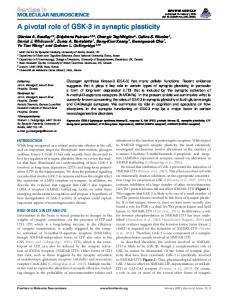
activation by the brainstem, it is the circuits and neurons of the spinal CPG (Buchanan and Cohen, 1982) that create the complex synergy that rhythmically activates the locomotor musculature (Grillner et al., 2008). The structure of descending commands to spinal CPGs and the synaptic connectivity of the spinal network itself provides an opportunity to understand how dendritic activation within behaviorally relevant circuits underlies the astonishing complexity of vertebrate behavioral patterns. The circuitry of the lamprey CPG is well understood (Grillner et al., 2000, 2008) including the identities of the key neurons (Rovainen, 1974; Buchanan and Cohen, 1982), their neuronal targets, and neuropharmacology (Alford et al., 2003). However, in common with most neurons, these circuit components possess a complex dendritic morphology (Figure 1), yet we understand little of the spatiotemporal profile of dendritic activation within these neurons and the role that such patterns of activation might play in the physiological activity of the neurons during behavior. This lack of understanding is true for simple inputs, but particularly during goal-directed locomotion. This is partly because tracing the spatial distribution of physiological targets of neurons is challenging, but also because most studies of CPGs, whether in simple systems like the lamprey, or in more complex systems such as mammals, use isolated spinal cords and activate the networks pharmacologically (Sigvardt et al., 1985; Rossignol et al., 1998; Kyriakatos et al., 2011). This undoubtedly obscures the precise physiologically relevant spatiotemporal activation patterns of dendritic synapses that would otherwise drive these behaviors in vivo. In studies of spinal motor activity this has been largely overlooked perhaps due to the strong resemblance of electrophysiological output (i.e., fictive locomotion), or even actual movement, to observed locomoting animals. Despite this similarity, it is crucial to understand how physiological patterns of synaptic input and intrinsic membrane electrodynamics generate rhythmic behaviors.
THE SYNAPTIC CONNECTIVITY OF THE SPINAL CPG NETWORK DRIVES RHYTHMIC NETWORK OSCILLATIONS The very fluid, controlled nature of lamprey locomotion is produced after RS axons activate the local circuit neurons within the spinal ventral horn (Figure 2). Among these neurons, collectively referred to as ventral horn neurons (VHNs), the best characterized neurons responsible for pattern generation are excitatory interneurons (EINs) that provide ipsilateral, glutamatergic excitation (Buchanan and Grillner, 1987; Buchanan et al., 1989), while crossed caudally projecting interneurons (CCINs) provide contralateral, glycinergic inhibition (Grillner and Wallén, 1980; Alford and Williams, 1989). Motor neurons are the final common output neuron of each segment, which bundle into ventral roots (VRs) as they leave the spinal cord, before synapsing directly onto myotomal cells of the trunk musculature (Buchanan and Cohen, 1982). The precise, synaptic connectivity of the VHNs within and between individual segments serves to ipsilaterally excite (i.e., EINs), while simultaneously delivering contralateral inhibition (i.e., CCINs; Buchanan and Grillner, 1987). This reciprocally inhibited network ensures that within each segment, when one side of the trunk musculature contracts, the contralateral
Frontiers in Cellular Neuroscience
FIGURE 1 | Lamprey spinal motoneurons have a complex dendritic architecture. (A) Schematic representation of an isolated lamprey brain and spinal cord. Spinal motoneurons and their complete dendritic architecture can be retrogradely labeled through an intramuscular injection of a dextran-conjugated fluorescent dye. Labeling (green) is visible on the side and segment of injection through axons converging into ventral roots (VRs) and to their respective neurons. Expansion shows a single spinal segment with multiple motoneurons labeled as in (B). (B) A 3D reconstruction of motoneurons labeled from one spinal ventral root to emphasize the complexity of their structure and dendritic trees. Neurons were labeled by injecting the muscle wall of an animal with 5 µL of 5 mM Oregon Green 488 BAPTA1 Dextran. After 24 h the animal was sacrificed and the spinal cord fixed and cleared. A confocal stack was imaged to generate the full extent of the motoneurons in one spinal segment (Viana di Prisco and Alford, 2004).
side is inhibited. Lateral interneurons, which project ipsilaterally to inhibit CCINs, facilitate the relief of reciprocal inhibition (Buchanan, 1982). However, the importance of lateral interneurons in maintaining network rhythmicity has been less emphasized because alternating, rhythmic bursting can persist in their absence as demonstrated by computer simulation (Wallén et al., 1992). Work in lamprey (Grillner et al., 1981; Brodin et al., 1985, 1988; Brodin and Grillner, 1985; Buchanan and Grillner, 1987), Xenopus tadpoles (Dale and Roberts, 1984; Roberts and Alford, 1986), rats (Kudo and Yamada, 1987), and cats (Douglas et al., 1993) demonstrates that spinal glutamate receptor-mediated transmission activates and maintains locomotion. These data are supported by recordings of excitatory postsynaptic potentials (EPSPs) onto motoneurons and premotor interneurons
www.frontiersin.org
September 2014 | Volume 8 | Article 290 | 2
Alford and Alpert
A synaptic mechanism for network synchrony
FIGURE 2 | Schematic representation of the lamprey spinal central pattern generator. (A) Spinal CPG neurons receive both ipsilateral glutamatergic (red) input from excitatory interneurons (EINs, red) and contralaterally projecting glycinergic inhibition (blue) from reciprocally inhibiting, crossed glycinergic interneurons (CCINs, blue). Output of the CPG occurs from motoneurons (green), which directly synapse onto myotomal cells of the trunk musculature to cause muscle contraction producing
(Dale and Roberts, 1985; Brodin et al., 1988; Noga et al., 2003) and pharmacological manipulation of the resultant behaviors (Brodin and Grillner, 1985; Dale and Roberts, 1985; Cazalets et al., 1992; Chau et al., 2002; Rybak et al., 2006). This neurotransmission both directly excites neurons of the CPG, and also activates complex non-linear membrane interactions, or oscillations, in these neurons mediated by NMDAR voltage-dependency and Ca2+ permeability coupled to the activation of Ca2+ -dependent currents. The cellular processes underlying such oscillations are believed to be central to the coordination of locomotor behavior. In lampreys the identity of the descending glutamatergic RS command neurons is well-defined (Dubuc et al., 2008) and similarly spinal neurons that release glutamate locally within the spinal ventral horn (i.e., EINs) have been identified (Buchanan et al., 1989;
Frontiers in Cellular Neuroscience
rhythmic locomotion. (B) Output pattern recorded using glass suction electrodes from paired, contralateral (top vs. bottom traces) VRs showing alternating bursting of the spinal network during rhythmic locomotion. The reciprocally connected network described in (A) prevents excitation of the contralateral spinal cord when the ipsilateral side is bursting for each cycle (burst-to-burst), leading each side of the spinal cord to be precisely 180◦ out-of-phase from the other (Alford et al., 2003).
Buchanan, 1993) as has their network role (Wallén et al., 1992). One prominent feature of the spinal network is that it transforms unpatterned, exogenous glutamatergic input into a patterned, rhythmic output. The details of synaptic connectivity responsible for this phenomenon have been substantially explored in the lamprey (Wallén and Grillner, 1987; Grillner et al., 2001; Grillner, 2006) and the Xenopus embryo (Dale and Roberts, 1984, 1985). More recently, work in higher vertebrates (Masino et al., 2012) has emphasized how well conserved this network motif is throughout the vertebrate subphylum including lampreys, fishes, amphibians, chelonids and mammals (Dale and Roberts, 1984; Sigvardt et al., 1985; Kudo and Yamada, 1987; Hernandez et al., 1991; Guertin and Hounsgaard, 1998; Gabriel et al., 2009; Masino et al., 2012). After complete
www.frontiersin.org
September 2014 | Volume 8 | Article 290 | 3
Alford and Alpert
A synaptic mechanism for network synchrony
spinal transection (Cohen and Wallén, 1980; Brodin et al., 1985), the lamprey swimming network can still generate the electrophysiological correlates of swimming. While recording output from pairs of contralateral VRs using glass suction electrodes, excitatory amino acid (EAA) receptor agonists, such as kainate, D-glutamate, or N-methyl-D-aspartic acid (NMDA; Brodin et al., 1985; Brodin and Grillner, 1985; Wallén and Grillner, 1987), bath-applied to an isolated spinal cord (devoid of muscle or any other surrounding tissue) generates antiphasic bursts of activity across the spinal midline—the phase relationship across sides of the spinal cord is enforced by glycinergic inhibition (Cohen and Wallén, 1980; Alford and Williams, 1989)—and the same rostro-caudal phase lag as seen in intact behavior (Wallén and Williams, 1984). This network behavior, termed “fictive locomotion”, refers to the electrical output of the spinal CPG. Thus, the network acts as a CPG, a term that refers collectively to centrally located, local circuit spinal neurons that provide precise rhythmic output from spinal motoneurons. The spinal CPG operates in the absence of both sensory feedback from the spinal dorsal roots or descending networks and is found in all vertebrates (Kahn and Roberts, 1978; Forssberg et al., 1980; Roberts et al., 1981; Sholomenko and Steeves, 1987; Delvolvé et al., 1997; Field and Stein, 1997; Masino and Fetcho, 2005). Thus, the ability to generate rhythmic output via network oscillations is inherent to the spinal network itself and does not require supraspinal control.
SINGLE NEURONS ARE INTRINSICALLY RHYTHMIC The study of spinal neurons offers a unique insight into how properties of neural networks emerge from membrane activity at the cellular level and provides a straightforward behavioral context— locomotion—in which to place this activity. EAA agonists, like NMDA, cause the membrane potential (Vm ) of individual VHNs in isolated spinal cords to undergo repetitive oscillations that are in-phase with the ipsilateral VR of the corresponding hemisegment (Sigvardt et al., 1985; Wallén and Grillner, 1987). During the depolarized phase, the cells can fire multiple action potentials (APs) before the cell is repolarized. This finding demonstrates how electrical properties of single cells within a network scale to direct the behavior of the network at large. Most VHNs oscillate in NMDA driven by phase-appropriate synaptic excitation from EINs and subsequent hyperpolarization from CCINs (Buchanan and Cohen, 1982). However, when tetrodotoxin (TTX) is applied, spiking is abolished, while the underlying Vm oscillation persists (Wallén and Grillner, 1987). Since TTX pharmacologically isolates the recorded neuron by preventing synaptic communication within the network, the cell then oscillates with tonic exposure to NMDA. This phenomenon, termed NMDA-dependent, TTXresistant oscillations (NMDA-TTX oscillations), is seen in most lamprey VHNs. This demonstrates that spinal neurons show intrinsic membrane properties that are capable of hyperpolarizing the cell during constant depolarizing challenge from an agonist. The net effect is to produce V m oscillations. Removal of Mg2+ from the perfusing Ringer’s solution abolishes the oscillation and causes the neurons to remain at depolarized potentials because Mg2+ confers voltage-sensitivity to the NMDAR (Wallén and Grillner, 1987). Thus, the intrinsic membrane property of
Frontiers in Cellular Neuroscience
spinal neurons that causes oscillations is subject to the voltagedependency of Mg2+ block of the NMDAR. More generally within the nervous system, NMDARs have been well characterized as non-specific cation channels permeable to Na+ , K+ , and Ca2+ (MacDermott et al., 1986; Ascher and Nowak, 1988). More recently, NMDAR-dependent Ca2+ entry has been demonstrated to be integral to dendritic computation (Branco et al., 2010) through regenerative “NMDA spikes” in pyramidal neurons (Schiller and Schiller, 2001; Larkum et al., 2009) with roles spanning from the induction of synaptic plasticity (Alford et al., 1993) to behavior (Smith et al., 2013b). In lamprey VHNs, removal of Ca2+ from the ringer and replacement with Ba2+ (an equivalent divalent cation which can also permeate Ca2+ ionophores) during NMDA-TTX oscillations causes the cell to become similarly trapped at a depolarized Vm . Thus, Ca2+ is necessary to hyperpolarize the cell from the depolarized state. Ca2+ activates myriad Ca2+ -dependent proteins. In particular, VHNs contain Ca2+ -dependent K+ channels (El Manira et al., 1994; Wall and Dale, 1995; Han et al., 2007; Li and Bennett, 2007), which upon binding Ca2+ , rapidly open a K+ channel that hyperpolarizes the cell. This “excitation-inhibition coupling” is a mechanism that effectively allows the cell to “turn off ” autonomously following activation. The Ca2+ -dependent K+ channel of the KCa 2 subtype (formerly SK2 (Wei et al., 2005)) participates in two distinct processes in lamprey VHNs both of which are integral to the behavioral locomotor output of the spinal cord. Its most well-described role follows the AP when depolarization activates N- and P/Qtype (Wikström and El Manira, 1998) voltage-gated Ca2+ channels (VGCCs) and the entering Ca2+ activates KCa 2 channels to cause an afterhyperpolarization (AHP; Figure 3; Hill et al., 1992; Meer and Buchanan, 1992). The AHP can be divided into fast, medium and slow subcomponents, of which the medium AHP (mAHP) is mediated by KCa 2 channels (Bond et al., 2004). Due to slow kinetics (decay time constant of ∼200 ms), the mAHP mediates spike frequency adaptation, the reduction in spike frequency from repeated spiking, by raising the relative threshold for subsequent AP generation due to an increase in K+ conductance. Blockade of KCa 2 channels with the selective antagonist, apamin, increases spike frequency from intracellular current pulses (Meer and Buchanan, 1992; Díaz-Ríos et al., 2007; Jones and Stuart, 2013). KCa 2 channels are extremely important for regulating neuronal firing, conserved among different species and cell types (Meer and Buchanan, 1992; Sah and Bekkers, 1996; Marrion and Tavalin, 1998; Wikström and El Manira, 1998; Faber and Sah, 2002; Bloodgood and Sabatini, 2007; Jones and Stuart, 2013). The second role for KCa 2 lies in the plateau termination and membrane repolarization during NMDA-TTX oscillations (Figure 3). The ionic mechanism driving Vm oscillations is well-characterized and is hypothesized to proceed as: (1) NMDAR activation depolarizes VHNs; (2) increasing NMDAR conductance by ejecting Mg2+ from the pore; (3) causing further depolarization and Ca2+ entry via the NMDAR as the Vm plateaus; (4) which activates KCa 2 channels to hyperpolarize the cell; and (5) ending the depolarized plateau to repolarize the cell where it can repeat the cycle (Wallén and Grillner,
www.frontiersin.org
September 2014 | Volume 8 | Article 290 | 4
Alford and Alpert
A synaptic mechanism for network synchrony
FIGURE 3 | Continued regular, alternating bursts (control) follows after a puff of glutamate into the MLR (arrow, glutamate). Blockade of KCa 2 channels with the selective antagonist, apamin, decreases the burst frequency and disrupts the alternating locomotor rhythm. This demonstrates the necessity of KCa 2 channels for correct alternation and regularity of the locomotor rhythm (Nanou et al., 2013). (B) The effect of KCa 2 channel blockade on locomotion can be explained by the role the channel plays at the cellular level. Within VHNs, KCa 2 currents may be evoked either at synapses (top left) whereby synaptic release of glutamate activates NMDAR-mediated Ca2+ entry and thereby closely located KCa 2 channels. It is this KCa 2-mediated current that is critical for the termination of NMDA-TTX oscillations (blue portion of trace) shown below recorded from somatic microelectrode recordings. KCa 2-mediated currents are also responsible for the mAHP seen following action potential firing shown at bottom left. However, this current is activated following Ca2+ entry from VGCCs.
1987). Selective blockade of KCa 2 channels with apamin (El Manira et al., 1994) or UCL 1684 (Alpert and Alford, 2013) prolongs the oscillation, and can even abolish the oscillation completely. The cell becomes trapped in a depolarized state, similar to extracellular Ca2+ removal, the substitution of Ca2+ for Ba2+ , or non-specific blockade of K+ channels (Grillner and Wallén, 1985; Grillner et al., 2001). Thus, KCa 2 channels are necessary for rhythmogenesis (Figure 3) in lamprey VHNs by supplying a cell-autonomous repolarization, or “off signal”, without the need of network inhibition (Nanou et al., 2013).
FIGURE 3 | Repolarizing KCa 2 channels are spatially segregated in lamprey spinal VHNs according to their function and mechanism of activation. (A) Top: An isolated lamprey CNS can be used to study the brain and spinal circuits controlling locomotion. Pressure-ejection of L-glutamate into the lamprey mesencephalic locomotor region (MLR) induces short episodes of fictive locomotion, the electrophysiological correlate of locomotion. Using a dual-pool recording chamber, pharmacological agents can be selectively applied to the spinal cord, without interfering with descending commands originating in the brainstem that initiate and maintain locomotion. Locomotor bursts are recorded directly from left and right VRs. Bottom: A long locomotor episode with (Continued)
Frontiers in Cellular Neuroscience
DENDRITIC Ca2+ SIGNALING IS DYNAMIC AND DETERMINED BY CELLULAR AND MICROCIRCUIT PROPERTIES KCa 2 channels within a single neuron have more than one distinct computational role. Two have been identified in lamprey VHNs, both subject to intracellular Ca2+ dynamics. Such a functional sub-specialization may be explained both by distinct spatial locations of channel expression and the adequate spatial and functional coupling to distinct sources of Ca2+ contributing to KCa 2 activation (Figure 3). Indeed, N- and P/Q-type (Wikström and El Manira, 1998) VGCCs are activated during the AP in lamprey, triggering Ca2+ entry that activates KCa 2 channels underlying the mAHP. However, the mAHP activated by somatic current injection is unaffected by NMDA application (Hill et al., 1989). This distinct separation between mAHP activation and NMDA-TTX oscillation repolarization can be explained by NMDAR-generated Ca2+ entry occurring in spatially distinct cellular sub-regions from VGCC-generated Ca2+ entry during the AP. Across different species and neuron types, the precise subtypes of VGCCs can differ, but to mediate the mAHP, KCa 2 channels must be sufficiently close to VGCCs to be activated by their Ca2+ permeation. Similarly, KCa 2 channels mediating repolarization during NMDATTX oscillations should be coupled to a distinct Ca2+ source, or a Ca2+ source in a distinct subcellular location. The two likely candidates for the latter are NMDARs and VGCCs (Wallén and Grillner, 1987)—located separately from those responsible for the mAHP (Hill et al., 1989)—while Ca2+ released from internal stores might also contribute. NMDAR activation is necessary to initiate oscillations, but as they lead to membrane depolarization, this may subsequently activate VGCCs. However, release from
www.frontiersin.org
September 2014 | Volume 8 | Article 290 | 5
Alford and Alpert
A synaptic mechanism for network synchrony
internal stores likely contributes little because their depletion has no effect on NMDA-induced swimming (Krieger et al., 2000)— a behavior to which NMDAR-dependent intrinsic oscillations contribute. The subcellular location of KCa 2 channels responsible for the repolarization may also be critical because physiological NMDAR activation requires the presynaptic release of glutamate, which occurs only at synapses. Determining the route of Ca2+ entry for repolarization of the oscillation is important for understanding how distinct Ca2+ domains and their coupling to KCa 2 channels impacts computation both within individual neurons and between synaptically connected neurons. The spatial and temporal patterning over which dendritic Ca2+ signaling occurs in spinal motor system VHNs during locomotion in lamprey (or in other vertebrate systems) is unknown. Do many dendrites receive synchronous input from their various synaptic partners? Does input occur in discrete spatial locations? The location and timing of synaptic input is crucial for the transmission of potentials arriving at the soma, which will greatly influence neuronal output (Larkum et al., 1999; Stuart and Häusser, 2001; Jarsky et al., 2005). Indeed, dendritic mechanisms that are location-dependent and rely on clustered NMDAR-dependent input generate plateau potentials and can change the mode of cell firing (Major et al., 2008; Augustinaite et al., 2014; Grienberger et al., 2014). Elucidating this pattern within lamprey spinal neurons will inform how the location and timing of Ca2+ entry leads to KCa 2 channel activation, and furthermore, how synaptic activity distributed across a dendritic tree is integrated to produce cell rhythmicity. This, in turn, will facilitate our understanding of how intrinsic membrane properties combined with synaptic input causes synchronization between neurons of the CPG. Neuronal Ca2+ signaling can have distinct spatial components, easily identifiable using Ca2+ imaging. APs will lead to Ca2+ entry wherever VGCCs are driven above threshold, and can cause many regions of a cell (e.g., soma and proximal dendrites) to show synchronized increases in intracellular Ca2+ (Ca2+ i ). In contrast, local Ca2+ signaling domains (i.e., micro- and nano-domains) in dendrites can occur following neurotransmitter receptor activation (e.g., NMDAR), but also from VGCCs following depolarization from local synaptic potentials (Augustine et al., 2003). Synaptic signals are remarkably localized, confined to individual dendritic spines or discrete areas in dendritic shafts. For this reason, Ca2+ imaging can directly identify active synapses. Each type of Ca2+ signaling domain may be considered to be a distinct processing unit within a neuron because Ca2+ signals can regulate local Ca2+ -dependent processes precisely where free Ca2+ levels transiently escape local buffering. However, Ca2+ signals exceeding this local threshold are transient—Ca2+ is rapidly buffered by Ca2+ -binding proteins, and then extruded via membrane pumps, or sequestered in intracellular stores (Augustine et al., 2003; Berridge, 2006). This places temporal and spatial restrictions on diffusion of Ca2+ within neurons and is an important consideration when assessing the degree of localization. Dendritic morphology, like the presence of spines (∼1 µm in length), is a large determinant for the extent of spread of Ca2+ because diffusion is restricted at the spine neck (Nimchinsky et al., 2002). Lamprey spinal neuron dendrites lack spines, but still posses fine compartments along dendritic shafts (∼10 µm, see Figure 1; Viana di Prisco and Alford, 2004;
Frontiers in Cellular Neuroscience
Alpert and Alford, 2013) that may theoretically serve a similar purpose—the local restriction of the flow of ions and intracellular messengers (Svoboda et al., 1996). Thus, morphology and the intrinsic properties of the dendritic membrane impacts Ca2+ dynamics and the integration of electrical and chemical signals. The functional distinction between global and local Ca2+ signals and their associated topography is integral to single neuron computation necessary to generate rhythmic activity. The synaptic localization of Ca2+ signals may represent the encoding of distinct presynaptic information. Global, synchronized Ca2+ signals can be generated by back-propagating action potential (bAP)-driven VGCC activation in dendrites (Schiller et al., 1997; Stuart et al., 1997; Svoboda et al., 1997). When Ca2+ i is elevated during these events, the number of parallel computations being performed by the dendritic arbor is effectively reduced. In contrast, local and spatially distributed NMDAR-dependent synaptic Ca2+ signals reflect multiple discrete, simultaneous computations (Chen et al., 2011). Each synapse can thus be understood to be its own computational unit, capable of being selectively tuned to support distinct information arriving within a network. Multiple, distinct routes can lead to Ca2+ entry. In behaving neurons within some networks, these mechanisms may work in concert, leading to nonlinear interactions between ion channels and Ca2+ sources when occurring simultaneously. For instance, following presynaptic release of glutamate, AMPA receptors (AMPARs), NMDARs and metabotropic glutamate receptors (mGluRs) may be activated in the postsynaptic compartment. AMPARs are responsible for fast depolarization, and can locally activate nearby VGCCs to cause Ca2+ entry. Local depolarization, or depolarization induced from bAPs can alleviate Mg2+ block of the NMDAR, facilitating Ca2+ influx during concurrent and subsequent release of glutamate at that synapse (Yuste and Denk, 1995; Nevian and Sakmann, 2004; Bloodgood and Sabatini, 2007). During bAPs, layer 5 pyramidal neurons have been shown to require tight spatial coupling between Ca2+ entry through R-type channels and KCa 2 channels in proximal dendrites and spines (Jones and Stuart, 2013). Group I mGluR activation can lead to the release of Ca2+ from internal stores (Frenguelli et al., 1993; Kettunen et al., 2002; Larkum et al., 2003; Topolnik et al., 2009; Plotkin et al., 2013). Release from internal stores has been shown to activate Ca2+ -dependent K+ channels in many neurons and species (Kawai and Watanabe, 1989; Akita and Kuba, 2000; Yamada et al., 2004; Faber, 2010; Nakamura and Yokotani, 2010). It is unknown if such combinatory mechanisms are present in lamprey spinal neurons, but lamprey neurons do possess all the necessary components. Indeed, specific agonists and antagonists acting on discrete components have well-described cellular and network effects (Alford et al., 2003). Any modulation of Ca2+ entry, either increasing or decreasing, within close proximity to KCa 2 channels, could impact subsequent channel activation and particular effects on the locomotor behavior. For example, an enhancement of Ca2+ could lead to early burst termination— an effect that, if it were to occur within many neurons simultaneously, would scale to the behavioral level to terminate muscle contraction earlier within the locomotor cycle. Upon repeated enhancements in Ca2+ , during rhythmic activity, this could
www.frontiersin.org
September 2014 | Volume 8 | Article 290 | 6
Alford and Alpert
A synaptic mechanism for network synchrony
facilitate a fast swimming rhythm. Defining their roles while acting in concert is necessary to delineate how Ca2+ entry and KCa 2 activations drives oscillation generation. The location of Ca2+ entry and the distance to its secondary effectors determines the efficacy with which Ca2+ will reach its target (Marrion and Tavalin, 1998). If the site of Ca2+ entry is located far from KCa 2 channels, then the probability of Ca2+ binding to a KCa 2 channel is diminished compared to its binding to other endogenous buffers that are located more proximally or are cytosolic and diffusible. Thus, a larger Ca2+ signal will be necessary to outcompete endogenous buffers. Conversely, if KCa 2 channels are located close to the site of Ca2+ entry, then depolarization will be quickly and locally counteracted by K+ activation. For KCa 2 channels to generate the mAHP, they must be sufficiently close to the site of Ca2+ entry generated by AP-driven VGCC activation. This functional coupling has been demonstrated in numerous species and cell types (Sah and Bekkers, 1996; Marrion and Tavalin, 1998; Wikström and El Manira, 1998; Faber and Sah, 2002; Bloodgood and Sabatini, 2007; Jones and Stuart, 2013). At present, the distance between the site of Ca2+ entry and KCa 2 channels can only be estimated based on differences between BAPTA and EGTA-mediated occlusion of KCa 2 activation in lamprey spinal VHNs. The range has been estimated to be between 20 and >200 nm in multiple CNS neuron types depending on the target’s affinity for Ca2+ (Fakler and Adelman, 2008). A recent measurement has suggested that KCa 2 channels activated following APs exhibit weak coupling to VGCCs, as they are occluded by EGTA, the slow Ca2+ buffer (Kforward = 1.5 × 10⁶ M−1 s−1 ) (Roberts, 1993), placing the separation at greater than ∼100 nm in neocortical pyramidal neurons (Jones and Stuart, 2013). Occlusion of KCa 2 channel activation from NMDAR-dependent Ca2+ entry using BAPTA, the fast Ca2+ buffer (Kforward = 6 × 10⁸ M−1 s−1 ) (Roberts, 1993), demonstrates a narrow range of 20–50 nm (Ngo-Anh et al., 2005), with experiments in lamprey suggesting similar degree of coupling (Alpert and Alford, 2013; Nanou et al., 2013). For KCa 2 channels to repolarize NMDA-TTX Vm oscillations, they must be activated by NMDAR-dependent Ca2+ entry. The subcellular expression of ion channels, including KCa 2 channels, is unknown in lamprey, while some spatial information has been detailed for mammalian hippocampal neurons. KCa 2 channel immunoreactivity demonstrates channel expression on dendritic spines in CA1 pyramidal neurons (Sailer et al., 2004; Ballesteros-Merino et al., 2012) in addition to shafts and soma in cultured mice hippocampal neurons (Ngo-Anh et al., 2005). Recently, however, using single-molecule atomic force microscopy with unprecedented spatial resolution (