한국전문물리치료학회지 제 15권 제 4호 PTK
Vol. 15
No. 4
2008.
The Effects of EEG Power and Coherence on Cognitive Function in Normal Elderly, Non-Demented Elderly With Mild Cognitive Impairment, and Demented Elderly During Working Cognition Task
Dong-wook Han, Ph.D., P.T.
Dept. of Physical Therapy, Silla University
Abstract 1) The purpose of this study was to find out the effects of electroencephalograph (EEG) power and coherence on cognitive function in normal elderly, non-demented elderly with mild cognitive impairment, and demented elderly during working cognition tasks. Forty elderly women (19 demented elderly, 10 non-demented elderly with mild cognitive impairment, 11 normal elderly) participated in this study. All subjects performed working cognition tasks with Raven's CPM while EEG signal was recorded. EEGs were measured continuously at rest and during the working cognition task. EEG power and coherence was computed over 21 channels: right and left frontal, central, parietal, temporal and occipital region. We found that there were more correct answers among normal elderly women than in other groups. During the working cognition task, θ wave at Fp1, Fp2 and F8, a wave at Fp2, ß wave at Fp1, Fp2, F4 and F8 of the frontal region was increased significantly in the demented elderly group. On the other hand, θ wave at Fp1, Fp2 and F7, ß wave at Fp1, Fp2, F3 and F7 of the frontal region was increased significantly in the group of non-demented elderly with mild cognitive impairment. In contrast, in the normal elderly group, all of the θ wave and ß wave at Fp1, Fp2, F3, F4, F7 and F8 of the frontal region (except ß wave at F3) was increased significantly. These results suggest that the nerves in prefrontal and right hemisphere regions were most active in the demented elderly group during problem solving, and the nerves in the prefrontal and left hemisphere lobe were most active in the group of non-demented elderly with mild cognitive impairment. In contrast, the majority of nerves in the frontal region were active in the normal elderly group.
Key Words:
Demented elderly; Electroencephalograph; Non-demented elderly with mild cognitive impairment; Normal elderly; Working cognition task.
Cognitive function refers to memory, visual-spatial
Introduction
capacity, frontal lobe performance, and linguistic and Failing of brain function with aging generally oc-
calculation ability. A person who once performed nor-
curs with decrease in the number of neurons, changes
mal daily activities with normal cognitive function may
of neurotransmitter and synaptic transmission in neu-
suffer from a decrease in cognitive function resulting
rons, biochemical changes, such as oxidization and in-
from acquired degenerative changes or vascular prob-
flammation, and pathological changes, such as amyloid
lems, and these overall symptoms are called dementia
deposition. Symptoms of failing of brain function with
(Choi, 2005). Accordingly, dementia is a cognitive
aging include decline in logical thinking, processing
function disorder which is chronic and progressive and
speed, and short-term memory (Compton et al, 2000).
includes damage to memory, thinking, understanding,
Corresponding author: Dong-Wook Han
[email protected] This work was supported by the Korea Research Foundation Grant funded by the Korean Government (MOEHRD, Basic Research Promotion Fund) (KRF-2007-331-E00188).
- 70 -
한국전문물리치료학회지 제 15권 제 4호 PTK
Vol. 15
No. 4
2008.
orientation, learning, judgment, and linguistic ability
Methods
(Mckhan and Drchman, 1984). Mild cognitive impairment, which develops into dementia, is defined to show
S tudy Design
a level of cognitive function, somewhere between nor-
Subjects were divided into three groups of normal
mal and demented (Jiang, 2005). Mild cognitive impair-
elderly, non-demented elderly with mild cognitive
ment does not cause great inconvenience in daily and
impairment, and demented elderly. To identify elec-
social activities (Korea Dementia Association, 2006).
troencephalogram changes in each group at rest and
Generally, people who suffer from mild cognitive im-
during
pairment have clear and logical thinking ability and
perimental design and pretest-posttest nonequivalent
lead normal activities of daily living, but they have a
control group design were employed. The study cov-
significantly
ered the period from January 2 to February 28, 2008.
poorer
memory,
especially
short-term
the
working
cognition
task,
quasi-ex-
memory, when compared with people of the same age who received same level of education (Lee, 2005).
S ubj ects
There are several methods which are used to di-
Researchers randomly selected three geriatric in-
agnose dementia. Recent studies show that electro-
stitutions in the Busan area. Subjects were selected
encephalography (EEG) is useful for evaluating brain
from the female elderly in those institutions who volun-
function, as changes in brain function can be exam-
tarily agreed to participate in the study after having
ined while the brain is performing cognitive activities
listened to purpose of the study. The subjects were able
(Kikuchi et al, 2002; Hwang et al, 2005). Moreover,
to perform normal daily activities, without any impair-
analysis of electroencephalogram helps classify the
ment in vision, audition, and language communication.
group of mild cognitive impairment (Jelic et al, 2000).
The subjects were also free from any kind of encephal-
EEG is more useful than other methods in some
opathy, such as a cerebrovascular accident (CVA), and
aspects. While single-photon emission computed to-
they
scored
under
21
on
the
Korean-Geriatric
mography (SPECT) and positron emission tomog-
Depression Scale (K-GDS) (Jung et al, 1998), signifying
raphy (PET), which use isotope in measurement, are
they do not suffer serious depression. Forty subjects
relatively slow in measurement of speed. EEG, which
were divided into three groups: dementia (19 persons,
uses electric signals in measuring brain activation, is
average age: 79.3 yrs), mild cognitive impairment (10
faster than other methods (Chance et al, 1993).
persons, average age: 81.5 yrs), and normal (11 persons,
Functional magnetic resonance imaging (fMRI) and
average age: 80.3 yrs).
PET are useful for determining the focuses, but lack time accuracy (more than one second). In other words, they capture images of the brain after some changes
Experiment Method
the brain. In the case of EEG, however, it is possible
Tools for Subject Selection and Selection Standard
to identify real time changes (Dale and Halgren, 2001).
Subjects were divided into three groups based on
This study is designed to identify electroencephalo-
the results of Mini-Mental State Examination-Korean
gram differences in normal elderly, non-demented eld-
(MMSE-K), Clinical Dementia Rating (CDR), and
erly with mild cognitive impairment, and demented
Global Deterioration Scale (GDS). At Mini-Mental
elderly at rest and during a working cognition task.
State Examination-Korean (Park and Kwon, 1989),
The results are to be used as a basic resource in di-
those who scored more than 24 points were classi-
agnosis of dementia and mild cognitive impairment and
fied as normal, 20 to 23 points as having mild cog-
in developing effective cognition rehabilitation methods.
nitive impairment, and under 19 points as having
occur, so it is difficult to grasp real time changes in
- 71 -
한국전문물리치료학회지 제 15권 제 4호 PTK
Vol. 15
No. 4
2008.
dementia. For Clinical Dementia Rating (Korean
Electroencephalography (EEG)3)
Association for Geriatric Psychiatry, 2003), point 0
A
was normal, .5 was mild cognitive impairment, and 1
used
to 3 was dementia. As for the result of Global
electroencephalography was performed by a neuro-
Deterioration Scale (GDS) (Reisberg et al, 1982),
physiologist who works at D hospital in Busan, and
GDS 1 and GDS 2 were classified as normal, GDS 3
has
and GDS 4 as mild cognitive impairment, and GDS 5
encephalograms (Figure 2). In accordance with the
to GDS 7 as dementia. To be classified into one of
ten-twenty electrode system, the electrodes were at-
the three groups of dementia, mild cognitive impair-
tached to Fp1, Fp2, Fpz, F3, F4, F7, F8, Cz, C3, C4,
ment, and normal, the subjects had to get the same
T3, T4, T5, T6, Pz, P3, P4, O1, O2, and ear lobes
results from at least two of the three measurement
(Kim et al, 2005). Fp1, Fp2 fall on the prefrontal
scales mentioned above.
lobe, Fpz on the median frontal lobe, F3, F4 on the
wireless for
portable
electroencephalograph2)was
electroencephalogram
extensive
experience
in
measurement;
measuring
the
electro-
median and rear frontal lobe, F7, F8 on the rear frontal lobe and front temporal lobe, Cz on the me-
Instruments
Cognitive Function Test Computerized 1)
(CNT)
dian central part, C3, C4 on the central part, T3, T4 on the median temporal lobe, T5, T6 on the rear
2)
Neurocognitive
Function
Test
temporal lobe, Pz on the median parietal lobe, P3, P4
was used for the cognitive function test of
on the parietal lobe, and O1, O2 on the occipital lobe
this study. It is designed to examine various neuro-
(Kim and Choi, 2001).
cognitive functions, such as attentiveness, memory, cognition-movement coordination, and comprehensive
Procedures
cognitive thought. Among 18 cognition measurement
Electroencephalography was carried out on normal
programs, the study used Raven's CPM (colored
elderly, non-demented elderly with mild cognitive
progressive matrices), which can be applied to any-
impairment, and demented elderly at resting state.
one regardless of literacy (Figure 1). Raven's CPM
Before
consists of 36 items, and the assessment was made
asked to take a rest on a comfortable chair for five
based on the number of correct answers.
minutes with their eyes closed. Electroencephalogram
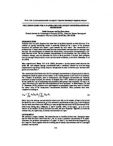
Figure 1.
Cognitive function test.
Figure 2.
the electroencephalography,
Electrodes position.
Figure 3.
Cognitive function test
and EEG. 1) CNT, MaxMedica Inc., Korea. 2) EEG, Nihonkhoden Inc., Japan.
- 72 -
subjects were
한국전문물리치료학회지 제 15권 제 4호 PTK
Vol. 15
No. 4
2008.
was measured for five minutes at this resting state.
normal group (p