Jun 6, 1991 - Universitat Polittcnica de Catalunya, 08028 Barcelona, Spain. H. Rix is with the ...... of variable latency neuroelectric signals." Med. Bid.
57 1
IEEE TRANSACTIONS ON BIOMEDICAL ENGINEERING. VOL. 38. NO. 6. JUNE 1991
Alignment Methods for Averaging of HighResolution Cardiac Signals : A Comparative Study of Performance Raimon Jank, Hem6 Rix, Member, IEEE, Pere Caminal, Member, IEEE, and Pablo Laguna
Abstract-Accurate signal estimation by means of coherent averaging techniques needs temporal alignment methods. A known low-pass filtering effect is yielded when alignment errors are present. This is very critical in the estimation of low-level high-frequency potentials in high-resolution ECG analysis. A comparative study of the performance of three alignment methods (the double-level method, a new time-delay estimation method based on normalized integrals, and matched filtering) is presented in this paper. A real signal and additive random noise for several signal-to-noise ratios (SNR’s) are selected to make an ensemble of computer-simulated beats. The relation between the standard deviation of temporal misalignment versus SNR is discussed. A second study with real ECG signals is also presented. Several morphologies of QRS and P waves are tested. The results are in agreement with the computer simulation study. Nevertheless, the power spectrum of the noise process can affect the results. Matched filter estimation has been tested in the presence of power line interference (50 Hz), with poor results. An application of the three alignment methods as a function of the SNR is proposed. The new time-delay estimation method has been observed to he robust, even in the presence of nonwhite noise.
I.
INTRODUCTION
S
IGNAL averaging is a classical method for the recovery of low-amplitude potentials in the analysis of biological signals. Such a technique improves the signal-tonoise ratio (SNR), and is based on the time relationship between a reference wave or a stimulus, and the potential that is hidden in the noise. Several applications of signal averaging to evoked responses have been presented by other authors [ 11, [ 2 ] .Signal averaging of cardiac signals, in particular, has allowed high-resolution ECG analysis for noninvasive detection of cardiac micropotentials. Different low-amplitude waves have been studied using this method. Among the most investigated ones are those related to His bundle activity [ 3 ] , [4] and ventricular late potentials [ 5 ] , [6].
Manuscript received January 26, 1989; revised July 26, 1990. This work was supported in part by grants from CICYT (ref. TIC88-204); CIRlT (ref. EE86/2-35); the Ministerio de Educacion y Ciencia (AI-FE no. 156), Spain; and the Ministere des Affaires Etrangeres (AI-HF 291120. 1988), France. R. Jane, P. Caminal, and P. Laguna are with the lnstitut de Cibernetica, Universitat Polittcnica de Catalunya, 08028 Barcelona, Spain. H . Rix is with the Laboratoire de Signaux et Systemes. URA 1376 CNRS Universite de Nice, Nice, France. IEEE Log Number 9144682.
A review of coherent averaging techniques published by Rompelman and Ros [7], [8] presents a model of the averaging process, the improvement of SNR, and the effect of trigger jitter. A continuation of the work, more oriented towards late potential detection, has been produced by Craelius er al. [9]. The low levels of sensitivity and specificity obtained in clinical studies are studied by them, and it is suggested that these results are due to alignment errors and other technical limitations. In coherent averaging estimation of micropotentials, the accuracy depends mainly on the accurate definition of a fiducial point, and on the constancy of time interval between this point and the signal to be extracted. In particular, in the study of micropotentials linked to P or T waves, the QRS complex alignment does not avoid the jitter effect due to PR or ST segment variability. It is therefore very useful to develop algorithms allowing direct alignment of these P or T waves in spite of their poor SNR. In signal processing of evoked potentials, an algorithm to align the individual components of the waveform that uses the technique of latency corrected average (LCA) was developed by McGillem et al. [ 101. As our study is more oriented to cardiac signals, methods allowing direct alignment of waves have been preferred, because ECG structure is better known than evoked potentials. This paper deals with alignment methods in signal averaging of cardiac signals and their performance for accurate signal estimation, enlarging the work of Koeleman er al. [ 111. The first part presents three alignment methods: the double-level method, a new time delay estimation method based on normalized integrals, and matched filtering. Several algorithms, directly applying these methods or combining some of them, are then derived. The second part reports on the accuracy of these algorithms for different SNR’s in simulated cases. The third part shows the application to real ECG signals with different morphologies and several types of noise. 11. ALIGNMENT METHODS
Precise synchronization of heartbeats in the process of signal averaging is essential for correct estimation of micropotentials. The existence of trigger jitter in synchronization provokes a low-pass filtering effect in the estimated signal, which seriously limits detection of high-
0018-9294/91/0600-0571$01 .OO @ 1991 IEEE
Authorized licensed use limited to: Universidad de Zaragoza. Downloaded on July 12,2010 at 09:57:34 UTC from IEEE Xplore. Restrictions apply.
IEEE T R A N S A C T I O N S O N BIOMEDICAL E N G I N E E R I N G , VOL. 38, N O . 6 . J U N E 1991
512
frequency low-amplitude components. Below, we present three alignment methods used for the definition of a temporal point of reference for ECG beats in the process of signal averaging. A . Double-Level Method The double-level (DL) alignment method is based on a previously fixed threshold level (1). The temporal point of alignment (t(,)for each beat x ( r ) is defined as the mean point between the first crossing ( r , ) of the upward slope of the signal and the last crossing ( t 2 )of the downward slope through the reference level 1. We thus obtain I,, = (tl
+
t2)P
(1)
wherex(tl) = x ( t 2 ) = 1. This method has been chosen for its easy computation and for its better performance when compared with other simple methods such as the single level method (121, and because it is more robust in the face of signal variations due to respiration. We now describe certain details relating to the implementation of this method. The selected value of the threshold level is 1 = 0.6V,,
(2)
where V,, is the highest positive or negative peak value of the signal. We have experimentally found that this value permits a precise definition of t(,. Time instants t i and t7 are selected after the definition of a temporal window and a subsequent symmetrical search for the crossover points with reference to the level from each end of the window. This solves certain limitations, in problematical morphologies for this method, which had been noticed in previous studies [ 1 11.
B. Normalized Integrals Method The normalized integrals method (NI) was proposed by Rix and Jesus [13] and has been applied to ECG signals [ 141. It is based on calculation of the delay between two signals by measuring the integral of the differences between their normalized integrals. The principle of the method is as follows. Given a positive signal s ( t ) and its integral
51,”
s ( t ) dt = A # 0
(3)
its normalized integral is defined as S(t)
=
-
A
S‘
~ ( 7d7 )
-m
(4)
If s ( t ) is a reference signal and ~ ( tis) another signal of the form t i ( t ) = k * ~ ( -t d ) (5) where k is a constant, the delay d of v(t) in comparison to s ( t ) can be computed by the formula d
=
5
f m
( S ( t ) - V ( t ) )dt -00
(6)
where S ( t ) and V ( t ) are the normalized integrals of s ( r ) and U ( t ) , respectively. This relationship constitutes the basis of the NI method. In the case when the signal s ( t ) is not positive for all t , the method can be applied to a positive function of s ( t ) like s ( t ) , defined by +
s(r),
s (t) =
s(r)
L
s(t)
E 0.12
-0 os
-0 2 2
I 0
I
38
I 75
I 113
I 150.
I
I88
I
I 225.
263.
I 300
m*
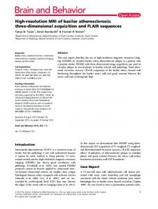
Fig. 1. Compute simulation for QRS complex. (a) The deterministic signal for the simulation (extracted from patient P l ) . (b) A simulated beat with a SNR of 20 dB. Signals estimated by averaging 80 beats (SNR = 20 dB) with (c) perfect synchronization and (d) and (e) with delays normally distributed with zero mean and standard deviations of U = 5 ms and U = I ms, respectively.
In order to observe the filtering effect produced by the synchronization errors on signal averaging, 80 signals were generated with an SNR of 20 dB and a variable delay d with respect to a point of reference, according to a normal distribution with zero mean and known standard deviation (@(,). The averaging was then carried out without applying the synchronization methods. The estimated signals for od = 5 ms and ad = 1 ms are shown in Fig. l(d) and (e) for QRS complex and in Fig. 2(d) and (e) for P wave, respectively. In order to analyze the low-pass filtering effect due to synchronization errors on signal averaging, we consider a record y , ( t ) of a random process { y ( t ) } . It can be expressed by y,(t) = s ( t
-
d,),
i
=
1,
*
9
N
(11)
where d is a random variable and s ( t ) is the deterministic signal. Then the estimation of s ( t ) is obtained by the es-
Authorized licensed use limited to: Universidad de Zaragoza. Downloaded on July 12,2010 at 09:57:34 UTC from IEEE Xplore. Restrictions apply.
>E
-
0.64
0 I4
0.47
0.08
-
-
1 -
-
0 29
-
-
0 02
0.12
-
-
I
> E
I
)
I
Authorized licensed use limited to: Universidad de Zaragoza. Downloaded on July 12,2010 at 09:57:34 UTC from IEEE Xplore. Restrictions apply.
JANE
CI
575
d . . A L I G N M E N T METHODS
TABLE I MEANVALUE A N D
s.1 A N D A R I I DtViATlON OF OBIAINtD D ~ L A YI N SSIMULATFI) CASE (QRS COMPLEX A N D P WAVE) FOR T H E DIFFERENT ALIGNMENT ALGORITHMS Signal-to-Noise Ratio (SNR)
- 1.062
DL NI-P NI-SQ MF-DL MF-NI-P MF-NI-SQ MF-IS
-0.187 0.101 0.000 0.000 0.000 0.000 0.000
0.390 0.302 0.000 0.000 0.000 0.000 0.000
-0.212 1.240 0.595 -0.175 1.225 0.625 0.062
0.585 0.860 0.516 0.441 0.446 0.484 0.242
2.456 1.506 -0.912 2.525 1.512 0.075
DL NI-P NI-SQ MF-DL MF-NI-P MF-NI-SQ MF-IS
1.862 0.101 0.341 2.000 0.000 0.000 1.000
0.627 0.302 0.474 0.000 0.000 0.000 0.000
0.825 0.5.57 1.430 0.825 0.112 1.075 0.950
1.948 0.823 0.806 0.380 0.316 0.263 0.218
0.325 1.063 2.671 -0.412 0.500 2.212 0.850
and SNR’s of 20, 10, 5 , 0, and -5 dB. Next, the synchronization methods were applied to each QRS and P wave, and then the estimated delays, referred to the point of perfect alignment, were calculated. Evaluation of errors introduced by these methods could thus be carried out for each SNR. In order to apply the NI method, the signal s ( t ) defined by (7) and the squared value s’(r) were successively considered (NI-P and NI-SQ algorithms. respectively). For the MF method, several signals estimated by the previous algorithms were taken as the ideal signal s ( t ) . Thus we have the results obtained by the algorithms MF-DL, MF-NI-P, and MF-NI-SQ, as defined in Section 11-C. The deterministic signal itself was also taken as the ideal signal (MF-IS algorithm) to show the limiting performance of the method. Obviously this situation is not possible in practice. Table I presents the results of the mean value ( p , / ) and standard deviation (U,/) of computed delays d for each of these situations. These values show the errors produced by application of the alignment algorithms. The U(, obtained in each case is related to the quality of the estimated signal, and indicates the smoothing effect in the estimation. Therefore, a comparative analysis of performance of the alignment methods for each SNR can be carried out by studying the U,/ values. The interpretation of the cutoff frequency, as a function of U,/ expressed in milliseconds, is obvious from (16). The influence of the jitter on averaging can also be appreciated using relative values of U,/ defined by U,. = u , , / w \ , where w,\ is the half-width at half-amplitude of the signals. The parameter U,. can be considered as an estimation of the
2.431 1.456 0.926 0.778 0.741 0.774 0.608
-2.525 4.696 4.341 -2.800 4.612 4.375 0.000
7.471 2.861 2.433 1.453 I .280 1.198 0.975
0.097 8.139 14.539 0.737 10.662 12.701 -0.087
13.049 10.937 10.041 2.412 2.230 2.629 1.818
3.577 1.353 1.403 2.131 0.880
-0.187 2.481 5.860 -3.050 0.875 4.250 -0.062
9.875 2.449 2.699 3.777 3.124 2.883 2.860
1.837 5.481 14.610 -4.700 3.812 I I .275 - 1.575
20.087 4.564 7.270 6.61 I 5.518 5.463 4.888
1.114
0.421
mean resolution between two signals randomly picked in the averaged series. The performances evaluated in this study can be compared to previous works [13] using this parameter. For the QRS wave selected in the simulation study, MI, is 7.5 ms. Thus, for example, applying DL and SNR = 0 dB (U(/ = 7.471) we can compute a relative U,. = 0.996.
C. Remarks The different alignment methods proposed were evaluated by applying them to a collection of QRS and P waves simulated with SNR in the range of -5 to 20 dB. The results obtained are different for the two waves. For the QRS complex, we should stress that for high SNR values of 20 and even 10 dB, all the three methods show good results. For lower SNR’s, the DL method begins to show rather unacceptable results, while NI and MF prove to be robust until SNR = 0 dB. For SNR values below 0 dB, only the MF method achieves good results. For the P wave, the DL method performs poorly below 10 dB. The NI and MF methods have good performance even below 0 dB. For these low SNR’s, NI-P gives slightly better results than MF. Finally, the goodness of the ideal signal estimation for the MF method has been shown to be an important factor for low SNR. IV. ANALYSIS OF ALIGNMENT FOR REALSIGNALS A . Signals Six pathological ECG signals (PI, . . , P6) were selected for evaluating the performance of the different
Authorized licensed use limited to: Universidad de Zaragoza. Downloaded on July 12,2010 at 09:57:34 UTC from IEEE Xplore. Restrictions apply.
516
IEEE TRANSACTIONS ON BlOMEDlCAL ENGINEERING. VOL. 38. NO. 6. JUNE 1991
alignment methods as applied to real signals. Diverse signal morphologies were taken, both for the QRS complexes and for the P waves, contaminated with noise of different spectral characteristics (muscular noise and 50-Hz interference) (Fig. 3). The objective was to obtain a precise estimation of the P and QRS waves, by means of signal averaging, from a prolonged ECG recording. The results obtained using the various alignment methods were then compared.
B. Signal Processing The signals were recorded at the Coronary Unit of the Hospital de la Santa Creu i Sant Pau. The instrumentation used was a Mingograf electrocardiograph, a low-pass analog filter with a cutoff frequency of 200 Hz, and a digital data acquisition system based on a PC. The sampling frequency selected w a s x = 1000 Hz with a resolution of 12 b. The total gain of the system was 8000. Once the signal had been digitized, the following processing phases were carried out: a) detection of heart beats, b) high-pass filtering, c) selection of windows corresponding to the P and QRS waves, d) application of the synchronization methods, and e) calculation of the estimated signal by averaging. A software QRS wave detector was used for detection of beats, this being an adaptation of the algorithm proposed by Pan and Tompkins [ 171. It is based on analysis of the slope, amplitude, and duration of the signal, and includes band-pass filtering, differentiation, and integration with a moving window. False detections are thus reduced, so low thresholds may be used and the detection sensitivity increased. This algorithm showed itself to be very robust, and permitted QRS selection even in the presence of strong variations in the baseline, muscular noise, and 50-Hz interference. High-pass filtering was carried out first to eliminate baseline variations. A first-order Lynn filter [18], with a cutoff frequency o f f , = 1 Hz, was selected. The occurrence time of the QRS complex ( t , ) was defined as the maximum absolute value of the band-pass filtered signal. This point was used as the reference for opening the temporal windows of the P and QRS waves used in the subsequent synchronization process. The point t,, was also the reference for calculation of the standard deviation of the delays obtained with the methods tested. The objective was to find a criterion for quantitative comparison of the performance of the methods. A collection of windows was defined with a duration of 200 ms for the QRS and P waves. The QRS windows were centered around the point defined by the QRS detector, while the P wave windows were defined according to the average P-R distance for each subject studied. The different alignment methods DL, NI, and MF were tested on a group of 80 beats for each patient. Averaging was carried out with respect to the temporal point of alignment for each beat. The results obtained for each method follow. 1) The estimated signal after 80 beats.
-100. O-
i
Fig. 4. Definition of the alignment times: the occurrence time of QRS complex ( I ~ given ) by the QRS detector, the absolute alignment time ( I < , ) , and the relative alignment time ( 7 ) .
2) The mean pL7and standard deviation a, of the random delay 7 defined as 7 = t(, - tit,, where t,, is the absolute alignment time. In fact, only the variable 7 is measurable instead oft,,, because a relative time basis (with respect to r,,,) is used for each beat (Fig. 4). Assuming that t,, and rl,, are uncorrelated, we can write U',
= a;,
+ uf,.
(17)
The performance of the alignment methods (indicated by U,(() may then be studied through the or values, given that U,,, is the same in all methods tested.
C. Results 1) QRS Waves: In general, the QRS complexes obtained by averaging with the different alignment methods present a similar morphology, although the signals obtained by NI and MF algorithms present better characteristics with respect to amplitude and high-frequency components. Table I1 compares a7 obtained by these methods for the six patients. Fig. 5 shows a comparison of the estimated signals for the patient P3. These signals and their corresponding a, can be classified in the same order of performance as in the simulated study. 2) P Waves: The P waves present a very much lower SNR than the QRS waves. For this reason, we may expect to obtain much greater differences between the different methods than those obtained in the QRS case. In order to synchronize P waves, the DL method was also applied to the QRS wave of the same beat as the P wave (DL-QRS). This algorithm provides good results if the P-R interval is
Authorized licensed use limited to: Universidad de Zaragoza. Downloaded on July 12,2010 at 09:57:34 UTC from IEEE Xplore. Restrictions apply.
JANE
('I U / . :
577
A L I G N M E N T METHODS
TABLE I1 MEANVALUE A N D STANDARD DEVIATION O F DELAYS I N REAL CASE (QRS COMPLEX) RELATED TO ALIGNMENT ALGORITHMS
POINTS
DEFINED B Y QRS DETECTOR FOR THE DIFFERENT
Patients
DL NI-P NI-SQ MF-DL MF-NI-P MF-NI-SQ
0.525 1.025 0.493 0.575 1.025 0.500
0.547 0.841 0.593 0.608 0.689 0.570
6.862 5.886 0.987 6.687 5.612 0.925
0.802 1.862 0.849 0.515 0.512 0.494
9.683 0.615 0.734 10.162 0.537 0.775
3.733 1.619 1.177 0.431 0.499 0.446
-2.000 -1.519 -0.152 -2.025 -1.412 -0.212
1.173 0.912 0.618 0.632 0.585 0.585
-0.700 -3.177 -1.215 -0.762 -3.150 -1.187
1.100 1.260 1.659 0.939 0.910 0.909
-0.150 0.215 -0.126 -0.212 0.225 -0.175
0.421 0.609 0.512 0.438 0.499 0.41 I
D. Remarks The results obtained on real signals agree with those obtained in the simulation studies. Thus for low SNR's, the signal estimations follow the hierarchy obtained in the simulation. The importance of the power spectral density of the noise is also apparent in the results obtained for each alignment method. Thus for cases in which there is a strong presence of 50 Hz, the matched filtering method does not achieve good results. It is necessary to make a general observation with respect to the calculated o, values. These take as a reference the relative delays obtained (7)with respect to the absolute temporal position defined by the QRS detector (tM,), used for definition of the windows. For this reason they constitute only a comparative result. The values of (T, are a good index to evaluate the relative performance of the alignment methods, given that (T,,~is the same in all tested methods. For the P wave signals, the P-R distance was considered constant. Thus the same QRS wave reference was used. In spite of some limitations, a study of these standard deviations provides a good quantitative representation of the performance of the alignment methods as applied to real signals. The conclusions presented in this paper are in accordance with the recent published results [ 191, where an application of these methods for obtaining late potentials was presented. I
I
I
I
0.
25.
so.
7s.
I
100.
I
11s
I 150
I
175
I 200
ml
Fig. 5. Signals estimated (QRS complex) by averaging 80 beats from patient P3, using the following alignment algorithms. (a) DL. (b) MF-DL. (c) NI-SQ. (d) MF-NI-SQ.
constant. The signals estimated in this way have been used for the MF method (MF-DL-QRS). Table I11 shows the (T, obtained for the six patients using different algorithms. Fig. 6 shows the estimation after averaging 80 beats for signals from patient P1. Patient P1 shows a clear presence of a 50-Hz interference, which causes estimated signals based on the MF method to have a very distorted shape [Fig. 6(d)-(f))].
V. DISCUSSION A N D CONCLUSION A comparative study of the performance of three alignment methods has been presented. Computer-simulated waves and real signals have been considered. Real deterministic signals and additive white noise were selected for the simulation study, and pathological ECG signals were used in the real-signal study. Several conclusions can be drawn from the simulation study. 1) The double level method shows poor performance for SNR below 10 dB, while the normalized inte-
grals and matched filtering methods give good results down to 0 dB.
Authorized licensed use limited to: Universidad de Zaragoza. Downloaded on July 12,2010 at 09:57:34 UTC from IEEE Xplore. Restrictions apply.
IEEE TRANSACTIONS ON BIOMEDICAL ENGINEERING, VOL. 38. NO. 6, JUNE 1991
57R
TABLE 111 MEANVALUE A N D STANDARD DEVIATION OF DELAYS I N REALCASE ( P WAVE) RELATED TO POINTS DEFINED B Y QRS DETECTOR FOR T H E DIFFERENT ALIGNMENT ALGORITHMS Patients
P2
PI
P Wave
P4
P3
Alignment Methods
P , (ms)
DL DL-QRS NI-P NI-SQ MF-DL MF-DL-QRS MF-NI-P MF-NI-SQ
0.925 -2.000 -0,354 -0.570 -1,300 -3.287 -1.575 -1.687
-4.262 0.525 1.873 1.253 -10.625 -0.725 1.062 0.187
4.155 0.547 1.898 1.906 7.774 4.390 3.355 4.120
4.587 6.862 -1.367 -1.215 4.512 6.625 -1.550 -1.425
12.218 9.683 -1.443 0.190 12.187 10.139 -1.387 0.062
2.742 0.802 0.732 0.687 0.724 0.640 0.669 0.647
3.083 3.733 2.243 1.822 1534 1477 1.561 1.638
P6
P5
U,
(ms)
8.264 1.173 1.387 1.620 1.584 1.811 1.849 1.907
P,
(ms)
-4.375 -0.700 -1.380 -1,076 -4.787 -1.350 -2.062 -1.762
(ms) 3.641 1.100 2.552 2.569 2.644 2.460 2.482 2.556
4.687 -0.150 -1.709 - 1.709 4.537 -0.325 -2.087 -2.087
2.194 0.421 1.692 1.692 1.710 1.253 1.325 1.325
The shape of the synchronized wave is an important feature affecting the results obtained in very low SNR’s. The normalized integrals and matched filtering methods present similar results for alignment of P waves. On the other hand, for QRS waves, only the matched filtering method achieves good performance. A combination of the matched filtering-normalized integrals methods gives the best performance. This result proves the influence of the estimated template signal in the application of the matched filtering method. The following additional conclusions have also been reached from the real-signal study.
4) Alignment methods applied to real signals contaminated by white noise produce similar results to those obtained in the simulation case. 5 ) For real signals with a strong presence of 50 Hz, the performance is different, and matched filtering does not achieve good results. Finally, a ranking of the three alignment methods has been obtained as a function of SNR and the type of wave considered (QRS or P wave). These results will be very useful for obtaining low-amplitude potentials in the averaging of high-resolution real cardiac signals. ACKNOWLEDGMENT The authors thank Dr. A. Bayes de Luna and Dr. J . M. Dominguez (Cardiology Service of the Hospital de la Santa Creu i Sant Pau in Barcelona) for their medical advice and their help in ECG signal acquisition. The authors also thank the reviewers for their helpful comments.
\ I 0.
I 25
I SO
I
1
I
I
100.
125.
150.
175.
I
J5
I
REFERENCES
200
ms
Fig. 6. Signals estimated (P wave) by averaging 80 beats from patient P I , using the following alignment algorithms. (a) DL. (b) DL-QRS. (c) NI-P. (d) MF-DL. ( e ) MF-DL-QRS. ( f ) MF-NI-P. The estimated signal shows a clear presence of a 50 Hz interference when MF method is used [(d), (e), and (f)].
N. W. Perry and D. G . Childers. The Huwuiri Visuul E i w k c d Recponsr. Springfield. 1L: Charles C . Thomas. 1969. E. R . John. D. S. Ruchkin. and J . J . Vidal, “Measurement ofeventrelated potentials,” in Ei~e/ir-Re/ritc~dPorc~,iriri/.c in M m , E. Callaway, P. Tueting and S . H . Koslow. Eds. New York: Academic, 1978, pp. 93-138.
Authorized licensed use limited to: Universidad de Zaragoza. Downloaded on July 12,2010 at 09:57:34 UTC from IEEE Xplore. Restrictions apply.
JAN^ ('r
o i . : A L I G N M E N T METHODS
519
131 E. J . Berbari. B. J . Scherlag. and R. Lazzara, "A computerized technique to record new components of the electrocardiogram." Proc. IEEE, vol. 65, pp. 799-802. 1977. (41 H. H . Ros, A. S. M. Koeleman. and T. J . v . d. Akker, "The techniquc of signal averaging and its practical application in the separation of atrial and His Purkinje activity." in Sigricrl AivruginR T e c h rriyuc, in Clirricnl Currliology, Ed. V. Hombach and H. H. Hilger. Eds. F. K. Schattauer Verlag. 1981. pp. 3-14. [SI G. Breithardt, R. Becker. L. Seipel, R. Abendroth, and J . Ostermeyer. "Non-invasivc detection of late potentials in man-a new markerforventriculartachycardia," Europ. H e u r t J . , pp. 1 - 1 I . 1981. M . B. Simson. "Use of signals in the terminal QRS complex to identify patients with ventricular tachycardia after myocardial infarction." Circuluriorr, vol. 64, pp. 235-242. 1981. 0 . Rompelman and H. H. Ros. "Coherent averaging technique: A tutorial review. Part I : Noise reduction and the equivalent filter." J . B i ~ ~ ~E in gd. .. vol. 8. pp. 24-29. 1986. -, "Coherent averaging technique: A tutorial review. Part 2: Triggcr jitter. overlapping responses and non-pcriodic stimulation," J . Eionled. Eng., vol. 8 - pp. 30-35. 1986. W. Craelius, M. Restivo, M. A. Assadi, and N . El-Sherif, "Criteria for optimal averaging of cardiac signals.'' IEEE Trurrs. Biowrrd. Errg. . vol. BME-33. pp. 957-966, 1986. C. D. McGillem. J . 1. Aunon and C. A. Pomalaza, "Improved waveform estimation procedures for event-related potentials," IEEE Trurrs. Biomed. Eirg., vol. BME-32. pp. 371-379, 1985. A. S . M. Koelcman. T. J . van den Akker. H. H . Ros. R . J . Janssen, and 0. Rompelman. "Estimation accuracy of P wave and QRS complex occurrence times in the ECG: The accuracy for simplified theoretical and computer simulated waveforms." Sigrrul Proc.esirrg, vol. 7 , pp. 389-405, 1984. G . J. H . Ui,jen. J . P. C. de Weerd, and A. J . H. Vcndrik. "Accuracy of QRS detection in relation to the analysis of high-frequency coniponents in the electrocardiogram," M c d . Biol. Er7g. Conrp., vol. 17. pp. 492-502. 1979. H. Rix and S . Jesus. "Estimation du retard entre signaux de m&me forme,'' C. R . Acudkrrrie des Scirricrs. t . 229 serie 11, pp. 399-404, 1984. S . Jesus and H. Rix. "High resolution ECG analysis by an improved signal averaging method and comparison with a beat-to-beat approach." J . Eiomecl. Errg., vol. I O . pp. 25-32. 1988. L. E. Franks, Signul Theory. William L. Everitt. Ed. Englewood CliRs. N J : Prentice-Hall. 1969. C. D. Woody. "Characterization of an adaptive filter for the analysis of variable latency neuroelectric signals." Med. Bid. Enx. Comp., vol. 5 . pp. 539-553. 1967. J . Pan and W . J . Tompkins, "A real-time QRS detection algorithm." lEEE Trurrs. Bio/ned. Errg.. vol. BME-32. pp. 230-236. 1985. P. A. Lynn. "Online digital filters for biological signals: Some fast designs for a small computer." Med. Biol. E q . Cornp., vol. 15, pp. 534-540, 1977. R. Jane, P. Caminal. H. Rix. E. Thierry, and P. Laguna. "Improved alignment methods in ECG signal averaging: Application to late potentials detection." in Cor?inutrr.\ irr Curdiolouv. Washington. DC: IEEE Computer Society Press. 1989, pp. 481-484.
the Master and Doctoral programs in Biomedical Engineering, at the Polytechnic University of Catalonia. His current research interests are in digital signal processing applied to biomedical signals and in particular to highresolution ECG.
Her& Rix (A'88) received the M . S . degree in applied mathematics in 1967, and the Speciality Doctoral degree in astrophysics in 1970 from the University of Nice, France, and the Ph.D. degree in 1980. He was an Assistant Professor at the Institute of Technology and worked in the field of signal processing with application to chromatography. Since 1981 he has been with the Department of Signals and Systems (LASSY) associated to French National Research Center (CNRS). Currently, he is Professor at the University of Nice (Institute of Technology). He teaches mathematics and signal processing. His research interests are in signal processing, mainly applied to Biomedical Signal and particularly to high-resolution electrocardiography.
Pere Caminal (M'83) was born in 1952 in Barcelona, Spain. He obtained the M.S. and Ph.D. degrees in mechanical engineering in 1974 and 1980, respectively, all from the Polytechnic University of Catalonia (U.P.C.), Spain. Since 1982 he has been Associate Professor of Automatic Control in the Department of Control Engineering at the same university. He is presently head of the Biomedical Engineering Division of the Institut of Cybernetics (U.P.C.C.S.I.C.). and director of the Master Program in Biomedical Engineering at the Polytechnic University of Catalonia. His areas of interest are modeling and simulation of biological systems, and biosignals processing.