Computational Methods for Cardiac Electromechanics ROY C. P. KERCKHOFFS, SARAH N. HEALY, TARAS P. USYK, AND ANDREW D. McCULLOCH
Invited Paper
Computational modeling provides a potentially powerful way to integrate structural properties measured in vitro to physiological functions measured in vivo. Focusing on the various scales (cell–tissue–organ–system), we give an overview of the importance and applications of numerical models of ventricular anatomy, electrophysiology, mechanics, and circulatory models. The integration of these models in one multiscale model of cardiac electromechanics is discussed in the light of applications to hypothesis generation, diagnosis, surgery (planning, training, and outcome of interventions), and therapies. Special attention is paid to practical use in terms of computational demand. Because of growing computer power and the development of efficient algorithms, we expect that real-time simulations with multiscale models of cardiac electromechanics become feasible in 2008 (despite the increasing complexity of models due to data accumulation on molecular and cellular mechanisms). Keywords—Cardiac electrophysiology, cardiac mechanics, cell, circulatory, heart modeling, multiscale, organ, system, tissue.
I. INTRODUCTION Because there are many interacting subsystems in basic physiological processes within and between spatial scales, there is a need for structural and functional integration in computer modeling. Integrative models help in understanding the influences of the various subsystems on
Manuscript received June 27, 2005; revised November 13, 2005. This work was supported in part by a grant from the Defense Advanced Research Projects Agency executed by the U.S. Army Medical Research and Material Command/TATRC Cooperative Agreement under Contract W81XWH-04-2-0012, in part by the National Biomedical Computation Resource under NIH Grant P41 RR08605, and in part by the National Science Foundation under Grant BES-0096492 and Grant BES-0506252. The work of S. N. Healy was supported by a Predoctoral Fellowship from the American Heart Association. This work was conducted in a facility constructed with support from Research Facilities Improvement Program under Grant C06 RR-017588-01 from the National Center for Research Resources, National Institutes of Health. A. D. McCulloch is a cofounder of Insilicomed Inc., a licensee of UCSD-owned software used in this research. The authors are with the Department of Bioengineering, Whitaker Institute for Biomedical Engineering, University of California, San Diego, La Jolla, CA 92093-0412 USA (e-mail:
[email protected]; shealy@bioeng. ucsd.edu;
[email protected];
[email protected]). Digital Object Identifier 10.1109/JPROC.2006.871772
whole-system behavior [1]–[5]. For example, on the tissue scale in cardiac physiology, a propagating depolarization wave activates mechanical contraction of the cardiac walls (functional integration). Across spatial scales (structural integration), contracting sarcomeres in the myocardium make the heart eject blood, working against the resistance of the vasculature tree. Computational models in cardiac physiology are valuable in understanding the normal and diseased heart, and as data accumulate on molecular and cellular mechanisms, the potential for powerful predictive mechanistic models is growing rapidly [2]. Various parameters are measured in vitro, such as morphology and biochemistry, and then this information is used to implement these models in silico. The results are then validated in vivo, leading to iterative model refinement. Already, computer models of cardiac electrophysiology and/or mechanics have helped in assessing size and location of ischemic regions in heart patients [6], [7], in developing diagnostic measures [8], in developing virtual surgery training platforms [9]–[12], in assessing the effects of heart surgery [13]–[15], of electric shocks [16]–[18], and the effects of pacing normal [19]–[21] and failing hearts [22]. Furthermore, numerical models have been used to test hypotheses [23]–[25], which were supported only later by experiments [26]–[28]. In this paper the importance of numerical cardiac modeling is addressed. A review of mathematical models of cardiac electrophysiology and mechanics, as well as the combination of cardiac electromechanics is presented. With the purpose of making (usage of) modeling of cardiac (electro)mechanics more accessible to a wider audience, we will first describe cardiac physiology in which we focus on the heart as a system, built from cell to tissue to whole-organ scale and the place of the heart in the circulation. Similarly, focusing on the various scales, we give an overview of the usefulness/applications of the four minimally needed components of modeling cardiac electromechanics: anatomy, electrophysiology, mechanics, and circulatory models. Finally, the integration of these four components in one
0018-9219/$20.00 © 2006 IEEE PROCEEDINGS OF THE IEEE, VOL. 94, NO. 4, APRIL 2006
769
multiscale model of cardiac electromechanics is reviewed and the feasibility of real-time computing. II. CARDIAC PHYSIOLOGY A. Cell Level Rod-shaped cardiac myocytes contain numerous myofibrils. Neighboring myofibrils have their sarcomeres aligned, which gives them a striated appearance. The sarcomere is considered to be the basic contractile unit of cardiac muscle, consisting of a three-dimensional (3-D) array of the contractile myofilaments actin and myosin. Myofibrils and mitochondria make up almost 85% of the myocyte volume [29]. Myofiber contraction is initiated by depolarization of the outer cell membrane. During diastole, a negative resting potential exists in the cell, with more potassium K intracellular than extracellular and more calcium Ca and sodium Na extracellular than intracellular. An action potential is generated in a resting myocyte when it gets activated by adjacent depolarizing cells. The initial upstroke of the action potential is caused by a large inward current of sodium. Next, calcium enters the cell through L-type calcium channels, which contributes to the action potential plateau [30]. Calcium entry triggers calcium release through the ryanodine receptors from the sarcoplasmatic reticulum (which stores most of the intracellular calcium). The combination of calcium influx and release raises the free intracellular calcium concentration, allowing it to bind to the regulatory protein troponin C, located on the actin filament. The resulting conformational change of the regulatory proteins tropomyosin and troponin on the actin filament exposes myosin binding sites. A bond between actin and myosin, a crossbridge, is formed when myosin heads attach to the actin binding sites. Depending on the loading of the myocyte, actin filaments slide along the myosin filaments, causing shortening of the sarcomere. Hence, calcium plays an important role in both electrophysiology and mechanics. Intracellular calcium also triggers calcium-activated potassium channels that generate outward potassium currents which end the action potential. Concentrations of ions are actively restored by pumps and exchangers during diastole. Based on electrophysiological characterization, some investigators have suggested the presence of three different subtypes of working myocardial working cells in the ventricular walls, named after their location in the myocardium: endocardial, M-, and epicardial cells [31], [32]. These cells produce action potentials of different shape and length, due to different expressions of ion channels (especially those responsible for the transient outward and delayed rectifier K currents), different calcium fluxes [27], [33], and hence potentially different active mechanical behavior [27]. B. Tissue Level The myocardium is largely composed of myofibers. Myofibers are situated within the extracellular matrix, which 770
consists, among others, of networks of collagen fibers. Passive material properties of cardiac muscle are largely determined by the composition and structure of this collagen (and by the supporting myofilament protein titin in the myocyte), and have been reported to vary transmurally [34]. Locally the myofibers are aligned approximately in parallel, thus defining a local mean myofiber orientation with dispersion about the mean of 12–15 [35]. Subepicardial myofibers follow a left-handed helical locus parallel to the wall. Near the apex, myofiber loci may have a significant transverse component, and continue in a right-handed helical pathway at the subendocardium. Near the base, myofiber orientations cross over toward the subepicardium again. Close to midwall the myofiber orientation is predominantly circumferential. Furthermore, cardiac myofibers are organized into laminar sheets about four to six cells thick. It has been observed that sheets tend to coincide with planes of maximum systolic shear and that sheet orientation may exist in two distinct populations [36]. In a normal heart, depolarization starts in the sinoatrial (SA) node. This node is a cluster of modified myocytes located at the endocardium of the right atrium [29]. Resting heart rates vary with species, gender, and age. The resulting cellular transmembrane action potential propagates as a wave over neighboring cells, thus depolarizing the atria. The atrio-ventricular (AV) node, located near the septum between the atria and the ventricles, slows down the depolarization wave. Distal to the AV node, the depolarization wave is conducted fast through the His bundle, splitting in a left and right branch. These branches bifurcate into many small branches, thus forming the fast conducting Purkinje system, located in the subendocardium of the left and right ventricle. These Purkinje fibers are insulated with connective tissue. Purkinje fibers enable a fast propagation of depolarization, typically 3–4 m/s [37]. The Purkinje system ends in the Purkinje–muscular junctions. Here, the depolarization wave enters the ventricular myocardium. In the myocardium, propagation is much slower [38]–[40], where the wave propagates mainly from endocardium to epicardium. Hooks et al. [41] have shown numerically that orthotropic conductivities result from sheet structure (as opposed to myofiber transverse isotropy), but whether this is of significant importance has been questioned by another numerical study by Colli-Franzone et al. [42]. Myocardial propagation velocities of 0.6–1.0 m/s and 0.2–0.5 m/s have been reported parallel and perpendicular to myofibers [38]–[40]. From the moment of leaving the Purkinje fibers, the whole human ventricular myocardium is depolarized within 50 ms [43]. For tissue mechanics, sheet structure seems to be more important [44]. Strain distributions were in better agreement with experiments when orthotropic passive properties were included in a model of cardiac mechanics as opposed to transversely isotropic properties [45]. Even active contraction seems to be anisotropic, where active tension is generated perpendicular to the myofiber [46]. Strain distributions as computed by models of cardiac mechanics showed better agreement with experiments when this biaxial PROCEEDINGS OF THE IEEE, VOL. 94, NO. 4, APRIL 2006
active behavior was included [45], [47], but the role of sheets here remains unclear. C. Organ Level A few billion myocytes make up the cardiac walls. The contracting cardiac walls pump blood through the (lower pressurized) pulmonary and (higher pressurized) systemic circulation. In fact, the heart consists of two pumps, located side-by-side. Both sides contain an atrium and a ventricle. The atria collect venous blood that returns to the heart and facilitate filling of the ventricles. The right ventricle (RV) maintains the pulmonary circulation, where blood receives oxygen and disposes of carbon dioxide in the lungs. The left ventricle (LV) maintains the systemic circulation, where blood delivers oxygen and nutrients to the rest of the body and receives carbon dioxide and waste products. The wall of the RV is thinner and less powerful than that of the LV. The RV is crescent-shaped in cross section, wrapped partially around the (ellipsoid-shaped) LV. The interventricular septum, which anatomically belongs to the LV, separates the LV and RV. From the status of the valves (opened or closed), four cardiac phases can be distinguished for the ventricles (diastole: isovolumic relaxation and filling phases, systole: isovolumic contraction and ejection phases). In brief, a cardiac cycle is described for a human body at rest with typical orders of magnitude for time and pressure [29]. For the LV, during the filling phase, the mitral valve between the left atrium and LV is open and the aortic valve between ventricle and aorta is closed. After 500 ms, the isovolumic contraction phase is entered when generated myofiber stress in the LV wall will increase the pressure in the ventricle and close the mitral valve ( 1.5 kPa, 11 mmHg). Pressure rises quickly ( 50 ms) and when LV pressure exceeds aortic pressure ( 10 kPa, 75 mmHg) the aortic valve will open and eject blood into the aorta during the ejection phase (reaching a maximum pressure of 16 kPa or 120 mmHg). After 250 ms, LV pressure becomes low enough and reversal of flow will close the aortic valve and the isovolumic relaxation phase is entered ( 80 ms). When LV pressure drops below left atrial pressure ( 0 kPa), the cycle starts over again with the filling phase. Equivalent RV valves are the tricuspid and pulmonary valves for the mitral and aortic valves, respectively. Diastolic pressures for the RV are in the same order of LV pressures, while systolic pressures are typically 4–5 times smaller. D. System Level The heart is part of an integrated system which compensates for a fall in blood pressure as a result of exercise (due to decreased arteriolar resistance in skeletal muscles), blood loss [48], and also during heart failure [29]. When arterial blood pressure falls, neurohumoral stimulation increases cardiac output, water and salt is retained in the kidneys (to increase preload), and blood supply is redistributed throughout the body. Feedback from the baroreceptors in the aorta and carotids increases -adrenergic stimulation, which increases beating
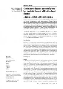
Fig. 1. The four most important components for models of cardiac electromechanics: anatomy, electrophysiology, mechanics, and hemodynamics. Ventricular anatomy is fitted to morphological measurements. The anatomical representation is then appropriately refined in space and time to reflect the physics of the specific part of the problem. For example, the spatial and temporal resolution required to model impulse propagation is smaller than that required for the analysis of regional mechanics, but the spatial refinement of the mechanics problem is nonhomogeneous.
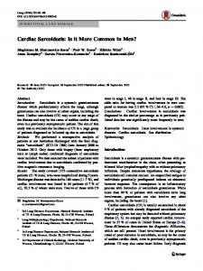
frequency and myocardial contractility. Integrated numerical models of signal transduction [49], cardiac electromechanics, and the hemodynamics of the circulation will allow a more complete investigation of regulation of myocardial contractility by -adrenergic stimulation on a multilevel scale. III. MODELING CARDIAC ELECTROMECHANICS In this section, an overview will be given of the four most important components (Fig. 1) of a whole-heart model of cardiac electromechanics, namely, anatomic models, models of electrophysiology from cell to organ, of mechanics from cell to organ, and circulatory models at the system level. Then, from cell to organ, combined models of cardiac electromechanics are reviewed, followed by a section on strategies for multiscale modeling and high-performance computing. A. Ventricular Anatomic Models Anatomically detailed 3-D models of geometry and myofiber orientation for different species have been described in literature (Fig. 2): for mouse [50], rabbit [51], dog [21], [52], [53], pig [54], sheep [47], and human [55]. Geometries of these models have been obtained from histological measurements [51]–[54], CT [55], or MRI scans [21], [47]. Myofiber orientations have been reconstructed from histological measurements [51]–[54], diffusion weighted MRI [56], [57], or by calculations [58]. In the latter study, myofiber orientation was calculated by using the hypothesis that myofibers are oriented such that shortening during ejection is as homogeneous as possible. Sheet orientations have been obtained by histological measurements [59] or can be obtained by calculations [36], but there is recent evidence that sheets can also be measured with diffusion weighted MRI [60]. In addition, Usyk et al. [61] included Purkinje fibers to their model, which were fitted to measurements.
KERCKHOFFS et al.: COMPUTATIONAL METHODS FOR CARDIAC ELECTROMECHANICS
771
Fig. 2. Anatomic models of: (a) rabbit [51]; (b) dog [53]; (c) pig [54]; and (d) dog [21].
B. Models of Cardiac Electrophysiology 1) Cell Level: Cellular models of cardiac electrophysiology typically describe the action potential either phenomenologically (e.g., with FitzHugh–Nagumo equations [62]), or—more biophysically detailed—as a result of changing transmembrane ionic currents [63], [64]. Most of these ionic models are based on the formalism originally established by Hodgkin and Huxley for the squid nerve [65]. In these models currents are described according to Ohm’s law, where (for example, for a voltage-gated channel) the conductance is dependent on time and voltage and is typically described as a function of the open-probability of gates (in the form of ordinary differential equations, or ODEs). Later models (from 1977 [66]) also include ion concentrations. Ionic models have become increasingly complex, through iterative interactions between experiment and simulation [63], describing more ion channels and ion concentrations, the latter in different compartments of the cell [67], [68]. Recent models also include transmural heterogeneity of channel expression [69]–[72] or describe certain channels in a more complex manner with so-called Markov models [68], [73], reflecting channel behavior more realistically than Hodgkin–Huxley formulations. These latter models typically have 30 ODEs and are computationally expensive. Ionic models have been developed for different species, for mouse [50], rat [70], guinea pig [67], rabbit [74], dog [68], [75], and human [66], [71], [76]. 772
2) Tissue Level: Cellular-level models can be incorporated into larger scale models—either as a discrete set of cells or tissue bundles or as a continuum. Creating a model that treats each cell as discrete unit results in what is called a network, or discrete model, and is the approach used by [77]–[79]. Tissue models with over 140 000 cells have been produced using this approach [80]. An alternative to this type of modeling is to treat the tissue as a syncytium and assume that at any given point there exists a transmembrane electrical potential (so-called monodomain models) or that any point given there exists both an intracellular and extracellular space (so-called bidomain models [81], [82]), for which the electrical potentials are computed. With tissue models of cardiac electrophysiology, investigators have studied, e.g., sinus node [83], dynamics of lifethreatening spiral waves [84]–[88] and the termination of them [85], [89], and ischemia [86]. Recently, Saucerman et al. [90] coupled a mechanistic signaling model to a cellular ionic and monodomain propagation model in a rabbit ventricular wedge to investigate the proarrhythmic consequences of a genetic mutation underlying long QT syndrome. One of the drawbacks of traditional finite-element implementations of monodomain and bidomain equations is that they fail to represent the discontinuous nature of electrical propagation. Trew et al. [91] have overcome this by using a finite volume technique that explicitly represents microstructural features such as the eletrically nonconducting cleavage PROCEEDINGS OF THE IEEE, VOL. 94, NO. 4, APRIL 2006
planes between sheets of myocardial fibers. They demonstrate that these discontinuities have important implications for defibrillation. As mentioned in Section II-A, isolated single cells from different transmural ventricular locations show different electrophysiological properties. However, in intact tissue, gradients of electrophysiologic properties (i.e., dispersion of action potential duration, APD) are much smaller due to electrotonic coupling [92]. With a numerical tissue model of cardiac electrophysiology it has been shown that electrotonic coupling has a diminishing effect on heterogeneity in the intact myocardium which strongly depends on the extent of intercellular coupling [93]. 3) Organ Level: Bidomain and monodomain models have been used to examine a wide variety of cardiac tissues and phenomena in the whole heart, including investigations of Purkinje fiber [94], ischemia [18], [95], electrotonic coupling [96], reentrant ventricular activity [78], [97]–[99], and defibrillation [100], [101]. Because of computational demand at the whole-heart level, these models either use simplified ionic or phenomenological models in hearts of larger mammals (e.g., dog and human) [94], [99], solve only parts of the cardiac cycle [95], or use more detailed ionic models yet in hearts of smaller mammals (like mouse and rabbit) [18], [96], [99]–[101]. C. Models of Cardiac Mechanics 1) Cell Level: According to Rice and de Tombe [102], the development of models of cellular cardiac mechanics have lagged behind the development of models of cellular cardiac electrophysiology, due to (lack of) available solving algorithms (and computer power) and controversies about basic mechanisms of force generation in myofilaments. A number of models of active tension development in cardiac muscle have been proposed. In essence they may be grouped into three categories: 1) time-varying elastance models that include the essential dependence of cardiac active force development on muscle length and time [44], [103], [104]; 2) “Hill” models, in which the active fiber stress development is modified by shortening or lengthening according to the force-velocity relation, so that fiber tension is reduced by increased shortening velocity [105], [106]; and 3) fully history-dependent models, either based on Huxley’s crossbridge theory [107]–[109], which yields a system of partial differential equations as functions of time and crossbridge position, or on myofilament activation models [110]–[116], which yields a system of (less computationally expensive) ordinary differential equations as functions of time and shortening velocity. Many of the early models were based on skeletal muscle models of Hill [117]. However, Hill’s model considers tetanic contraction only, and hence is inappropriate for describing cardiac muscle mechanics [118]. Wong generalized Huxley’s model of the skeletal muscle cross-bridge to partial and length-dependent activation. Panerai [109], using Huxley’s original model, incorporated length-dependent activation in a first order kinetic equation
describing Ca -troponin C interaction. Instead of considering individual myofilaments, Tozeren [119] proposed a “continuum” model of cardiac muscle contraction. Tozeren generalized Hill’s equation to partial activation to describe active fiber tension as a function of fiber strain, strain rate, and time after onset of contraction. In these studies, model predictions were validated by experimental length-tension or force-velocity relations during contractions in which overall muscle length was controlled. Panerai accounted for the appreciable internal shortening that occurs during isomeric contractions at the expense of lengthening in the damaged muscle at the clamped ends [120]. 2) Tissue Level: Two different approaches have been used to develop 3-D constitutive models of resting ventricular myocardium: mechanical testing of myocardial tissue specimens under prescribed homogeneous loading conditions gives stress–strain relationships directly. Alternatively, measurements of regional tissue deformations in isolated or intact whole hearts subject to prescribed loading conditions can be used together with the solution of a boundary value problem to estimate material constants of an assumed constitutive law in a semi-inverse analysis. The simplest and most common test in the former approach is the uniaxial tension test [121]. However, such uniaxial stress–strain data are unable to characterize the 3-D constitutive behavior of the myocardium even when independently conducted along different structural axes (which is very difficult in any case). Biaxial tests measure force and displacement (stress and strain) along orthogonal fiber and cross fiber axes, and the results of such tests in myocardium were reported first by Demer and Yin [122]. It remains uncertain how biaxial properties measured in isolated tissue slices are related to the properties of the intact ventricular wall. The irregularity of isolated myocardial tissue and variation of fiber direction through the specimen complicate in vitro measurements. Moreover, shearing deformations are an important component of 3-D mechanics in the intact heart [123], but the usual biaxial protocols do not test responses to shearing with respect to fiber and crossfiber axes. A recent study by Dokos et al. [124] describes a novel shear testing device that is capable of applying simultaneous shear loads in two orthogonal directions to small isolated specimens of ventricular myocardium while the resultant forces are measured. Guccione et al. [125] used a different approach to identify constitutive parameters. They sought a material law that was representative of the 3-D loading conditions of the intact heart by prescribing the deformations as measured from isolated arrested canine hearts in a cylindrical model to reproduce axial, circumferential, and torsional strains [126]. They then used a nonlinear least-squares optimization algorithm to identify the parameters of a transversely isotropic exponential strain energy function that minimized the errors between computed boundary tractions and the known pressure loading conditions on the isolated heart. The resulting model was able to reproduce 3-D transmural strain distributions subsequently measured with radiopaque markers [127], even though the original data used in the optimization were only
KERCKHOFFS et al.: COMPUTATIONAL METHODS FOR CARDIAC ELECTROMECHANICS
773
two-dimensional (2-D) strains at one point on the epicardial surface. The finite-element method is particularly well suited for this inverse analysis due to its ability to incorporate the 3-D geometry, fibrous tissue architecture, pressure boundary conditions, and nonlinear material properties of the tissue. If it can be shown that a model accurately describes experimentally measured 3-D strains, then this provides confidence in the estimated stresses, which are important for understanding myocardial growth and remodeling in physiological and pathophysiological conditions. Alternatively, identifying the source of discrepancies between estimated and measured strains by carefully testing the assumptions of the model can provide a valuable means of gaining insight into the essential features which determine 3-D cardiac mechanical function. For an overview of different constitutive equations for passive mechanics, see Usyk and McCulloch [128]. 3) Organ Level: Mathematical models of cardiac mechanics in the whole heart [45], [104], [107], [129]–[132] have been developed to understand the distribution of myofiber stress and strain under a variety of circumstances. They have been used to investigate the effect of (pathological) myofiber orientation [133], [134] and regional ischemia [135]–[137] on myofiber stress and strain. Furthermore, models of cardiac mechanics have also been used as tools in developing diagnostic measures, in surgical planning and guidance, and in predicting outcomes of surgical interventions. Qualitative measurement of left ventricular endocardial wall motion with real-time 3-D ultrasound is a sensitive predictor of coronary artery disease [138]. Improvement of this diagnostic approach should be accomplished by quantitative measurement of wall motion. To formulate and test candidate quantitative measures of endocardial wall motion, Herz et al. [8] have successfully used a model of cardiac mechanics of the regionally ischemic canine left ventricle [136]. It is expected that models of cardiac (electro)mechanics will be a helpful tool in formulating new quantitative diagnostic measures [8]. Common preparation for heart surgery is often performed with 2-D images of the patient. A more effective preparation for surgery, however, would result in less patient trauma, shorter hospital stays and reduced costs [10]. To address the lack of 3-D planning and guidance, researchers are developing virtual cardiac surgery planning platforms [9], [11], [12], [139] in which a patient-specific 3-D model of heart-inchest is visualized and used for planning and guidance. Dynamic models of the heart are being incorporated for a more realistic environment [10]. Looking further ahead, 3-D patient-specific models of cardiac electromechanics, embedded in the chest wall, could become helpful tools, such that cardiovascular surgeons can virtually practice any intervention beforehand. Effects of a variety of surgical interventions have been investigated using 3-D numerical models of the heart or parts thereof. Using such models, it has been shown, for example, that ventricular volume reduction surgery in dilated hearts, in which a slice of the left ventricle is removed to restore a 774
normal cavity volume to wall volume ratio, mean myofiber stress is reduced (and hence results in less oxygen consumption) but, unfortunately, also stroke volume [13]. Similar results (both a decrease in stress and stroke volume) were found for partial removal of infarcted tissue, with or without the application of patches [15]. The application of a transcavitary myosplint, a device to change left ventricular shape, also decreased myofiber stress, but (fortunately) did not significantly reduce stroke volume [14]. D. Circulatory Models To simulate a complete cardiac cycle, a circulatory (or—more limited—a separate pre- and afterload) model is needed to ensure realistic diastolic filling and ventricular–vascular coupling. Using such a design, the effects of changes at the cellular (neurohormonal, drugs, heart failure) or tissue (abnormal conduction) levels to cardiac output can be investigated and—depending on the complexity of the circulatory model—feedback pathways from the baroreceptors in the large vessels to -adrenergic stimulation. Of the models of cardiac (electro-)mechanics coupled to some type of afterload model [19]–[22], [24], [61], [131], [140]–[142], only one model—to the authors’ knowledge—contained a realistic preload [142]. Although zero-dimensional varying elastance cardiac models have been embedded in a closed circulation [143]–[146], no studies are known yet to have embedded a 3-D finite-element model of the heart in a closed circulation. Perhaps the best known afterload models are the windkessel models (Fig. 3). In 1899, Otto Frank published the description of a two-element windkessel model [147]. This model relates pressure to flow using two parameters: arterial compliance, representing the extensibility of the major arteries, and peripheral resistance, the resistance to flow encountered by the blood as it flows from the major arteries to the capillaries. However, this model produces unrealistic wave forms, mainly due to the poor medium- to high-frequency representation [148]. More realistic waveforms are obtained by the three-element windkessel model [149], [150], by adding a characteristic impedance of the aorta to the two-element version. This third term accounts for the local inertia and local compliance of the proximal aorta. Although yielding realistic waveforms, estimates of compliance and impedance values, after fitting, are different from values obtained from measurements. A fourth parameter in the four-element windkessel model, the inertia of the whole arterial system, resolves this problem [148]. The circulation can be represented in more detail by identifying individual components and assembling them in a more complex lumped circulatory model. By adding even more complexity (e.g., baroreceptors [144], airways mechanics [151], [152], blood gas handling [151], [153]), the influence of specific pathologies, (surgical) interventions, or trauma on heart function can be investigated [143], [144]. These latter models describe more than blood flows, pressures, and volumes alone, representing a more integrated system, and are therefore also referred to as highly PROCEEDINGS OF THE IEEE, VOL. 94, NO. 4, APRIL 2006
Fig. 3. Electrical circuit analogues of two-, three-, and four-element windkessel models (A, B, C, respectively). C : arterial compliance [ml/mmHg], L: arterial inertance [mmHg 1 s =ml], P : arterial pressure [mmHg], P : left ventricular pressure [mmHg], q : aortic flow [ml/s], R : peripheral resistance [mmHg 1 s/ml], Z : aortic impedance [mmHg 1 s/ml].
integrated physiology models. The main problem with these models is the high number of parameters, some of which need to be estimated or collected from different species. An elegant approach to reduce this problem is making use of a limited number of adaptation rules and actually have the model compute the parameter values [145]. E. Integrated Models of Cardiac Electromechanics So far the integration across biophysical scales has been discussed in the context of incorporation of cellular models into models of tissues and the whole heart. This has been done for electrophysiology and mechanics separately. However, the electrophysiological function of the heart does not exist separately from its mechanical function. The incorporation of mechanical function into such simulations is of fundamental interest. Contraction itself occurs within sarcomeres at the cellular level and depends on the interaction with calcium. Furthermore, electrophysiology of the cell experiences feedback from mechanical deformations [mechanoelectric feedback (MEF)] [154], [155] through sarcomerelength-dependent myofilament calcium sensitivity [156], and through stretch-activated ion channels (SACs) [157]. From a clinical standpoint, understanding of electromechanical interaction is becoming more relevant,
for example, because of asymmetric hypertrophy induced by chronic pacing [158] and the rapidly increasing interest in cardiac resynchronization [159], [160]. The most common approach in designing models of cardiac electromechanics is coupling existing models of cellular electrophysiology and active mechanics. Naturally, testing of these models is done by experimental validation in which both electrical and mechanical variables are involved, such -force relations [115], stimulation frequency-force as Ca [115], interval-force [161], transmural electromechanical heterogeneity [162], and relations between electrical activation and strain [19], [20], [24], [162], [163]. 1) Cell Level: Sachse et al. [115] have coupled a model of human cellular electrophysiology [76] for normal and failing myocytes to a model of myofilament activation [114] and further modified it, making it suitable for nonisometric twitches. Calcium transients from the former model served as input for the latter model. Rice et al. [161] have focused on interval-force relations by coupling their model of canine electrophysiology [68] to their model of myofilament activation [114] in which feedback was introduced by the dependence of affinity of troponin for Ca on force. Thus, the level of developed force can also alter the Ca transient. Model assembly of Solovyova et al. [162] was similar to that of Rice et al. [114], but the former group introduced a second type of myocyte with different mechanical properties and thus coupled a “slow” and “fast” muscle in series. They showed that when the slow muscle was activated 30 ms earlier than the fast muscle, total force development was more optimal and dispersion of action potential duration was significantly decreased. An ionic model of rabbit cell electrophysiology was recently extended to include transmurally varying stretch-activated and stretch-modulated currents. The model predicted that endocardial and midmyocardial cells are the most sensitive to stretch-induced changes in action potential duration. Inclusion of a stretch-sensitive transmurally varying outwardly rectifying K+ current may reduce repolarization gradients in intact myocardium caused by intrinsic ion channel densities, nonuniform strains, and electrotonic effects [72]. 2) Tissue Level: A similar effect on action potential duration has been shown in a 2-D model by Smith et al. [164]. In this study, an ionic model [165] was tightly coupled to a model of active mechanics [44]. Embedding the heart in a torso model enabled the calculation of an ECG. Due to action potential duration shortening as a result of contraction, the ECG showed a leftward shift of the T wave. An example of MEF solely due to deforming tissue (i.e., deformation affecting a propagating wave) is shown in the study of Nash and Panfilov [166]. Using a combination of finite differences and finite elements to solve a FitzHugh–Nagumo excitation model coupled to active stress generation (which means in this case there was not any feedback on calcium behavior), on a 2-D domain, they showed that tissue mechanics contributed significantly to the dynamics of electrical propagation. The use of FitzHugh–Nagumo equations (which consists of two ODEs)
KERCKHOFFS et al.: COMPUTATIONAL METHODS FOR CARDIAC ELECTROMECHANICS
775
enabled a relatively fast simulation of spiral waves traveling through a contracting medium. 3) Organ Level: Whole-heart models of cardiac electromechanics can and already have been used to study, e.g., the influence of local disturbances in electrical propagation or cellular electrophysiological pathologies on regional contraction and global heart function [19]–[22], [24], [55], [61], [140]–[142], [163], [167], [168]. Usyk et al. [19], [22], [61] and Kerckhoffs et al. [20], [21], [24], [140] have developed whole-heart finite-element models of canine electromechanics to simulate normal and paced hearts, in which either a two-element [19], [22], [61] or three-element windkessel model [20], [21], [24], [140] served as an afterload. In the studies of Usyk et al., modified FitzHugh–Nagumo equations and a monodomain propagation model [169]) were used for the electrical part. Kerckhoffs et al. used an eikonal-diffusion model [170], [171] to solely compute electrical activation times. Both groups completed the electromechanical coupling through activation times, hence MEF was not included. Calculated myofiber strain patterns suggested that the transmural distribution of time-delay between depolarization and onset of active stress generation might be heterogeneous [24], as supported by experimental evidence [27]. The latter experimental study also showed transmural heterogeneous active mechanical properties in single cells, which had been suggested by the previously mentioned numerical study of Solovyova et al. [162]. Watanabe et al. [142] have coupled FitzHugh–Nagumo equations and a monodomain propagation model to a 4 state myofilament activation model [116]. In addition to these, another functional scale was introduced, namely, left ventricular flow (blood viscosity was assumed Newtonian) and the interaction of fluid with the cardiac walls, applying an arbitrary Lagrange–Euler method. A pulmonary circulation model and active atrial contraction served as a preload, enabling a realistic simulation of diastolic filling, while a threeelement windkessel served as an afterload. Nickerson et al. [163] presented a tightly coupled model of (normal) cardiac electromechanics on the whole-heart level. In this study a relatively simple—yet biophysically based—ionic model consisting of three ionic currents [88] and monodomain model were coupled to a model of active mechanics [44]. A calcium transient was calculated from the slow inward current and drove the mechanics model of active tension. The more refined mesh needed to solve electrical activation was embedded in the coarser mesh for mechanics. Results from the electrical activation patterns were interpolated at the Gauss points in the mechanics mesh. Electrical activation was computed on the deformed mesh (as was also done in [166]), hence both excitation-contraction coupling and mechanoelectrical feedback were included. Temporal resolution for the electrical model was 100 times finer than the mechanical model. Despite the low number of ODEs, it took about three weeks to compute 1 s of a heartbeat on eight parallel processors of a shared memory machine. Vetter et al. [172] and Kohl et al. [173] have investigated the influence of strains on electrophysiology in the whole 776
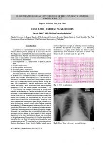
Fig. 4. Simulation speeds (in calculated beats per second) for models of cardiac electromechanics, designed in our lab, throughout the years using different versions of our electromechanics solver Continuity (http://www. continuity.ucsd.edu/). Note that, despite the fact that models are becoming more complex (shown here by the number of ODEs), real-time calculations of whole-heart electromechanics could be reached in 2008 (as is also independently predicted in [163]).
heart through stretch activated channels. In the latter study, a local region in the left ventricular wall with a higher compliance (aneurysm) generated a propagating action potential, showing that MEF can be pro-arrhythmic. F. Implementation Issues Although computer memory and speed continue to grow with advances in technology (Fig. 4), simulations of 3-D cardiac electromechanics problems remain large and time consuming and continue to grow rapidly along with advances in biology. Issues of convergence and parameter sensitivity are also of increasing concern as the number of variables increases. Therefore, Bassingthwaighte et al. [174] have proposed a strategy for developing practical multiscale models, wherein the multiscale model is designed in several steps. The core of their method includes building optimized reduced-form modules of detailed models (where applicable), validating individual levels and the systems model against multiple experimental data sets, and defining physiological ranges in which the model is valid to operate in. Obviously, each model is built for a specific purpose. Such memory and computationally intensive models require high performance computing techniques using distributed and shared memory parallel computing architectures. Parallelization techniques can be roughly divided into two categories: data parallelization and control parallelization. In data parallel applications, the same computations are performed on data that is partitioned among several processors. An example of this is in the solution of the ODEs representing the local cellular processes. If local values of membrane potential are known, the currents passing through the ion channels of a cell in one location can be calculated independently of those occurring within a cell in another location. Distributing these calculations over many processors makes feasible the integration of sophisticated ionic models (with large numbers of ODEs) into tissue and organ scale models. As the ODE calculation time grows PROCEEDINGS OF THE IEEE, VOL. 94, NO. 4, APRIL 2006
Fig. 5. Modeling scheme for cardiac electromechanics. The colors denote the different temporal and spatial levels (level 1 is the largest). Solid and dashed arrows represent data flows within and between levels, respectively.
with the sophistication of the ionic model, the solution of the linear systems do not change size or complexity. Thus, for complex ionic models, the data parallel solution of the ordinary differential equations portion of the problem is also where improvements in speed are most needed. In some instances, such as for the linear solution of partial differential equations, the data is not independent and either algebraic or geometric parallelization can be employed. In algebraic parallelization of the linear solve, the global matrix is factorized on several processors, via a special parallel linear solver, such as the distributed memory version of SuperLU [175], whereas in geometric parallelization methods, as the name suggests, regions of the full mesh are distributed over the processors. The main challenge of such domain decomposition techniques is the assignment of appropriate boundary constraints to the inner nonphysical boundaries. This technique requires iteration toward a converged solution with frequent updates at the boundaries. In control parallelization, instructions (not data) are partitioned to separate processors. This can be used to help overcome the computational hurdles for creating a functionally integrated model. These problems are typically solved with different time and space scales [20], [22], [163] (Fig. 5) and therefore require schemes for synchronizing calcula-
tions and translating meshes. For smaller spatial scales, as needed for electrophysiological problems, linear hexahedral elements in the finite-element method are sufficient for convergence, since higher order elements (e.g., cubic Hermite) are specifically designed to gain enough accuracy with few elements in a mesh and for the solution of higher order differential equations. Control parallelization can be exploited to allow the electrical and mechanical portions of the problem to be solved on different (groups of) processors with carefully developed communication between instances of these major problem classes. There are many important considerations to optimizing the performance of parallel code. Ideally, all processors should have an equal workload; however, the nature of propagation problems is that local areas of tissue near the wave front are the same areas experiencing fast ion kinetics. Conversely, in locations far from the wave front, kinetics are relatively slow. Parallel programming that balances the calculation load could lead to even further speedups. Load balancing works by keeping processors busy, for example, while one processor calculates an ODE integration for a point near the wave front requiring many small time steps to cover global time increment, another processor might calculate ordinary differential equation integration for several
KERCKHOFFS et al.: COMPUTATIONAL METHODS FOR CARDIAC ELECTROMECHANICS
777
points away from the wave front that each require few local time steps to cover the same global time increment. Adaptive meshing techniques that resize elements can also be used for improving efficiency. These techniques use smaller elements to discretize the domain in regions near the wave front and larger elements in areas away from the wave front. For adaptive meshing and load balancing technique the location of the wave front must be identified. In addition, for adaptive meshing, the wavefront location must also be predicted so that it will not reach elements that are too large in a single time step leading to a divergent solution. Various adaptive meshing techniques have been applied to cardiac electrophysiology [176]–[178] and elsewhere. Results from a recently developed space-time adaptive mesh refinement algorithm (AMRA) for simulating anisotropic 3-D excitable media suggest that this method will be able to simulate the 3-D electrical dynamics of canine ventricles quantitatively (for a biophysically detailed ionic model [67]) for 1 s using 32 1-GHz Alpha processors in approximately 9 h [179]. Recently, Fenton et al. [99] have presented a new algorithm, based on a phase-field approach, to efficiently solve wave propagation in the whole-heart using finite differences. In the rabbit heart [51], using a three-variable ionic model [88] and a monodomain approach, 1 s of simulation time took approximately 1.5 h on a single 1-GHz Alpha processor. Since the phase-field method is suited to handle complex moving boundaries, they expect their algorithm to be an attractive alternative to finite-element methods in whole-heart models of cardiac electromechanics. In examining the solution of cardiac propagation problems, separate algorithm portions can be implemented in separate software components. For example, the integration of the ordinary differential equations for the cellular ionic model may be carried out by an implicit Runge–Kutta solver suitable for stiff problems, but there are many equally effective choices of solvers [180], [181]. The best choice may depend on exactly which ionic model has been chosen. Similarly, the solution of linear systems for the partial differential equations may be performed by a range of linear equation solvers. The best solver may depend on the number of mesh elements or the computational platform being used, i.e., some solvers use matrix free methods that can solve the linear system without assembling a global system resulting in less memory use while other solvers may be specialized for solving linear systems on a distributed memory multiprocessor system. The nonlinear elasticity equations of heart mechanics are solved most efficiently with a modified Newton approach that only updates the global stiffness matrix when slow convergence occurs [182]. When reasonable convergence is maintained, quasi-Newton methods usually outperform the regular Newton’s method on efficiency. In summary, integration across multiple physical scales and biological functions in simulating cardiac electrophysiology and mechanics is now feasible in current cardiac models (we estimate real-time computing in 2008). This paves the way for integration of structurally and functionally integrated models of cardiac electromechanical function that 778
combine data-intensive cellular systems models with compute-intensive anatomically detailed multiscale simulations. Open source software licenses and government supported academic community software development projects such as the National Biomedical Computation Resource will facilitate wider distribution of modeling tools, but the tremendous effort of software engineering required to develop robust and user-friendly tools is often underestimated. Numerical models help in generating hypotheses in an iterative manner together with experiments and are on the threshold of becoming helpful tools in the clinic. REFERENCES [1] P. J. Hunter and T. K. Borg, “Integration from proteins to organs: The Physiome Project,” Nature Rev. Mol. Cell Biol., vol. 4, pp. 237–243, 2003. [2] A. D. McCulloch, “Functionally and structurally integrated computational modeling of ventricular physiology,” Jpn. J. Physiol., vol. 54, pp. 531–539, 2004. [3] J. B. Bassingthwaighte and K. C. Vinnakota, “The computational integrated myocyte—A view into the virtual heart,” in Cardiac Engineering: From Genes and Cells to Structure and Function. New York: New York Acad. Sci., 2004, vol. 1015, Annals of the New York Academy of Sciences, pp. 391–404. [4] R. L. Winslow, D. F. Scollan, A. Holmes, C. K. Yung, J. Zhang, and M. S. Jafri, “Electrophysiological modeling of cardiac ventricular function: From cell to organ,” Annu. Rev. Biomed. Eng., vol. 2, 2000, pp. 119-+. [5] P. Kohl, D. Noble, R. L. Winslow, and P. J. Hunter, “Computational modeling of biological systems: Tools and visions,” Philos. Trans. R. Soc. Lond. Series A, Math. Phys. Eng. Sci., vol. 358, pp. 579–610, 2000. [6] F. W. L. Aelen, T. Arts, D. G. M. Sanders, G. R. P. Thelissen, A. M. M. Muijtjens, F. W. Prinzen, and R. S. Reneman, “Relation between torsion and cross-sectional area change in the human left ventricle,” J. Biomech., vol. 30, pp. 207–212, 1997. [7] A. Van der Toorn, P. Barenbrug, G. Snoep, F. H. Van der Veen, T. Delhaas, F. W. Prinzen, J. Maessen, and T. Arts, “Transmural gradients of cardiac myofiber shortening in aortic valve stenosis patients using MRI tagging,” Amer. J. Physiol., Heart Circulat. Physiol., vol. 283, pp. H1609–H1615, 2002. [8] S. L. Herz, C. M. Ingrassia, S. Homma, K. D. Costa, and J. W. Holmes, “Parameterization of left ventricular wall motion for detection of regional ischemia,” Ann. Biomed. Eng., vol. 33, pp. 912–919, 2005. [9] R. Friedl, M. B. Preisack, W. Klas, T. Rose, S. Stracke, K. J. Quast, H. A. , and O. Godje, “Virtual reality and 3-D visualizations in heart surgery education,” in Proc. Heart Surgery Forum 2005, vol. 5, pp. E17–21. [10] M. Wierzbicki, M. Drangova, G. Guiraudon, and T. Peters, “Validation of dynamic heart models obtained using nonlinear registration for virtual reality training, planning, and guidance of minimally invasive cardiac surgeries,” Med. Image Anal., vol. 8, pp. 387–401, 2004. [11] A. M. Chiu, D. Dey, M. Drangova, W. D. Boyd, and T. M. Peters, “3-D image guidance for minimally invasive robotic coronary artery bypass,” in Proc. Heart Surgery Forum 2000, vol. 3, pp. 224–231. [12] E. Coste-Maniere, L. Adhami, F. Mourgues, and C. A. , “Planning, simulation, and augmented reality for robotic cardiac procedures: The STARS system of the ChIR team,” Semin. Thorac. Cardiovasc. Surg., vol. 15, pp. 141–156, 2003. [13] J. M. Guccione, S. M. Moonly, A. W. Wallace, and M. B. Ratcliffe, “Residual stress produced by ventricular volume reduction surgery has little effect on ventricular function and mechanics: A finite element model study,” J. Thorac. Cardiovasc. Surg., vol. 122, pp. 592–599, 2001. [14] J. M. Guccione, A. Salahieh, S. M. Moonly, J. Kortsmit, A. W. Wallace, and M. B. Ratcliffe, “Myosplint decreases wall stress without depressing function in the failing heart: A finite element model study,” Ann. Thorac. Surg., vol. 76, pp. 1171–1180, 2003.
PROCEEDINGS OF THE IEEE, VOL. 94, NO. 4, APRIL 2006
[15] A. B. C. Dang, J. M. Guccione, P. Zhang, A. W. Wallace, R. C. Gorman, J. H. Gorman, and M. B. Ratcliffe, “Effect of ventricular size and patch stiffness in surgical anterior ventricular restoration: A finite element model study,” Ann. Thorac. Surg., vol. 79, pp. 185–193, 2005. [16] A. Sambelashvili and I. R. Efimov, “Dynamics of virtual electrode-induced scroll-wave reentry in a 3-D bidomain model,” Amer. J. Physiol., Heart Circulat. Physiol., vol. 287, pp. H1570–H1581, 2004. [17] F. Aguel, J. Eason, and N. Trayanova, “Advances in modeling cardiac defibrillation,” Int. J. Bifurcat. Chaos, vol. 13, pp. 3791–3803, 2003. [18] B. Rodriguez, B. M. Tice, J. C. Eason, F. Aguel, and N. Trayanova, “Cardiac vulnerability to electric shocks during phase 1A of acute global ischemia,” Heart Rhythm, vol. 1, pp. 695–703, 2004. [19] T. P. Usyk and A. D. McCulloch, “Relationship between regional shortening and asynchronous electrical activation in a three-dimensional model of ventricular electromechanics,” J. Cardiovasc. Electrophysiol., vol. 14, pp. S196–S202, 2003. [20] R. C. P. Kerckhoffs, O. Faris, P. H. M. Bovendeerd, F. W. Prinzen, K. Smits, E. R. McVeigh, and T. Arts, “Electromechanics of paced left ventricle simulated by straightforward mathematical model: Comparison with experiments,” Amer. J. Physiol., Heart Circulat. Physiol., vol. 289, pp. H1889–H1897, 2005. [21] R. C. P. Kerckhoffs, P. H. M. Bovendeerd, F. W. Prinzen, K. Smits, and T. Arts, “Intra- and interventricular asynchrony of electromechanics in the ventricularly paced heart,” J. Eng. Math., vol. 47, pp. 201–216, 2003. [22] T. P. Usyk and A. D. McCulloch, “Electromechanical model of cardiac resynchronization in the dilated failing heart with left bundle branch block,” J. Electrocardiol., vol. 36, pp. 57–61, 2003. [23] W. F. Bluhm, W. Y. W. Lew, A. Garfinkel, and A. D. McCulloch, “Mechanisms of length history-dependent tension in an ionic model of the cardiac myocyte,” Amer. J. Physiol., Heart Circulat. Physiol., vol. 43, pp. H1032–H1040, 1998. [24] R. C. P. Kerckhoffs, P. H. M. Bovendeerd, J. C. S. Kotte, F. W. Prinzen, K. Smits, and T. Arts, “Homogeneity of cardiac contraction despite physiological asynchrony of depolarization: A model study,” Ann. Biomed. Eng., vol. 31, pp. 536–547, 2003. [25] N. Wiener and A. Rosenblueth, “The mathematical formulation of the problem of conduction of impulses in a network of connected excitable elements, specifically in cardiac muscle,” Arch. Inst. Cardiol. Mexico, vol. 16, pp. 205–265, 1946. [26] B. V. Alvarez, N. G. Perez, I. L. Ennis, M. C. C. de Hurtado, and H. E. Cingolani, “Mechanisms underlying the increase in force and Ca2+ transient that follow stretch of cardiac muscle—A possible explanation of the Anrep effect,” Circulat. Res., vol. 85, pp. 716–722, 1999. [27] J. M. Cordeiro, L. Greene, C. Heilmann, D. Antzelevitch, and C. Antzelevitch, “Transmural heterogeneity of calcium activity and mechanical function in the canine left ventricle,” Amer. J. Physiol., Heart Circulat. Physiol., vol. 286, pp. H1471–H1479, 2004. [28] M. A. Allessie, F. I. M. Bonke, and F. J. C. Schopman, “Circus movement in rabbit atrial muscle as a mechanism of tachycardia,” Circulat. Res., vol. 33, pp. 54–62, 1973. [29] A. M. Katz, Physiology of the Heart, 3rd ed. Philadelphia, PA: Lippincott, 2001. [30] D. M. Bers, “Cardiac excitation-contraction coupling,” Nature, vol. 415, pp. 198–205, 2002. [31] C. Antzelevitch, S. Sicouri, S. H. Litovsky, A. Lukas, S. C. Krishnan, J. M. Didiego, G. A. Gintant, and D. W. Liu, “Heterogeneity within the ventricular wall—Electrophysiology and pharmacology of epicardial, endocardial, and M-cells,” Circulat. Res., vol. 69, pp. 1427–1449, 1991. [32] D. Fedida and W. R. Giles, “Regional variations in action—Potentials and transient outward current in myocytes isolated from rabbit left-ventricle,” J. Physiol. Lond., vol. 442, pp. 191–209, 1991. [33] K. R. Laurita, R. Katra, B. Wible, X. P. Wan, and M. H. Koo, “Transmural heterogeneity of calcium handling in canine,” Circulat. Res., vol. 92, pp. 668–675, 2003. [34] V. P. Novak, F. C. P. Yin, and J. D. Humphrey, “Regional mechanical-properties of passive myocardium,” J. Biomech., vol. 27, pp. 403–412, 1994.
[35] W. L. Karlon, Influence of Myocardial Fiber Organization on Ventricular Function. La Jolla: Univ. California San Diego Press, 1998. [36] T. Arts, K. D. Costa, J. W. Covell, and A. D. McCulloch, “Relating myocardial laminar architecture to shear strain and muscle fiber orientation,” Amer. J. Physiol., Heart Circulat. Physiol., vol. 280, pp. H2222–H2229, 2001. [37] R. J. Myerburg, H. Gelband, and K. Nilsson, “Physiology of canine intraventricular conduction and endocardial excitation,” Circulat. Res., vol. 30, pp. 217–243, 1972. [38] D. W. Frazier, W. Krassowska, P. S. Chen, P. D. Wolf, N. D. Danieley, W. M. Smith, and R. E. Ideker, “Transmural activations and stimulus potentials in three-dimensional anisotropic canine myocardium,” Circulat. Res., vol. 63, pp. 135–146, 1988. [39] D. E. Roberts and A. M. Scher, “Effect of tissue anisotropy on extracellular potential fields in canine myocardium insitu,” Circulat. Res., vol. 50, pp. 342–351, 1982. [40] B. Taccardi, E. Macchi, R. L. Lux, P. R. Ershler, S. Spaggiari, S. Baruffi, and Y. Vyhmeister, “Effect of myocardial fiber direction on epicardial potentials,” Circulation, vol. 90, pp. 3076–3090, 1994. [41] D. A. Hooks, K. A. Tomlinson, S. G. Marsden, I. J. LeGrice, B. H. Smaill, A. J. Pullan, and P. J. Hunter, “Cardiac microstructure—Implications for electrical, propagation and defibrillation in the heart,” Circulat. Res., vol. 91, pp. 331–338, 2002. [42] P. Colli-Franzone, L. Guerri, and B. Taccardi, “Modeling ventricular excitation: Axial and orthotropic anisotropy effects on wavefronts and potentials,” Math. Biosci., vol. 188, pp. 191–205, 2004. [43] D. Durrer, R. T. Vandam, G. E. Freud, M. J. Janse, F. L. Meijler, and R. C. Arzbaecher, “Total excitation of isolated human heart,” Circulation, vol. 41, pp. 899–912, 1970. [44] P. J. Hunter, A. D. McCulloch, and H. ter Keurs, “Modeling the mechanical properties of cardiac muscle,” Prog. Biophys. Mol. Biol., vol. 69, pp. 289–331, 1998. [45] T. P. Usyk, R. Mazhari, and A. D. McCulloch, “Effect of laminar orthotropic myofiber architecture on regional stress and strain in the canine left ventricle,” J. Elasticity, vol. 61, pp. 143–164, 2000. [46] D. H. S. Lin and F. C. P. Yin, “A multiaxial constitutive law for mammalian left ventricular myocardium in steady-state barium contracture or tetanus,” J. Biomech. Eng., Trans. ASME, vol. 120, pp. 504–517, 1998. [47] J. C. Walker, M. B. Ratcliffe, P. Zhang, A. W. Wallace, B. Fata, E. W. Hsu, D. Saloner, and J. M. Guccione, “MRI-based finiteelement analysis of left ventricular aneurysm,” Amer. J. Physiol., Heart Circulat. Physiol., vol. 289, pp. H692–H700, 2005. [48] K. Mackway-Jones, B. A. Foex, E. Kirkman, and R. A. Little, “Modification of the cardiovascular response to hemorrhage by somatic afferent nerve stimulation with special reference to gut and skeletal muscle blood flow,” J. Trauma Injury Infection Crit. Care, vol. 47, pp. 481–485, 1999. [49] J. J. Saucerman and A. D. McCulloch, “Mechanistic systems models of cell signaling networks: A case study of myocyte adrenergic regulation,” Prog. Biophys. Mol. Biol., vol. 85, pp. 261–278, 2004. [50] J. V. Tranquillo, J. Hlavacek, and C. S. Henriquez, “An integrative model of mouse cardiac electrophysiology from cell to torso,” Europace (Suppl.), vol. 7, pp. 56–70, 2005. [51] F. J. Vetter and A. D. McCulloch, “Three-dimensional analysis of regional cardiac function: A model of rabbit ventricular anatomy,” Prog. Biophys. Mol. Biol., vol. 69, pp. 157–183, 1998. [52] P. M. F. Nielsen, I. J. Legrice, B. H. Smaill, and P. J. Hunter, “Mathematical-model of geometry and fibrous structure of the heart,” Amer. J. Physiol., vol. 260, pp. H1365–H1378, 1991. [53] I. J. LeGrice, P. J. Hunter, and B. H. Smaill, “Laminar structure of the heart: A mathematical model,” Amer. J. Physiol., Heart Circulat. Physiol., vol. 41, pp. H2466–H2476, 1997. [54] C. Stevens and P. J. Hunter, “Sarcomere length changes in a 3-D mathematical model of the pig ventricles,” Prog. Biophys. Mol. Biol., vol. 82, pp. 229–241, 2003. [55] L. Xia, M. M. Huo, Q. Wei, F. Liu, and S. Crozier, “Analysis of cardiac ventricular wall motion based on a three.-dimensional electromechanical biventricular model,” Phys. Med. Biol., vol. 50, pp. 1901–1917, 2005. [56] D. F. Scollan, A. Holmes, R. Winslow, and J. Forder, “Histological validation of myocardial microstructure obtained from diffusion tensor magnetic resonance imaging,” Amer. J. Physiol., Heart Circulat. Physiol., vol. 44, pp. H2308–H2318, 1998.
KERCKHOFFS et al.: COMPUTATIONAL METHODS FOR CARDIAC ELECTROMECHANICS
779
[57] T. G. Reese, R. M. Weisskoff, R. N. Smith, B. R. Rosen, R. E. Dinsmore, and V. J. Wedeen, “Imaging myocardial fiber architecture in-vivo with magnetic-resonance,” Magn. Reson. Med., vol. 34, pp. 786–791, 1995. [58] J. Rijcken, P. H. M. Bovendeerd, A. J. G. Schoofs, D. H. van Campen, and T. Arts, “Optimization of cardiac fiber orientation for homogeneous fiber strain during ejection,” Ann. Biomed. Eng., vol. 27, pp. 289–297, 1999. [59] I. J. Legrice, B. H. Smaill, L. Z. Chai, S. G. Edgar, J. B. Gavin, and P. J. Hunter, “Laminar structure of the heart—Ventricular myocyte arrangement and connective-tissue architecture in the dog,” Amer. J. Physiol., Heart Circulat. Physiol., vol. 38, pp. H571–H582, 1995. [60] P. Helm, M. F. Beg, M. I. Miller, and R. L. Winslow, “Measuring and mapping cardiac fiber and laminar architecture using diffusion tensor MR imaging,” Ann. NY. Acad. Sci., vol. 1047, pp. 296–307, 2005. [61] T. P. Usyk, I. J. LeGrice, and A. D. McCulloch, “Computational model of three dimensional cardiac electromechanics,” Comput. Vis. Sci., vol. 4, pp. 249–257, 2002. [62] R. A. Fitzhugh, “Impulses and physiological states in theoretical models of nerve membrane,” Biophys. J., vol. 1, pp. 445–466, 1961. [63] D. Noble and Y. Rudy, “Models of cardiac ventricular action potentials: Iterative interaction between experiment and simulation,” Philos. Trans. R. Soc. Lond. Series A, Math. Phys. Eng. Sci., vol. 359, pp. 1127–1142, 2001. [64] M. E. Belik, T. P. Usyk, and A. D. McCulloch, “Computational methods for cardiac electrophysiology,” in Handbook of Numerical Analyses, Computational Models for the Human Body. Amsterdam, The Netherlands: Elsevier, 2004, vol. 12, p. 676. [65] A. L. Hodgkin and A. F. Huxley, “A quantitative description of membrane current and its application to conduction and excitation in nerve,” J. Physiol., vol. 117, pp. 500–544, 1952. [66] G. W. Beeler and H. Reuter, “Reconstruction of action potential of ventricular myocardial fibers,” J. Physiol. Lond., vol. 268, pp. 177–210, 1977. [67] C. H. Luo and Y. Rudy, “A dynamic-model of the cardiac ventricular action-potential. 1. Simulations of ionic currents and concentration changes,” Circulat. Res., vol. 74, pp. 1071–1096, 1994. [68] M. S. Jafri, J. J. Rice, and R. L. Winslow, “Cardiac Ca2+ dynamics: The roles of ryanodine receptor adaptation and sarcoplasmic reticulum load,” Biophys. J., vol. 74, pp. 1149–1168, 1998. [69] P. C. Viswanathan and Y. Rudy, “Pause induced early afterdepolarizations in the long QT syndrome: A simulation study,” Cardiovasc. Res., vol. 42, pp. 530–542, 1999. [70] S. V. Pandit, R. B. Clark, W. R. Giles, and S. S. Demir, “A mathematical model of action potential heterogeneity in adult rat left ventricular myocytes,” Biophys. J., vol. 81, pp. 3029–3051, 2001. [71] K. H. W. J. ten Tusscher, D. Noble, P. J. Noble, and A. V. Panfilov, “A model for human ventricular tissue,” Amer. J. Physiol., Heart Circulat. Physiol., vol. 286, pp. H1573–H1589, 2004. [72] S. N. Healy and A. D. McCulloch, “An ionic model of stretch-activated and stretch-modulated currents in rabbit ventricular myocytes,” Europace, vol. 7, pp. S128–S134, 2005. [73] C. E. Clancy and Y. Rudy, “Linking a genetic defect to its cellular phenotype in a cardiac arrhythmia,” Nature, vol. 400, pp. 566–569, 1999. [74] J. L. Puglisi and D. M. Bers, “LabHEART: An interactive computer model of rabbit ventricular myocyte ion channels and Ca transport,” Amer. J. Physiol., Cell Physiol., vol. 281, pp. C2049–C2060, 2001. [75] J. J. Fox, J. L. McHarg, and R. F. Gilmour, “Ionic mechanism of electrical alternans,” Amer. J. Physiol., Heart Circulat. Physiol., vol. 282, pp. H516–H530, 2002. [76] L. Priebe and D. J. Beuckelmann, “Simulation study of cellular electric properties in heart failure,” Circulat. Res., vol. 82, pp. 1206–1223, 1998. [77] F. J. van Capelle and D. Durrer, “Computer simulation of arrhythmias in a network of coupled excitable elements,” Circulat. Res., vol. 47, pp. 454–466, 1980. [78] L. J. Leon and B. M. Horacek, “Computer-model of excitation and recovery in the anisotropic myocardium .3. Arrhythmogenic conditions in the simplified left-ventricle,” J. Electrocardiol., vol. 24, pp. 33–41, 1991.
780
[79] N. Trayanova, “Discrete versus syncytial tissue behavior ina model of cardiac stimulation .1. Mathematical formulation,” IEEE Trans. Biomed. Eng., vol. 43, pp. 1129–1140, 1996. [80] B. J. Mullerborer, D. J. Erdman, and J. W. Buchanan, “Electrical coupling and impulse propagation in anatomically modeled ventricular tissue,” IEEE Trans. Biomed. Eng., vol. 41, no. 5, pp. 445–454, May 1994. [81] C. S. Henriquez, “Simulating the electrical behavior of cardiac tissue using the bidomain model,” Crit. Rev. Biomed. Eng., vol. 21, pp. 1–77, 1993. [82] D. B. Geselowitz and W. T. Miller, “A bidomain model for anisotropic cardiac muscle,” Ann. Biomed. Eng., vol. 11, pp. 191–206, 1983. [83] R. L. Winslow, D. M. Cai, A. Varghese, and Y. C. Lai, “Generation and propagation of normal and abnormal pacemaker activity in network models of cardiac sinus node and atrium,” Chaos Solitons Fractals, vol. 5, pp. 491–512, 1995. [84] I. R. Efimov, V. I. Krinsky, and J. Jalife, “Dynamics of rotating vortices in the beeler-reuter model of cardiac tissue,” Chaos Solitons Fractals, vol. 5, pp. 513–526, 1995. [85] Z. L. Qu, K. Kil, F. G. Xie, A. Garfinkel, and J. N. Weiss, “Scroll wave dynamics in a three-dimensional cardiac tissue model: Roles of restitution, thickness, and fiber rotation,” Biophys. J., vol. 78, pp. 2761–2775, 2000. [86] A. X. Xu and M. R. Guevara, “Two forms of spiral-wave reentry in an ionic model of ischemic ventricular myocardium,” Chaos, vol. 8, pp. 157–174, 1998. [87] K. Ten Tusscher and A. V. Panfilov, “Reentry in heterogeneous cardiac tissue described by the Luo-Rudy ventricular action potential model,” Amer. J. Physiol., Heart Circulat. Physiol., vol. 284, pp. H542–H548, 2003. [88] F. Fenton and A. Karma, “Vortex dynamics in three-dimensional continuous myocardium with fiber rotation: Filament instability and fibrillation,” Chaos, vol. 8, pp. 20–47, 1998. [89] T. Ashihara, T. Namba, T. Ikeda, M. Ito, K. Nakazawa, and N. Trayanova, “Mechanisms of myocardial capture and temporal excitable gap during spiral wave reentry in a bidomain model,” Circulation, vol. 109, pp. 920–925, 2004. [90] J. J. Saucerman, S. N. Healy, M. E. Belik, J. L. Puglisi, and A. D. McCulloch, “Proarrhythmic consequences of a KCNQ1 AKAPbinding domain mutation—Computational models of whole cells and heterogeneous tissue,” Circulat. Res., vol. 95, pp. 1216–1224, 2004. [91] M. Trew, I. Le Grice, B. Smaill, and A. Pullan, “A finite volume method for modeling discontinuous electrical activation in cardiac tissue,” Ann. Biomed. Eng., vol. 33, pp. 590–602, 2005. [92] P. Taggart, P. Sutton, T. Opthof, R. Coronel, and P. Kallis, “Electrotonic cancellation of transmural electrical gradients in the left ventricle in man,” Prog. Biophys. Mol. Biol., vol. 82, pp. 243–254, 2003. [93] C. E. Conrath, R. Wilders, R. Coronel, J. M. T. de Bakker, P. Taggart, J. R. de Groot, and T. Opthof, “Intercellular coupling through gap junctions masks M cells in the human heart,” Cardiovasc. Res., vol. 62, pp. 407–414, 2004. [94] A. E. Pollard and R. C. Bar, “Computer simulations of activation in an anatomically based model of the human ventricular conduction system,” IEEE Trans. Biomed. Eng., vol. 38, no. 10, pp. 982–986, Oct. 1991. [95] M. C. MacLachlan, J. Sundnes, and G. T. Lines, “Simulation of ST segment changes during subendocardial ischemia using a realistic 3-D cardiac geometry,” IEEE Trans. Biomed. Eng., vol. 52, no. 5, pp. 799–807, May 2005. [96] K. J. Sampson and C. S. Henriquez, “Electrotonic influences on action potential duration dispersion in small hearts: A simulation study,” Amer. J. Physiol., Heart Circulat. Physiol., vol. 289, pp. H350–H360, 2005. [97] O. Berenfeld and J. Jalife, “Purkinje-muscle reentry as a mechanism of polymorphic ventricular arrhythmias in a three-dimensional model of the ventricles,” Circulat. Res., vol. 82, pp. 1063–1077, 1998. [98] A. V. Panfilov, “Modeling of re-entrant patterns in an anatomical model of the heart,” in Computational Biology of the Heart, A. V. Panfilov and A. V. Holden, Eds. Chichester, U.K.: Wiley, 1997. [99] F. H. Fenton, E. M. Cherry, A. Karma, and W. J. Rappel, “Modeling wave propagation in realistic heart geometries using the phase-field method,” Chaos, vol. 15, 2005.
PROCEEDINGS OF THE IEEE, VOL. 94, NO. 4, APRIL 2006
[100] B. Rodriguez, L. Li, J. C. Eason, I. R. Efimov, and N. A. Trayanova, “Differences between left and right ventricular chamber geometry affect cardiac vulnerability to electric shocks,” Circulat. Res., vol. 97, pp. 168–175, 2005. [101] B. Rodriguez, J. C. Eason, and N. Trayanova, “Differences between left and right ventricular anatomy determine the types of reentrant circuits induced by an external electric shock. A rabbit heart simulation study,” Prog. Biophys. Mol. Biol., vol. 90, no. 1–3, pp. 399–413, 2005. [102] J. J. Rice and P. P. de Tombe, “Approaches to modeling crossbridges and calcium-dependent activation in cardiac muscle,” Prog. Biophys. Mol. Biol., vol. 85, pp. 179–195, 2004. [103] R. S. Chadwick, “Mechanics of the left ventricle,” Biophys. J., vol. 39, pp. 279–288, 1982. [104] L. A. Taber, M. Yang, and W. W. Podszus, “Mechanics of ventricular torsion,” J. Biomech., vol. 29, pp. 745–752, 1996. [105] E. Nevo and L. Y. , “Structural finite deformation model of the left ventricle during diastole and systole,” J. Biomech. Eng., vol. 111, pp. 342–349, 1989. [106] T. Arts, P. C. Veenstra, and R. S. Renehan, “Epicardial deformation and left ventricular wall mechanisms during ejection in the dog,” Amer. J. Physiol., vol. 243, pp. H379–H390, 1982. [107] M. Vendelin, P. H. M. Bovendeerd, T. Arts, J. Engelbrecht, and D. H. van Campen, “Cardiac mechanoenergetics replicated by cross-bridge model,” Ann. Biomed. Eng., vol. 28, pp. 629–640, 2000. [108] A. F. Huxley, “Muscle structure and theories of contraction,” Prog. Biophys. Chem., vol. 7, pp. 255–318, 1957. [109] R. B. Panerai, “A model of cardiac muscle mechanics and energetics,” J. Biomech., vol. 13, pp. 929–940, 1980. [110] A. Landesberg and S. Sideman, “Mechanical regulation of cardiac-muscle by coupling calcium kinetics with cross-bridge cycling—A dynamic-model,” Amer. J. Physiol., vol. 267, pp. H779–H795, 1994. [111] M. Regnier, C. Morris, and E. Homsher, “Regulation of the crossbridge transition from a weakly to strongly bound state in skinned rabbit muscle fibers,” Amer. J. Physiol., Cell Physiol., vol. 269, pp. C1532–C1539, 1995. [112] A. Landesberg, L. Livshitz, and H. ter Keurs, “The effect of sarcomere shortening velocity on force generation, analysis, and verification of models for crossbridge dynamics,” Ann. Biomed. Eng., vol. 28, pp. 968–978, 2000. [113] M. V. Razumova, A. E. Bukatina, and K. B. Campbell, “Stiffness-distortion sarcomere model for muscle simulation,” J. Appl. Physiol., vol. 87, pp. 1861–1876, 1999. [114] J. J. Rice, R. L. Winslow, and W. C. Hunter, “Comparison of putative cooperative mechanisms in cardiac muscle: Length dependence and dynamic responses,” Amer. J. Physiol., Heart Circulat. Physiol., vol. 276, pp. H1734–H1754, 1999. [115] F. B. Sachse, G. Seemann, K. Chaisaowong, and D. Weiss, “Quantitative reconstruction of cardiac electromechanics in human myocardium: Assembly of electrophysiologic and tension generation models,” J. Cardiovasc. Electrophysiol., vol. 14, pp. S210–S218, 2003. [116] J. N. Peterson, W. C. Hunter, and M. R. Berman, “Estimated time course of Ca-2+ bound to troponin C during relaxation in isolated cardiac-muscle,” Amer. J. Physiol., vol. 260, pp. H1013–H1024, 1991. [117] A. V. Hill, “Time heart of shortening and the dynamic constants of muscle,” Proc. R. Soc. Lond., vol. 126, pp. 136–195, 1938. [118] Y. C. Fung, Biomechanics: Mechanical Properties of Living Tissues, 2nd ed. New York: Springer-Verlag, 1993. [119] A. Tozeren, “Continuum rheology of muscle contraction and its application to cardiac contractility,” Biophys. J., vol. 47, pp. 303–309, 1985. [120] H. E. ter Keurs, W. H. Rijnsburger, R. van Heuningen, and M. J. Nagelsmit, “Tension development and sarcomere length in rat cardiac trabeculae. Evidence of length-dependent activation,” Circulat. Res., vol. 46, pp. 703–714, 1980. [121] I. Mirsky, “Assessment of passive elastic stiffness of cardiac-muscle—Mathematical concepts, physiologic and clinical considerations, directions of future research,” Prog. Cardiovasc. Dis., vol. 18, pp. 277–308, 1976. [122] L. L. Demer and F. C. P. Yin, “Passive biaxial mechanical-properties of isolated canine myocardium,” J. Physiol., Lond., vol. 339, pp. 615–630, 1983.
[123] L. K. Waldman, Y. C. Fung, and J. W. Covell, “Transmural myocardial deformation in the canine left-ventricle—Normal invivo three-dimensional finite strains,” Circulat. Res., vol. 57, pp. 152–163, 1985. [124] S. Dokos, I. J. LeGrice, B. H. Smaill, J. Kar, and A. A. Young, “A triaxial-measurement sheer-test device for soft biological tissues,” J. Biomech. Eng., Trans. ASME, vol. 122, pp. 471–478, 2000. [125] J. M. Guccione, A. D. McCulloch, and L. K. Waldman, “Passive material properties of intact ventricular myocardium determined from a cylindrical model,” J. Biomech. Eng., Trans. ASME, vol. 113, pp. 42–55, 1991. [126] A. D. McCulloch and J. H. Omens, “Nonhomogeneous analysis of three-dimensional transmural finite deformation in canine ventricular myocardium,” J. Biomech., vol. 24, pp. 539–548, 1991. [127] J. H. Omens, K. D. May, and A. D. McCulloch, “Transmural distribution of three-dimensional strain in the isolated arrested canine left-ventricle,” Amer. J. Physiol., vol. 261, pp. H918–H928, 1991. [128] T. P. Usyk and A. D. McCulloch, “Computational methods for soft tissue biomechanics,” in Biomechanics of Soft Tissue in Cardiovascular Systems, G. A. Holzapfel and R. W. Ogden, Eds. New York: Springer-Verlag, 2003, pp. 273–342. [129] T. Arts, “A model of the mechanics of the left ventricle,” Ann. Biomed. Eng., vol. 7, pp. 299–318, 1979. [130] M. P. Nash and P. J. Hunter, “Computational mechanics of the heart: From tissue structure to ventricular function,” J. Elasticity, vol. 61, pp. 113–141, 2000. [131] P. H. M. Bovendeerd, T. Arts, J. M. Huyghe, D. H. Vancampen, and R. S. Reneman, “Dependence of local left-ventricular wall mechanics on myocardial fiber orientation—A model study,” J. Biomech., vol. 25, pp. 1129–1140, 1992. [132] F. J. Vetter and A. D. McCulloch, “Three-dimensional stress and strain in passive rabbit left ventricle: A model study,” Ann. Biomed. Eng., vol. 28, pp. 781–792, 2000. [133] T. P. Usyk, J. H. Omens, and A. D. McCulloch, “Regional septal dysfunction in a three-dimensional computational model of focal myofiber disarray,” Amer. J. Physiol., Heart Circulat. Physiol., vol. 281, pp. H506–H514, 2001. [134] P. H. M. Bovendeerd, J. M. Huyghe, T. Arts, D. H. Vancampen, and R. S. Reneman, “Influence of endocardial-epicardial crossover of muscle-fibers on left-ventricular wall mechanics,” J. Biomech., vol. 27, pp. 941–951, 1994. [135] L. A. Taber and W. W. Podszus, “A laminated shell model of the infarcted left ventricle,” Int. J. Solids Struct., vol. 34, pp. 223–241, 1997. [136] R. Mazhari, J. H. Omens, J. W. Covell, and A. D. McCulloch, “Structural basis of regional dysfunction in acutely ischemic myocardium,” Cardiovasc. Res., vol. 47, pp. 284–293, 2000. [137] P. H. M. Bovendeerd, T. Arts, T. Delhaas, J. M. Huyghe, D. H. Vancampen, and R. S. Reneman, “Regional wall mechanics in the ischemic left ventricle: Numerical modeling and dog experiments,” Amer. J. Physiol., Heart Circulat. Physiol., vol. 39, pp. H398–H410, 1996. [138] D. R. Zwas, S. Takuma, S. Mullis-Jansson, A. Fard, H. Chaudhry, H. Wu, M. R. Di Tullio, and S. Homma, “Feasibility of real-time three-dimensional treadmill stress echocardiography,” J. Amer. Soc. Echocardiography, vol. 12, pp. 285–289, 1999. [139] C. Lanning, S. Y. Chen, A. Hansgen, D. Chang, K. C. Chan, and R. Shandas, “Dynamic three-dimensional reconstruction and modeling of cardiovascular anatomy in children with congenital heart disease using biplane angiography,” Biomed. Sci. Instrum., vol. 40, pp. 200–205, 2004. [140] R. C. P. Kerckhoffs, O. P. Faris, P. H. M. Bovendeerd, F. W. Prinzen, K. Smits, E. R. McVeigh, and T. Arts, “Timing of depolarization and contraction in the paced canine left ventricle: Model and experiment,” J. Cardiovasc. Electrophysiol., vol. 14, pp. S188–S195, 2003. [141] T. Usyk and R. C. P. Kerckhoffs, “Three dimensional electromechanical model of porcine heart with penetrating wound injury,” Stud. Health Technol. Inf., vol. 111, pp. 568–573, 2005. [142] H. Watanabe, S. Sugiura, H. Kafuku, and T. Hisada, “Multiphysics simulation of left ventricular filling dynamics using fluidstructure interaction finite element method,” Biophys. J., vol. 87, pp. 2074–2085, 2004. [143] K. Lu, J. W. Clark, F. H. Ghorbel, D. L. Ware, and A. Bidani, “A human cardiopulmonary system model applied to the analysis of the Valsalva maneuver,” Amer. J. Physiol., Heart Circulat. Physiol., vol. 281, pp. H2661–H2679, 2001.
KERCKHOFFS et al.: COMPUTATIONAL METHODS FOR CARDIAC ELECTROMECHANICS
781
[144] Y. Sun, M. Beshara, R. J. Lucariello, and S. A. Chiaramida, “A comprehensive model for right-left heart interaction under the influence of pericardium and baroreflex,” Amer. J. Physiol., Heart Circulat. Physiol., vol. 41, pp. H1499–H1515, 1997. [145] T. Arts, T. Delhaas, P. Bovendeerd, X. Verbeek, and F. W. Prinzen, “Adaptation to mechanical load determines shape and properties of heart and circulation: The CircAdapt model,” Amer. J. Physiol., Heart Circulat. Physiol., vol. 288, pp. H1943–H1954, 2005. [146] J. B. Olansen, J. W. Clark, D. Khoury, F. Ghorbel, and A. Bidani, “A closed-loop model of the canine cardiovascular system that includes ventricular interaction,” Comput. Biomed. Res., vol. 33, pp. 260–295, 2000. [147] O. Frank, “Die grundform des arteriellen pulses,” Zeitung fuer Biologie, vol. 37, pp. 483–586, 1899. [148] N. Stergiopulos, B. E. Westerhof, and N. Westerhof, “Total arterial inertance as the fourth element of the windkessel model,” Amer. J. Physiol., Heart Circulat. Physiol., vol. 276, pp. H81–H88, 1999. [149] P. Broemser and O. F. Ranke, “Ueber die messung des schlagvolume des herzens auf unblutigem weg,” Zeitung fuer Biologie, vol. 90, pp. 467–507, 1930. [150] N. Westerhof, G. Elzinga, and G. C. van den Bos, “Influence of central and peripheral changes on hydraulic input impedance of systemic arterial tree,” Med. Biol. Eng., vol. 11, pp. 710–723, 1973. [151] C. H. Liu, S. C. Niranjan, J. W. Clark, K. Y. San, J. B. Zwischenberger, and A. Bidani, “Airway mechanics, gas exchange, and blood flow in a nonlinear model of the normal human lung,” J. Appl. Physiol., vol. 84, pp. 1447–1469, 1998. [152] J. F. Golden, J. W. Clark, and P. M. Stevens, “Mathematical modeling of pulmonary airway dynamics,” IEEE Trans. Biomed. Eng., vol. BM-20, no. 6, pp. 397–404, Nov. 1973. [153] R. K. Dash and J. B. Bassingthwaighte, “Blood HbO(2) and HbCO(2) dissociation curves at varied O-2, CO2, pH, 2,3-DPG and temperature levels,” Ann. Biomed. Eng., vol. 32, pp. 1676–1693, 2004. [154] M. J. Lab, P. Taggart, and F. Sachs, “Mechano-electric feedback,” Cardiovasc. Res., vol. 32, pp. 1–2, 1996. [155] P. Kohl and U. Ravens, “Cardiac mechano-electric feedback: Past, present, and prospect,” Prog. Biophys. Mol. Biol., vol. 82, pp. 3–9, 2003. [156] D. G. Allen and S. Kurihara, “The effects of muscle length on intracellular calcium transients in mammalian cardiac muscle,” J. Physiol., vol. 327, pp. 79–94, 1982. [157] W. Craelius, “Stretch-activation of rat cardiac myocytes,” Exp. Physiol., vol. 78, pp. 411–423, 1993. [158] M. F. M. van Oosterhout, F. W. Prinzen, T. Arts, J. J. Schreuder, W. Y. R. Vanagt, J. P. M. Cleutjens, and R. S. Reneman, “Asynchronous electrical activation induces asymmetrical hypertrophy of the left ventricular wall,” Circulation, vol. 98, pp. 588–595, 1998. [159] D. A. Kass, C. H. Chen, C. Curry, M. Talbot, R. Berger, B. Fetics, and E. Nevo, “Improved left ventricular mechanics from acute VDD pacing in patients with dilated cardiomyopathy and ventricular conduction delay,” Circulation, vol. 99, pp. 1567–1573, 1999. [160] X. Verbeek, K. Vernooy, M. Peschar, T. Van der Nagel, A. Van Hunnik, and F. W. Prinzen, “Quantification of interventricular asynchrony during LBBB and ventricular pacing,” Amer. J. Physiol., Heart Circulat. Physiol., vol. 283, pp. H1370–H1378, 2002. [161] J. J. Rice, M. S. Jafri, and R. L. Winslow, “Modeling short-term interval-force relations in cardiac muscle,” Amer. J. Physiol., Heart Circulat. Physiol., vol. 278, pp. H913–H931, 2000. [162] O. Solovyova, N. Vikulova, L. B. Katsnelson, V. S. Markhasin, P. J. Noble, A. Garny, P. Kohl, and D. Noble, “Mechanical interaction of heterogeneous cardiac muscle segments in silico: Effects on Ca2+ handling and action potential,” Int. J. Bifurcat. Chaos, vol. 13, pp. 3757–3782, 2003. [163] D. P. Nickerson, N. Smith, and P. Hunter, “New developments in a strongly coupled cardiac electromechanical model,” Europace, vol. 7, pp. 5118–5127, 2005. [164] N. P. Smith, M. L. Buist, and A. J. Pullan, “Altered T wave dynamics in a contracting cardiac model,” J. Cardiovasc. Electrophysiol., vol. 14, pp. S203–S209, 2003. [165] D. Noble, A. Varghese, P. Kohl, and P. Noble, “Improved guinea-pig ventricular cell model incorporating a diadic space, I-Kr and I-Ks, and length- and tension-dependent processes,” Can. J. Cardiol., vol. 14, pp. 123–134, 1998.
782
[166] M. P. Nash and A. V. Panfilov, “Electromechanical model of excitable tissue to study reentrant cardiac arrhythmias,” Prog. Biophys. Mol. Biol., vol. 85, pp. 501–522, 2004. [167] F. Liu, W. X. Lu, L. Xia, and G. H. Wu, “The construction of three-dimensional composite finite element mechanical model of human left ventricle,” JSME Int. J. Series C, Mech. Syst. Mach. Elements Manuf., vol. 44, pp. 125–133, 2001. [168] F. Liu, L. Xia, and X. Zhang, “Analysis of the influence of the electrical asynchrony on regional mechanics of the infarcted left ventricle using electromechanical heart models,” JSME Int. J. Series A, Solid Mech. Mater. Eng., vol. 46, pp. 1–9, 2003. [169] J. M. Rogers and A. D. McCulloch, “A collocation-galerkin finite-element model of cardiac action-potential propagation,” IEEE Trans. Biomed. Eng., vol. 41, no. 8, pp. 743–757, Aug. 1994. [170] P. Colli-Franzone, L. Guerri, and B. Taccardi, “Spread of excitation in a myocardial volume—Simulation studies in a model of anisotropic ventricular muscle activated by point stimulation,” J. Cardiovasc. Electrophysiol., vol. 4, pp. 144–160, 1993. [171] K. A. Tomlinson, P. J. Hunter, and A. J. Pullan, “A finite element method for an eikonal equation model of myocardial excitation wavefront propagation,” Siam J. Appl. Math., vol. 63, pp. 324–350, 2002. [172] F. J. Vetter and A. D. McCulloch, “Mechanoelectric feedback in a model of the passively inflated left ventricle,” Ann. Biomed. Eng., vol. 29, pp. 414–426, 2001. [173] P. Kohl, P. Hunter, and D. Noble, “Stretch-induced changes in heart rate and rhythm: Clinical observations, experiments and mathematical models,” Prog. Biophys. Mol. Biol., vol. 71, pp. 91–138, 1999. [174] J. B. Bassingthwaighte, H. J. Chizeck, L. E. Atlas, and H. Qian, “Multiscale modeling of cardiac cellular energetics,” Ann. NY Acad. Sci., vol. 1047, pp. 395–426, 2005. [175] X. S. Li and J. W. Demmel, “SuperLU_DIST: A scalable distributed-memory sparse direct solver for unsymmetric linear systems,” ACM Trans. Math. Softw., vol. 29, pp. 110–140, 2003. [176] N. F. Otani, “Computer modeling in cardiac electrophysiology,” J. Comput. Phys., vol. 161, pp. 21–34, 2000. [177] Z. L. Qu and A. Garfinkel, “An advanced algorithm for solving partial differential equation in cardiac conduction,” IEEE Trans. Biomed. Eng., vol. 46, no. 9, pp. 1166–1168, Sep. 1999. [178] W. L. Quan, S. J. Evans, and H. M. Hastings, “Efficient integration of a realistic two-dimensional cardiac tissue model by domain decomposition,” IEEE Trans. Biomed. Eng., vol. 45, no. 3, pp. 372–385, Mar. 1998. [179] E. M. Cherry, H. S. Greenside, and C. S. Henriquez, “Efficient simulation of three-dimensional anisotropic cardiac tissue using an adaptive mesh refinement method,” Chaos, vol. 13, pp. 853–865, 2003. [180] J. Sundnes, G. T. Lines, and A. Tveito, “Efficient solution of ordinary differential equations modeling electrical activity in cardiac cells,” Math. Biosci., vol. 172, pp. 55–72, 2001. [181] ——, “ODE-solvers for a stiff system arising in the modeling of the electrical activity of the heart,” Int. J. Nonlinear Sci. Numer. Simul., vol. 4, pp. 67–80, 2003. [182] S. Linge, G. Lines, and J. Sundnes, “Solving the heart mechanics equations with Newton and quasi-Newton methods—A comparison,” Comput. Methods Biomech. Biomed. Eng., vol. 8, pp. 31–38, 2005.
Roy C. P. Kerckhoffs received the M.S. degree in mechanical engineering and the Ph.D. degree in biomedical engineering from Eindhoven University of Technology, Eindhoven, The Netherlands, in 1998 and 2003, respectively. Currently, he is working in the Cardiac Mechanics Research Group, University of California, San Diego, La Jolla. His research interests include multiscale modeling of cardiovascular mechanics and electrophysiology.
PROCEEDINGS OF THE IEEE, VOL. 94, NO. 4, APRIL 2006
Sarah N. Healy received the M.S degree in bioengineering from the University of California, San Diego, La Jolla, in 2003. She is currently working toward the Ph.D degree in the Cardiac Mechanics Research Group, University of California, San Diego. Her research interests include high-performance computing and multiscale modeling of cardiac electrophysiology.
Andrew D. McCulloch received the Ph.D. degree from the University of Auckland, New Zealand, in 1986. He is currently Professor and Chair of Bioengineering at the University of California, San Diego (UCSD), La Jolla. Prof. McCulloch is a Fellow of the American Institute for Medical and Biological Engineering and a cofounder of Insilicomed, Inc. a licensee of UCSD software used in this research.
Taras P. Usyk received the M.S. degree in engineering from Kharkov State University, Ukraine in 1993 and the Ph.D. degree in mechanical engineering from Institute of Mechanical Engineering Problems, Kharkov Ukraine in 1998. He is currently with the Cardiac Mechanics Research Group, University of California, San Diego, La Jolla. His research interests include modeling of cardiovascular mechanics and electrophysiology from cell to organ level.
KERCKHOFFS et al.: COMPUTATIONAL METHODS FOR CARDIAC ELECTROMECHANICS
783