ASL images were collected from 10 survivors in mining disaster with recent ..... stress disorder patients without re-exposure to accident-related stimuli,â Clin.
Altered resting-state functional connectivity in post-traumatic stress disorder: a perfusion MRI study Baojuan Li1,2, Jian Liu2, Yang Liu1, Hong-Bing Lu1,*, Hong Yin3,* 1
School of Biomedical Engineering, Fourth Military Medical University, Xi’an, Shaanxi, 710032, China; 2Department of Automatic Control, College of Mechatronics and Automation, National University of Defense Technology, Changsha, Hunan, 410073, P.R. China; 3Department of Radiology, Xijing Hospital, Xi’an, Shaanxi, China
ABSTRACT The majority of studies on posttraumatic stress disorder (PTSD) so far have focused on delineating patterns of activations during cognitive processes. Recently, more and more researches have started to investigate functional connectivity in PTSD subjects using BOLD-fMRI. Functional connectivity analysis has been demonstrated as a powerful approach to identify biomarkers of different brain diseases. This study aimed to detect resting-state functional connectivity abnormities in patients with PTSD using arterial spin labeling (ASL) fMRI. As a completely non-invasive technique, ASL allows quantitative estimates of cerebral blood flow (CBF). Compared with BOLD-fMRI, ASL fMRI has many advantages, including less low-frequency signal drifts, superior functional localization, etc. In the current study, ASL images were collected from 10 survivors in mining disaster with recent onset PTSD and 10 survivors without PTSD. Decreased regional CBF in the right middle temporal gyrus, lingual gyrus, and postcentral gyrus was detected in the PTSD patients. Seed-based resting-state functional connectivity analysis was performed using an area in the right middle temporal gyrus as region of interest. Compared with the non-PTSD group, the PTSD subjects demonstrated increased functional connectivity between the right middle temporal gyrus and the right superior temporal gyrus, the left middle temporal gyrus. Meanwhile, decreased functional connectivity between the right middle temporal gyrus and the right postcentral gyrus, the right superior parietal lobule was also found in the PTSD patients. This is the first study which investigated resting-state functional connectivity in PTSD using ASL images. The results may provide new insight into the neural substrates of PTSD. Keywords:rCBF, resting-state, functional connectivity, ASL, perfusion, posttraumatic stress disorder
1. INTRODUCTION Posttraumatic stress disorder (PTSD) is a mental condition that develops after exposure to terrifying and life-threatening events such as warfare1, accidents2, disasters3, 4, and physical assaults 5.The core symptoms of PTSD include re-experiencing, avoidance, and hyperarousal
6, 7
. Neuroimaging techniques have been used widely to detect structural
and functional biomarkers of PTSD. Decreased hippocampus volume has been consistently reported in previous studies8,9. Moreover, functional neuroimaging studies have shown aberrant brain activity in the anterior cingulate cortex10, 11, the medial prefrontal cortex12, 13, and amygdala14 in subjects with PTSD.
Medical Imaging 2013: Image Perception, Observer Performance, and Technology Assessment, edited by Craig K. Abbey, Claudia R. Mello-Thoms, Proc. of SPIE Vol. 8673, 867318 © 2013 SPIE · CCC code: 1605-7422/13/$18 · doi: 10.1117/12.2008168 Proc. of SPIE Vol. 8673 867318-1 Downloaded From: http://proceedings.spiedigitallibrary.org/ on 06/23/2014 Terms of Use: http://spiedl.org/terms
Recently, more and more researches have started to investigate functional connectivity in PTSD subjects using BOLD-fMRI15, 16. Functional connectivity analysis has been demonstrated to be a powerful approach to identify biomarkers of different brain diseases. The majority of studies investigating alterations in brain activity and connectivity in subjects PTSD are limited to BOLD-fMRI, very few attempts have been made to evaluated dynamic interactions between brain regions using arterial spin labeling (ASL) fMRI. As a completely non-invasive technique, ASL fMRI allows quantitative estimates of cerebral blood flow (CBF). Compared to BOLD-fMRI, ASL fMRI has many advantages. Firstly, low-frequency signal drifts in ASL-fMRI are less than BOLD-fMRI, which makes ASL-fMRI more suitable for the investigation of slow changes in neural activity 17. Secondly, ASL-fMRI can provide superior functional localization than BOLD-fMRI
17, 18
. Thirdly, it has been shown that the resting-state spatial patterns of low-frequency fluctuations
detected using perfusion imaging fit those obtained at the level of cerebral metabolic rate of oxygenation better than BOLD-fMRI 19. Finally, some researchers have suggested that functional connectivity patterns result from CBF images may be more physiologically relevant than those obtained from BOLD-fMRI
20
. Given these advantages of perfusion
imaging, brain connectivity analysis using ASL-fMRI images may provide new information that is not available in conventional BOLD-fMRI studies. In fact, some researches have started to establish the validity of functional connectivity analysis using ASL-fMRI images 21, 22. The current study aimed to illustrate resting-state rCBF and functional connectivity abnormities in patients with PTSD using ASL-fMRI. Ten male survivors of a coal mining flood disaster with recent onset PTSD and 10 trauma-exposed non-PTSD subjects participated in this study. Two hypotheses were tested: 1) the PTSD patients would demonstrate altered rCBF compared with trauma-exposed healthy controls; 2) Seed-based functional connectivity analysis would reveal abnormalities in resting-state functional connectivity in the PTSD group.
2. METHODS 2.1 Subjects
Participants were 20 male survivors of a coal mining flood disaster occurred in July 2007 in Henan province of China. During this disaster, 69 miners were trapped in the coal mine with milk and water supply. All the miners were rescued in 72 hours and none of the miners died. Six months later, 48 of the 69 survivors received a Structured Clinical Interview (SCID) and 17 subjects fulfilled DSM-IV criteria for PTSD. Ten survivors who were diagnosed with PTSD (the PTSD group) and 10 survivors without PTSD (the non-PTSD group) agreed to take part in the current study 9. Consent from the participants was obtained prior to the survey, in accord with the recommendations of the Ethics Committee of the Xijing Hospital of The Fourth Military Medical University. There were no significant differences in age, education, or symptom severity (assessed with the Chinese version of the Clinician-Administered PTSD Scale, CAPS) between survivors who participated in the current study and who did not.
Proc. of SPIE Vol. 8673 867318-2 Downloaded From: http://proceedings.spiedigitallibrary.org/ on 06/23/2014 Terms of Use: http://spiedl.org/terms
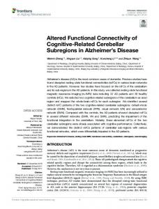
Figure 2 Altered functional connectivity in the PTSD group. Increased functional connectivity in the PTSD patients were marked with blue, while decreased functional connectivity were marked with red and yellow. STG, superior temporal gyrus; MTG, middle temporal gyrus; PG, postcentral gyrus.
4. DISCUSSION In the current study, we investigated alterations in rCBF in survivors of a mining disaster with recent onset PTSD using ASL fMRI images. Decreased rCBF in the right middle temporal gyrus, the right lingual gyrus, and the right postcentral gyrus was detected in PTSD patients compared to trauma-exposed non-PTSD subjects. We also tried to examine changes in resting-state functional connectivity of the PTSD patients using perfusion images. A region in the right middle temporal gyrus which showed decreased rCBF in the PTSD group was selected as the seed. Functional connectivity between the seed and all other voxels in the brain was calculated. A comparison between the PTSD group and the healthy trauma survivors showed increased functional connectivity between the right middle temporal gyrus and the right superior temporal gyrus, the left middle temporal gyrus. Meanwhile, decreased functional connectivity between the right middle temporal gyrus and the right postcentral gyrus, the right superior parietal lobule was also detected in the PTSD patients. Our result confirmed the involvement of the temporal cortices in the pathogenesis of PTSD and may provide new insight into the neural substrate of PTSD. 4.1 Decreased rCBF in the PTSD patients Decreased cortical rCBF was detected in brain regions including the right middle temporal gyrus, the right lingual gyrus, and the right postcentral gyrus in PTSD patients in this study. The results are consistent with previous neuroimaging studies that have reported abnormalities in structure and activation of the temporal cortices in PTSD patients. PTSD subjects have been shown to exhibit reduction in middle temporal gyrus gray matter volume in previous studies 23, 24. In addition, it has been reported that the volume of the temporal cortex is inversely correlated with the CAPS scores 23, the reexperiencing scores 24, and self-reported anxiety 25. Although there are only few perfusion imaging studies on PTSD, abnormal rCBF in temporal brain regions have also been observed 1, 5, 26, 27. In an early study performed by Sachinvala et al., increased regional cerebral perfusion was found in the right temporal regions in PTSD patients compared with
Proc. of SPIE Vol. 8673 867318-5 Downloaded From: http://proceedings.spiedigitallibrary.org/ on 06/23/2014 Terms of Use: http://spiedl.org/terms
Table 1 Regions that showed decreased rCBF in the PTSD group compared with non-PTSD survivors Brain region
L/R
BA
Cluster
T
X
Y
Z
middle temporal gyrus
R
37
172
4.55
55.5
-51.5
-12
lingual gyrus
R
62
4.56
21
-84.5
-4.5
postcentral gyrus
R
54
3.59
22.5
-32
60
3
Figure 1 Decreased rCBF in the right middle temporal gyrus, the right lingual gyrus, and the right postcentral gyrus in the PTSD group. MTG, middle temporal gyrus; LG, lingual gyrus; PG, postcentral gyrus.
Resting-state functional connectivity analysis revealed increased functional connectivity between the right middle temporal gyrus and the right superior temporal gyrus, the left middle temporal gyrus in the PTSD group. Meanwhile, we also found decreased functional connectivity between the right middle temporal gyrus and the right postcentral gyrus, the right superior parietal lobule in the PTSD patients (Figure 2, table 2, pNon-PTSD
Non-PTSD>PTSD
Proc. of SPIE Vol. 8673 867318-4 Downloaded From: http://proceedings.spiedigitallibrary.org/ on 06/23/2014 Terms of Use: http://spiedl.org/terms
Figure 2 Altered functional connectivity in the PTSD group. Increased functional connectivity in the PTSD patients were marked with blue, while decreased functional connectivity were marked with red and yellow. STG, superior temporal gyrus; MTG, middle temporal gyrus; PG, postcentral gyrus.
4. DISCUSSION In the current study, we investigated alterations in rCBF in survivors of a mining disaster with recent onset PTSD using ASL fMRI images. Decreased rCBF in the right middle temporal gyrus, the right lingual gyrus, and the right postcentral gyrus was detected in PTSD patients compared to trauma-exposed non-PTSD subjects. We also tried to examine changes in resting-state functional connectivity of the PTSD patients using perfusion images. A region in the right middle temporal gyrus which showed decreased rCBF in the PTSD group was selected as the seed. Functional connectivity between the seed and all other voxels in the brain was calculated. A comparison between the PTSD group and the healthy trauma survivors showed increased functional connectivity between the right middle temporal gyrus and the right superior temporal gyrus, the left middle temporal gyrus. Meanwhile, decreased functional connectivity between the right middle temporal gyrus and the right postcentral gyrus, the right superior parietal lobule was also detected in the PTSD patients. Our result confirmed the involvement of the temporal cortices in the pathogenesis of PTSD and may provide new insight into the neural substrate of PTSD. 4.1 Decreased rCBF in the PTSD patients Decreased cortical rCBF was detected in brain regions including the right middle temporal gyrus, the right lingual gyrus, and the right postcentral gyrus in PTSD patients in this study. The results are consistent with previous neuroimaging studies that have reported abnormalities in structure and activation of the temporal cortices in PTSD patients. PTSD subjects have been shown to exhibit reduction in middle temporal gyrus gray matter volume in previous studies 23, 24. In addition, it has been reported that the volume of the temporal cortex is inversely correlated with the CAPS scores 23, the reexperiencing scores 24, and self-reported anxiety 25. Although there are only few perfusion imaging studies on PTSD, abnormal rCBF in temporal brain regions have also been observed 1, 5, 26, 27. In an early study performed by Sachinvala et al., increased regional cerebral perfusion was found in the right temporal regions in PTSD patients compared with
Proc. of SPIE Vol. 8673 867318-5 Downloaded From: http://proceedings.spiedigitallibrary.org/ on 06/23/2014 Terms of Use: http://spiedl.org/terms
age-matched normal volunteers
26
. Later, another study detected decreased rCBF in the right temporal cortex in 8
survivors of torture suffering from PTSD28. In addition, Chung and co-authors reported decreased perfusion in the temporal regions in PTSD patients compared to normal controls 27. A recent study found increased rCBF in primarily right parietal and superior temporal cortices in male veterans with PTSD compared to male veterans without PTSD 1. Furthermore, Kim and the colleagues compared the functional neuroimaging pattern of resting glucose metabolism in female PTSD patients experienced sexual assault with healthy controls. Significantly lower cerebral glucosemetabolic activity in the superior temporal was found in the patients 5. Taken together, these findings may provide supports for the right temporal lobe syndrome hypothesis of PTSD 29. In this study, alterations in rCBF were largely located in the right middle temporal gyrus. Electrical stimulation of the right temporal cortices has been shown to elicit experiential response which is re-living or re-enacting of past experiences
30, 31
. Thus, decreased resting-state rCBF in the right middle
temporal gyrus may be associated with maladaptive re-experiencing of the traumatic events in PTSD patients. 4.2 Altered resting-state functional connectivity pattern in PTSD As far as we know, this is the first study that investigated alteration in resting-state functional connectivity of PTSD patients using ASL-fMRI data. Abnormal resting state functional connectivity pattern revealed by ASL-fMRI images is in line with previous studies that utilized BOLD-fMRI images
32-34
. Zhou et al. investigated resting-state functional
connectivity of the PTSD patients 2 days post-event. The results showed that functional connectivity between the posterior cingulate cortex and the left superior temporal gyrus was negatively correlated with the CAPS scores32. Moreover, Yin et al. found decreased ReHo in the right middle temporal gyrus in PTSD patients compared to non-PTSD trauma survivors of the 2008 Sichuan earthquake33. Qin et al. found that PTSD patients exhibited decreased functional connectivity between the PCC and the right middle temporal gyrus34. In the current study, we studied resting-state functional connectivity using ASL fMRI images and detected increased functional connectivity between the right middle temporal gyrus and the right superior temporal gyrus, the left middle temporal gyrus in the PTSD group. Moreover, we also found decreased functional connectivity between the right middle temporal gyrus and the right postcentral gyrus, the right superior parietal lobule in the PTSD patients. Although many studies have noticed changes in structure23, 24, activation1, 5, 26, 27 and functional connectivity32-34 of the right temporal gyrus in PTSD subjects, PTSD is conventionally considered to be a disorder of fear circuit29 and the majority of previous studies have focused on abnormalities in the amygdala12, 14, 35, anterior cingulate cortex10, 36 and hippocampus9, 37, 38. The contribution of the right temporal gyrus to the neurocircuitry of PTSD remains largely unknown 39. In this study, we focused on this region and evaluated the functional connectivity between the right middle temporal gyrus and all the other voxels in the brain in PTSD. This study expanded previous researches on the neurocircuitry of PTSD and our findings may provide new insight into the neural substrate of PTSD.
ACKNOWLEDGEMENTS
This work was supported in part by the National Natural Science Foundation of China under grant Nos. 81230035 and 81071220.
Proc. of SPIE Vol. 8673 867318-6 Downloaded From: http://proceedings.spiedigitallibrary.org/ on 06/23/2014 Terms of Use: http://spiedl.org/terms
REFERENCES [1]
Schuff, N., Zhang, Y., Zhan, W., Lenoci, M., Ching, C., Boreta, L. et al., “Patterns of altered cortical perfusion and diminished subcortical integrity in posttraumatic stress disorder: an MRI study,” Neuroimage, 54 Suppl 1, S62-8 (2011).
[2]
Nardo, D., Hogberg, G., Flumeri, F., Jacobsson, H., Larsson, S. A., Hallstrom, T., and Pagani, M., “Self-rating scales assessing subjective well-being and distress correlate with rCBF in PTSD-sensitive regions,” Psychol Med, 1-13 (2011).
[3]
Yin, Y., Jin, C., Eyler, L. T., Jin, H., Hu, X., Duan, L. et al., “Altered regional homogeneity in post-traumatic stress disorder: a restingstate functional magnetic resonance imaging study,” Neurosci Bull, 28(5), 541-9 (2012).
[4]
Liu, Y., Li, Y. J., Luo, E. P., Lu, H. B., and Yin, H., “Cortical thinning in patients with recent onset post-traumatic stress disorder after a single prolonged trauma exposure,” PLoS One, 7(6), e39025 (2012).
[5]
Kim, S. Y., Chung, Y. K., Kim, B. S., Lee, S. J., Yoon, J. K., and An, Y. S., “Resting cerebral glucose metabolism and perfusion patterns in women with posttraumatic stress disorder related to sexual assault,” Psychiatry Res, 201(3), 214-7 (2012).
[6]
Sripada, R. K., King, A. P., Garfinkel, S. N., Wang, X., Sripada, C. S., Welsh, R. C., and Liberzon, I., “Altered resting-state amygdala functional connectivity in men with posttraumatic stress disorder,” J Psychiatry Neurosci, 37(4), 241-9 (2012).
[7]
Rauch, S. L., Shin, L. M., and Phelps, E. A., “Neurocircuitry models of posttraumatic stress disorder and extinction: human neuroimaging research--past, present, and future,” Biol Psychiatry, 60(4), 376-82 (2006).
[8]
Nardo, D., Hogberg, G., Lanius, R. A., Jacobsson, H., Jonsson, C., Hallstrom, T., and Pagani, M., “Gray matter volume alterations related to trait dissociation in PTSD and traumatized controls,” Acta Psychiatr Scand, (2012).
[9]
Zhang, J., Tan, Q., Yin, H., Zhang, X., Huan, Y., Tang, L. et al., “Decreased gray matter volume in the left hippocampus and bilateral calcarine cortex in coal mine flood disaster survivors with recent onset PTSD,” Psychiatry Res, 192(2), 84-90 (2011).
[10]
Thomaes, K., Dorrepaal, E., Draijer, N., de Ruiter, M. B., Elzinga, B. M., Sjoerds, Z. et al., “Increased anterior cingulate cortex and hippocampus activation in Complex PTSD during encoding of negative words,” Soc Cogn Affect Neurosci, (2011).
[11]
Shin, L. M., Bush, G., Milad, M. R., Lasko, N. B., Brohawn, K. H., Hughes, K. C. et al., “Exaggerated activation of dorsal anterior cingulate cortex during cognitive interference: a monozygotic twin study of posttraumatic stress disorder,” Am J Psychiatry, 168(9), 979-85 (2011).
[12]
Shin, L. M., Rauch, S. L., and Pitman, R. K., “Amygdala, medial prefrontal cortex, and hippocampal function in PTSD,” Ann N Y Acad Sci, 1071, 67-79 (2006).
[13]
Shin, L. M., Orr, S. P., Carson, M. A., Rauch, S. L., Macklin, M. L., Lasko, N. B. et al., “Regional cerebral blood flow in the amygdala and medial prefrontal cortex during traumatic imagery in male and female Vietnam veterans with PTSD,” Arch Gen Psychiatry, 61(2), 168-76 (2004).
[14]
El Khoury-Malhame, M., Reynaud, E., Soriano, A., Michael, K., Salgado-Pineda, P., Zendjidjian, X. et al., “Amygdala activity correlates with attentional bias in PTSD,” Neuropsychologia, 49(7), 1969-73 (2011).
Proc. of SPIE Vol. 8673 867318-7 Downloaded From: http://proceedings.spiedigitallibrary.org/ on 06/23/2014 Terms of Use: http://spiedl.org/terms
[15]
Lanius, R. A., Williamson, P. C., Bluhm, R. L., Densmore, M., Boksman, K., Neufeld, R. W. et al., “Functional connectivity of dissociative responses in posttraumatic stress disorder: a functional magnetic resonance imaging investigation,” Biol Psychiatry, 57(8), 873-84 (2005).
[16]
Bluhm, R. L., Williamson, P. C., Osuch, E. A., Frewen, P. A., Stevens, T. K., Boksman, K. et al., “Alterations in default network connectivity in posttraumatic stress disorder related to early-life trauma,” J Psychiatry Neurosci, 34(3), 187-94 (2009).
[17]
Wang, Z., Aguirre, G. K., Rao, H., Wang, J., Fernandez-Seara, M. A., Childress, A. R., and Detre, J. A., “Empirical optimization of ASL data analysis using an ASL data processing toolbox: ASLtbx,” Magn Reson Imaging, 26(2), 261-9 (2008).
[18]
Luh, W. M., Wong, E. C., Bandettini, P. A., Ward, B. D., and Hyde, J. S., “Comparison of simultaneously measured perfusion and BOLD signal increases during brain activation with T(1)-based tissue identification,” Magn Reson Med, 44(1), 137-43 (2000).
[19]
Wu, C. W., Gu, H., Lu, H., Stein, E. A., Chen, J. H., and Yang, Y., “Mapping functional connectivity based on synchronized CMRO2 fluctuations during the resting state,” Neuroimage, 45(3), 694-701 (2009).
[20]
Zou, Q., Wu, C. W., Stein, E. A., Zang, Y., and Yang, Y., “Static and dynamic characteristics of cerebral blood flow during the resting state,” Neuroimage, 48(3), 515-24 (2009).
[21]
De Luca, M., Beckmann, C. F., De Stefano, N., Matthews, P. M., and Smith, S. M., “fMRI resting state networks define distinct modes of long-distance interactions in the human brain,” Neuroimage, 29(4), 1359-67 (2006).
[22]
Chuang, K. H., van Gelderen, P., Merkle, H., Bodurka, J., Ikonomidou, V. N., Koretsky, A. P. et al., “Mapping resting-state functional connectivity using perfusion MRI,” Neuroimage, 40(4), 1595-605 (2008).
[23]
Herringa, R., Phillips, M., Almeida, J., Insana, S., and Germain, A., “Post-traumatic stress symptoms correlate with smaller subgenual cingulate, caudate, and insula volumes in unmedicated combat veterans,” Psychiatry Res, 203(2-3), 139-45 (2012).
[24]
Kroes, M. C., Whalley, M. G., Rugg, M. D., and Brewin, C. R., “Association between flashbacks and structural brain abnormalities in posttraumatic stress disorder,” Eur Psychiatry, 26(8), 525-31 (2011).
[25]
Kroes, M. C., Rugg, M. D., Whalley, M. G., and Brewin, C. R., “Structural brain abnormalities common to posttraumatic stress disorder and depression,” J Psychiatry Neurosci, 36(4), 256-65 (2011).
[26]
Sachinvala, N., Kling, A., Suffin, S., Lake, R., and Cohen, M., “Increased regional cerebral perfusion by 99mTc hexamethyl propylene amine oxime single photon emission computed tomography in post-traumatic stress disorder,” Mil Med, 165(6), 473-9 (2000).
[27]
Chung, Y. A., Kim, S. H., Chung, S. K., Chae, J. H., Yang, D. W., Sohn, H. S., and Jeong, J., “Alterations in cerebral perfusion in posttraumatic stress disorder patients without re-exposure to accident-related stimuli,” Clin Neurophysiol, 117(3), 637-42 (2006).
[28]
Mirzaei, S., Knoll, P., Keck, A., Preitler, B., Gutierrez, E., Umek, H. et al., “Regional cerebral blood flow in patients suffering from post-traumatic stress disorder,” Neuropsychobiology, 43(4), 260-4 (2001).
[29]
Engdahl, B., Leuthold, A. C., Tan, H. R., Lewis, S. M., Winskowski, A. M., Dikel, T. N., and Georgopoulos, A. P., “Post-traumatic stress disorder: a right temporal lobe syndrome?,” J Neural Eng, 7(6), 066005 (2010).
[30]
Penfield, W., and Perot, P., “The Brain's Record of Auditory and Visual Experience. A Final Summary and Discussion,” Brain, 86, 595-696 (1963).
Proc. of SPIE Vol. 8673 867318-8 Downloaded From: http://proceedings.spiedigitallibrary.org/ on 06/23/2014 Terms of Use: http://spiedl.org/terms
[31]
Bancaud, J., Brunet-Bourgin, F., Chauvel, P., and Halgren, E., “Anatomical origin of deja vu and vivid 'memories' in human temporal lobe epilepsy,” Brain, 117 ( Pt 1), 71-90 (1994).
[32]
Zhou, Y., Wang, Z., Qin, L. D., Wan, J. Q., Sun, Y. W., Su, S. S. et al., “Early altered resting-state functional connectivity predicts the severity of post-traumatic stress disorder symptoms in acutely traumatized subjects,” PLoS One, 7(10), e46833 (2012).
[33]
Yin, Y., Jin, C., Eyler, L. T., Jin, H., Hu, X., Duan, L. et al., “Altered regional homogeneity in post-traumatic stress disorder: a resting-state functional magnetic resonance imaging study,” Neurosci Bull, 28(5), 541-9 (2012).
[34]
Qin, L. D., Wang, Z., Sun, Y. W., Wan, J. Q., Su, S. S., Zhou, Y., and Xu, J. R., “A preliminary study of alterations in default network connectivity in post-traumatic stress disorder patients following recent trauma,” Brain Res, 1484, 50-6 (2012).
[35]
Debiec, J., and LeDoux, J. E., “Noradrenergic signaling in the amygdala contributes to the reconsolidation of fear memory: treatment implications for PTSD,” Ann N Y Acad Sci, 1071, 521-4 (2006).
[36]
Hamner, M. B., Lorberbaum, J. P., and George, M. S., “Potential role of the anterior cingulate cortex in PTSD: review and hypothesis,” Depress Anxiety, 9(1), 1-14 (1999).
[37]
Thomaes, K., Dorrepaal, E., Draijer, N. P., de Ruiter, M. B., Elzinga, B. M., van Balkom, A. J. et al., “Increased activation of the left hippocampus region in Complex PTSD during encoding and recognition of emotional words: a pilot study,” Psychiatry Res, 171(1), 44-53 (2009).
[38]
Filipovic, B. R., Djurovic, B., Marinkovic, S., Stijak, L., Aksic, M., Nikolic, V. et al., “Volume changes of corpus striatum, thalamus, hippocampus and lateral ventricles in posttraumatic stress disorder (PTSD) patients suffering from headaches and without therapy,” Cent Eur Neurosurg, 72(3), 133-7 (2011).
[39]
Hou, C., Liu, J., Wang, K., Li, L., Liang, M., He, Z. et al., “Brain responses to symptom provocation and trauma-related short-term memory recall in coal mining accident survivors with acute severe PTSD,” Brain Res, 1144, 165-74 (2007).
Proc. of SPIE Vol. 8673 867318-9 Downloaded From: http://proceedings.spiedigitallibrary.org/ on 06/23/2014 Terms of Use: http://spiedl.org/terms