100 amino acids. AMINO ACIDS. ~ 700 amino acids. ▫ free compounds. ▫
structural units of peptides, proteins and other compounds structure amino group.
acids, and DNA as sources of carbon and nitrogen to marine bacteria. Niels 0. G. ~srgensen', Niels Kroer 2r*, Richard B. Coffin3, Xiao-Hua yang4,. Cindy Lee.
Apr 25, 2016 - the role of parent body processes and composition in the creation, ... Meteorites, the rocky remnants of asteroids or comets that land on Earth ...
Feb 28, 2013 - MU-Plovdiv, Medical College, Plovdiv, Bulgaria. ABSTRACT ..... Archives of The Balkan Medical Union 2011; 46, 4,supl:197-203. 18. Frau M ...
Fight off the free radicals in your body and slow down aging and common disorders with our amino acid or Glutathione drips. While we all need oxygen to live, did you know that too much oxygen can cause free radicals to develop in the body? These free
trypsinogen. Trypsin, chymotrypsin, and elastase cata- lyse the breakdown of proteins, polypeptides, and peptides into smaller peptides and amino acids in the ...
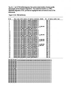
U L LL LLL LLL LLL LLL LLL LLLL. OYL LLL ... TK L LL LLLL LLLL LLLL LLLL ...... QQ AA. AA. ET SH. ET SH. ET. QRE. E E GRE R. E E ORE R. QRE. Î Î QRE.
Nov 28, 2014 - Reddy used a zinc mediated allylation of a chlorosilane as the key. Fig. 1. Silicon-containing ...... by distillation. Quenching the reactions with ...
Apr 21, 2003 - transferred to specific face to form only the L-isomer of glutamate. ... glutamine to oxaloacetate to form aspartate and α-ketoglutarate. There are ...
May 14, 2018 - scale to afford the α-hydroxy acid (S)-14f in 80% isolated yield and >99% ee (Scheme 4). The product ..... oxidase from Streptomyces coelicolor.
Jun 17, 2010 - tively augment the physiological adaptation to exercise. ... fat-free milk after resistance exercise, for 12 weeks, ...... Book of abstracts, ICST 5th Congress, Odense 2006. 90. Tipton KD, Elliott TA, Cree MG, Wolf SE, Sanford AP, ...
May 24, 2004 - Circuit in Alachua County, Florida on September 10, 2002. His .... The cerebral CT scan taken on August 2, 2000 at 1028 showed that Robert ...
fering 14C age, is taken up by organisms and incorporated in the bone .... detector set at 205 nm, and a fraction collector controlled by Star workstation PC ...
Lecture 2. Slide 1. Biochemistry 2000. Chapter 4: Amino Acids. Voet & Voet:
Pages 67 - 81. Any introductory Biochemistry textbook will have a chapter on
amino ...
28 Feb 2013 - recent analytical methods in the analysis of the essential amino acids .... important area of fluorescence spectroscopy. They include ... tryptophan, tyrosine and phenylalanine in proteins, vitamin A and B2, NADH derivatives of.
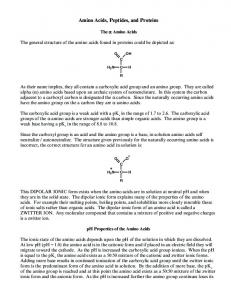
Amino Acids, Peptides, and Proteins. The α Amino Acids. The general structure of
the amino acids found in proteins could be depicted as: R. C. C. H. H2N. O.
Mar 10, 2017 - (1) The protein-wasting effect of hypocaloric (low carbohydrate) ..... Kumar, A.; Roberts, D.; Wood, K.E.; Light, B.; Parrillo, J.E.; Sharma, S.; ...
CopR is a HTH protein belonging to the lambda ... Here, we describe site-directed mu- ... characterize transcriptional repressors controlling lysis/ lysogeny of bacteriophages, e.g., (cI and Cro) or P22 (Arc, c2), and .... the PCR fragments 1 and 2 f
In this article, amino acids, its functions and associated diseases have been elaborated. ... acids. Adrenaline, nor-adrenaline and thyroxin are made up of single amino acid. Glutathione .... It is a genetic disorder that occurs due to deficiency of.
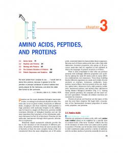
AMINO ACIDS, PEPTIDES,. AND PROTEINS. 3.1 Amino Acids 75. 3.2 Peptides
and Proteins 85. 3.3 Working with Proteins 89. 3.4 The Covalent Structure of ...
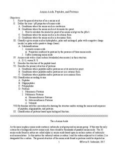
Amino Acids, Peptides, Proteins. Functions of proteins: Enzymes. Transport and
Storage. Motion, muscle contraction. Hormones. Mechanical support. Immune ...
a) Properties conferred to proteins by the presence of these amino acids. 2. ... A.
Conditions where peptides and/or proteins are at its isoelectric point.
283 –. Amin o. A cids. Derivatives. Amino Acids and Their. Derivatives. Boc-L-
Amino Acids …………………………………… 284. Boc-D-Amino Acids …
Brush border peptidases split oligopeptides of six or less amino acids in length. Many of the resulting di- and tripep- tides are transported into the enterocyte.
Amino Acids - Chap 03
3
12/3/03
12:24 pm
Page 41
Absorption of Amino Acids and Peptides C.R. Krehbiel1* and J.C. Matthews2
1Department
of Animal Science, Oklahoma State University, Stillwater, Oklahoma, USA; 2Department of Animal Sciences, University of Kentucky, Lexington, Kentucky, USA
Introduction Assimilation of dietary or microbial (ruminants) protein involves the interaction of a series of steps beginning in the stomach (non-ruminants), abomasum (ruminants), or proventriculus (poultry) and ending with the transport of amino acids and peptides from the basolateral membrane of the small intestine. In the glandular stomach, hydrochloric acid (HCl) denatures dietary protein and promotes proteolysis protein to large polypeptides via the action of pepsin. On entering the small intestine, pancreatic proteases principally hydrolyse large polypeptides and proteins into oligopeptides of six or less amino acid residues as well as free amino acids. Degradation of dietary protein continues by hydrolytic enzymes of the small intestine epithelia that are present in the luminal surface (apical membrane or brush border) of absorptive epithelial cells (enterocytes). Brush border peptidases split oligopeptides of six or less amino acids in length. Many of the resulting di- and tripeptides are transported into the enterocyte intact by a single H+-coupled transporter and then hydrolysed to free amino acids by cytosolic peptidases (primary) or transported across the basolateral membrane. In contrast, free amino acids are absorbed by a
variety of iron-dependent and -independent transporters. The fate of absorbed peptides is principally further hydrolysis to free amino acids by a variety of cytosolic peptidases, whether absorbed as free or peptide-bound amino acids, cytosolic amino acids are available as energy substrates, incorporation into constitutive protein, or transport across the basolateral membrane into blood. Ultimately, digested protein enters the hepatic portal circulation in the form of free amino acid and peptides. The working hypothesis for assimilation of luminal proteins by enterocytes is illustrated in Fig. 3.1. The model identifies gastric, luminal, glycocalyx/apical membrane, and intracellular hydrolytic digestion events, in addition to apical and basolateral membrane-mediated absorption events of peptidebound and free amino acids by specific transport proteins. Each component of the model is discussed in this chapter. Although poorly understood, and in contrast to the specificity of digestion and transport events, it is also important to note that the potential contribution to the absorption of amino acids by relatively non-specific transmembrane simple diffusion and paracellular flow events may be of nutritional significance. Despite anatomical differences in the digestive tracts among farm animal species,
BLOOD Fig. 3.1. The current model for the role of peptide uptake in protein assimilation, as adapted from Ganapathy et al. (1994). The relative contribution of peptide (di-, tri-) versus free amino acid (AA) to total protein assimilation through hydrolytic and transport events is indicated by the relative thickness of the lines. After hydrolysis by gastric proteases and luminal peptidases, oligopeptides are hydrolysed to small peptides and free amino acids by apical membrane-bound (1) peptidases. Peptides are absorbed across brush border membrane by PepT1 (2). Whereas a small proportion of absorbed peptides are then absorbed intact across the basolateral membrane by a H+-independent transport activity (3), the majority are hydrolysed by intracellular peptidases (4). The resulting free AA, plus those absorbed across the apical membrane by a complement of Na+-dependent and -independent amino acid transporters (5) are then transported across the basolateral membrane by a complement of Na+-independent and amino acid exchanger transport proteins (6). The extracellular–intracellular H+ gradient that drives PepT1 activity, is re-established by the combined function of the apical Na+/H+ exchanger (7) and the basolateral Na+/K+ ATPase (8), which re-establishes the extracellular–intracellular Na+ gradients diminished by both Na+/H+ exchanger and Na+-coupled free amino acid transport. The contribution to total protein assimilation by free AA uptake from the lumen is represented by a composite transporter model (7), representing AA transport by Na+-coupled, AA counterexchange, and/or facilitated transport proteins. The transepithelial passage of intact proteins is also indicated (dashed line). The mechanisms responsible for this relatively minor, but immunologically important process, have been reviewed by Gardner (1994).
enzyme and transporter expression and activity of the associated tissues is fundamentally similar. Although differences do exist, enzymes and transport proteins responsible for digestion and absorption have probably adapted to the nature of the food more than the type of animal (Lassiter and Edwards, 1982). Therefore, processes involved in mammalian protein digestion and peptide and amino acid absorption are generally common to all species. We will attempt to point out differences where known. Our goal in this chapter is to review some of the more recent findings regarding
peptide and amino acid net flux and transport processes. We begin with a brief review of processes involved in protein digestion in the glandular stomach. Pregastric digestion processes of ruminants and birds are topics of the other reviews.
Digestive Processes Gastric digestion Mammalian and avian protein digestion is initiated in the stomach (non-ruminants), abo-
Amino Acids - Chap 03
12/3/03
12:24 pm
Page 43
Absorption of Amino Acids and Peptides
masum (ruminants) or proventriculus (poultry) in the presence of HCl and pepsin. As discussed by Atasoglu and Wallace in Chapter 15, ruminant animals derive their amino acid supply from a mixture of feed protein that escapes ruminal degradation and microbial protein that is formed as a result of microbial fermentation in the reticulorumen. Microbial protein is readily digested by the host animal and constitutes a well-balanced array of essential amino acids for ruminants in many production systems. Gastric digestion involves the secretion of HCl by gastric parietal cells. Hydrochloric acid is required to initiate the conversion of pepsinogens into pepsins, and also to maintain pepsin activity. Pepsins are secreted as inactive precursors (i.e. pepsinogens) by chief cells in the stomach. In chickens, both gastric acid and pepsinogen are secreted by oxynticopeptic cells of the proventriculus (true stomach). Although HCI production is relatively high, little digestion occurs in the proventriculus as there is little storage capacity, and digesta transit rate is rapid. The synthesis and secretion of inactive precursors, known as zymogens or proenzymes, allow vertebrates to digest exogenous protein without destroying constituent protein in the stomach and pancreas. Once secreted and activated (pH