Nigerian Journal of Technology (NIJOTECH) Vol. 37, No. 2 April 2018, pp. 525 – 536 Copyright© Faculty of Engineering, University of Nigeria, Nsukka, Print ISSN: 0331-8443, Electronic ISSN: 2467-8821
www.nijotech.com http://dx.doi.org/10.4314/njt.v37i2.32
AN ADAPTIVE NEURO-FUZZY INFERENCE SYSTEM FOR THE PHYSIOLOGICAL PRESENTATION OF SEIZURE DISORDER O. O. E. Ajibola1,* and A. N. Aladefa2 1, 2, DEPARTMENT OF SYSTEMS ENGINEERING, UNIVERSITY OF LAGOS, AKOKA, YABA, LAGOS, LAGOS STATE. NIGERIA.
Email addresses: 1
[email protected]; 2
[email protected] ABSTRACT
Seizure is the clinical manifestation of an excessive, hypersynchronous discharge of a population of cortical neurons accompanied by indescribable "pins- and needles-like” bodily sensations, smells or sounds, fear or depression, hallucinations, momentary jerks or head nods, staring with loss of awareness, and convulsive movements (i.e., involuntary muscle contractions) lasting for some seconds to a few minutes. In this work, an attempt is made to promote a better understanding of seizure disorder by proposing an adaptive neuro-fuzzy simulation model as a tool for capturing the physiological presentation of the disorder. Decision making was performed in two stages, namely the feature extractions using Microsoft Excel for corresponding digital value of the waveform of the EEG recordings of a seizure and those of a non-seizure patient directly from the EEG machine, and the transient features are accurately captured and localized in both time and amplitude. This extracted data were used for our Adaptive Neuro-Fuzzy Inference System (ANFIS) training and the ANFIS was trained with the backpropagation gradient descent method in combination with the least squares method to establish the validity of our ANFIS. The result shows an accuracy of 90.7% of predictions as the number of epochs increase. Keywords: Adaptive Neuro-Fuzzy Inference System, Electroencephalogram, Seizure Disorder. 1. INTRODUCTION Derived from a Latin word sacire which literarily translate to “take possession of”, seizure is a discrete, time-limited alterations in the brain functions which involves interruption of motor activity and autonomic function (lack of consciousness). It is the second most common neurological disorder next to stroke, affecting about 50 million people worldwide. According to [1 & 2], 25 % of seizure patient do not respond to available therapies. In the official release of [3], at least 50 % of seizure cases begin at childhood or adolescence, and globally, there are about 2.4 million new cases every year. Sudden onset may also arise in geriatric population (people above the age of 65) [4]. According to the aforementioned world health organization (WHO) source, seizure patients are two or three times more likely to die prematurely as a result of the traumatic manifestations of the disease. Nevertheless, up to 10 % of the population of the world will experience a seizure during their lifetime. Epilepsy is a disorder of the central nervous system characterized by recurrence of seizures, unprovoked by an acute systemic or neurologic insult and is also "an occasional,
* Corresponding author, tel: +234 – 802 – 302 – 5053
an excessive and a disorderly discharge of nervous tissue" induced by any process involving the cerebral cortex that pathologically increases the likelihood of depolarization and synchronized firing of groups of neurons [5]. 1.1 Neurophysiological Analysis of Seizure A seizure is produced when neurons within an area of the brain are activated in an unusually synchronous manner. The focal activation of a group of neurons may subsequently spread to involve nearby or distant neurons in an abnormal activation pattern. Thus, any event or combination of events that disturbs the delicate balance between neuronal excitation and inhibition can produce a seizure. According to literature, neurons are interconnected with one another to form circuits and many neural circuits together form a neural system [6]. Neural circuits present information in the form of a pattern of action potentials (discovered by Emil du Bois-Reymond in 1848), and this is the means by which information is transmitted from one point to the next in the nervous system [7 & 8]. However, the basic mechanism of
AN ADAPTIVE NEURO-FUZZY INFERENCE SYSTEM FOR THE PHYSIOLOGICAL PRESENTATION OF SEIZURE DISORDER, O. O. E. Ajibola & A. N. Aladefa
neuronal excitability is the action potential and the excitable state can be caused by: increased excitatory synaptic neurotransmission, a decrease in inhibitory synaptic neurotransmission and an alteration in voltage-gated ion channels, or an alteration of intra- or extra-cellular ion concentrations in favor of membrane depolarization. Nonetheless, a faulty circuitry network system can cause excessive firing in a particular neighbourhood of the central nervous system [9]. In general, depolarization is mediated by synaptic currents generated by the excitatory neurotransmitters glutamate and aspartate [10]. Moreover, neuronal synchronization occurs through local enhancement of excitatory circuits and an increase in synaptic efficacy is thought to be due to recruitment of N-methyl-Daspartate (NMDA) receptors [11]. Furthermore, as more NMDA receptors are activated, further depolarization occurs, additional calcium enters the cell, and excitability is enhanced. However, as these excitatory processes increase, there may be a simultaneous reduction in the activity of inhibitory circuits that are down-regulated during high-frequency activation. Neurons can also be synchronized by extracellular currents that may reflect changes in the perineuronal environment, such as local edema, or changes in the extracellular potassium, calcium, or magnesium concentration [12]. Finally, neurons may also be synchronized by local ephaptic (nonsynaptic) contacts, which facilitate the development of excitatory circuits [13 – 145].
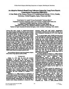
Source: www.pinterest.com/art
Figure 1: Neuro-excitability at synaptic cleft Excitatory amino acids are released from the presynaptic terminal and act on postsynaptic NMDA and non-NMDA receptors (NMDAR) to cause excitation. Gamma-aminobutyric acid (GABA) is an inhibitory neurotransmitter and acts on postsynaptic GABA receptors (GABAR). However, the glial cells play a central homeostatic role in the control of neuroNigerian Journal of Technology,
excitation by controlling extra-neuronal potassium concentration and by removing excitatory neurotransmitters such as glutamate (Glu). Neuronal excitability may also be influenced by ions such as magnesium. However, the onset of seizure appears to occur when a small group of abnormal neurons undergo a prolonged depolarization that is associated with the rapid firing of repeated action potentials. These abnormally discharging epileptic neurons recruit adjacent neurons with which they are connected into the process. On the other hand, a clinical seizure occurs when the electrical discharge of a large number of neurons become abnormally linked together, it thus create a storm of electrical activity in the brain. In spite of that, seizure may then spread to involve adjacent areas of the brain or through established anatomic pathways to other distant areas. 1.2 Adaptive Neuro-Fuzzy Inference System In machine learning and cognitive science, artificial neural networks (ANN) refers to an information processing paradigm that is inspired by the way biological nervous systems, such as the brain, process information (i.e. an eclectic simulation of biological neuron, and it consists of its own dendrites, synapses, cell body and axon terminals), [15]. Nonetheless, it receives stimulation from nearby cells, or from its environment, and generates a modified action potential or nerve signal. It’s a system of interconnected neurons working in unison to solve specific problems through a learning process and usually configured for a specific application. Because it has greater predictive power than signal analysis technique, ANN have been used as computational tools for pattern classification including diagnosis of diseases [16 – 19]. Wilson and Emerson [20] used ANN for automatic seizure detection. Xu, et al [21], used ANN to classify seizures using radial basis function. On the other hand, [22 & 23] detected epileptogenic transient waveforms by using wavelet coefficients as an input to a feed-forward neural network. In their studies, Guler and Ubeyli [24] and Yaunanghi, et al [25] both deployed extracted EEG to identify normal and abnormal EEG using back propagation algorithm. Fuzzy set theory introduced by [26] plays a vital role in dealing with uncertainty when making decisions in biomedical applications. However, Fuzzy logic and fuzzy set theory are employed to describe human thinking and reasoning in a mathematical framework. According to [27], fuzzy-rule based modeling is a qualitative modeling scheme where the system behavior is described using a natural language. Nevertheless, fuzzy sets have attracted the growing Vol. 37, No. 2, April 2018
526
AN ADAPTIVE NEURO-FUZZY INFERENCE SYSTEM FOR THE PHYSIOLOGICAL PRESENTATION OF SEIZURE DISORDER, O. O. E. Ajibola & A. N. Aladefa
attention and interest in modern information technology, production technique, decision making, pattern recognition, diagnostics and data analysis to mention but a few [28 & 29]. However, when the underlying physical relationships are not fully understood, the intelligent computational method of fuzzy logic comes to real advantage than conventional crisp modeling. Neural network and fuzzy logic can be incorporated into a single framework, thereby capturing the prowess of both fields. This however has given birth to a NeuroFuzzy system which actually eliminates the basic problem in fuzzy system design (i.e. obtaining a set of fuzzy if–then rules) by effectively using the learning capability of an ANN for automatic fuzzy if–then rule generation and parameter optimization. Neuro-Fuzzy utilizes linguistic information from the human expert as well as measured data during modeling. . However, this system can be used for signal processing, automatic control, information retrieval, database management, computer vision and data classification. Another approach in neuro-fuzzy development is the Adaptive Neuro-Fuzzy Inference System (ANFIS), which produced remarkable results in modeling nonlinear functions [30]. In ANFIS, the membership function parameters are extracted from a data set that describes the system behavior. The ANFIS learns features in the data set and adjusts the system parameters according to a given error criterion. However, in the field of Biomedical Engineering, ANFIS has been successfully implemented for: classification [19, 24, 30 – 32], modeling and controlling real systems, [33] and also in data analysis [34].
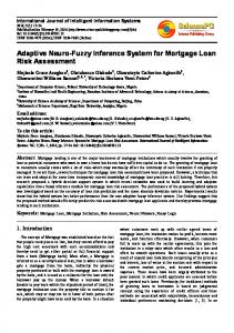
through electrodes placed on the scalp. EEG measures the electrical potentials of cortical neuronal dendrites near the brain's surface [35, 36]. Its recordings reveals the epileptiform discharge of a patient with generalized tonic clonic seizure. Conventionally, the electrodes are labeled and each electrode is attached to an individual connector for the onward transmission of signal to the machine. Basically, electrode locations and names are specified by the international 10 – 20 system (Figure 2) for most clinical and research applications except when high-density arrays are necessary. This system ensures that the naming of electrodes is consistent across laboratories, [37]. 2.1 Basis for formulating the Model The EEG data used in this study is a recording of a patient with generalized tonic clonic seizure and the outcome is compare with that of a non-seizure Patient. During the course of the study, the following bipolar channel were selected for analysis: F4-C4, C4-P4, P4O2, FP1-F3, F3-C3, C3-P3, P3 -O1, FP2-F8, F8 -T4, T4 T6, T6 -O2, P1-F7, F7 -T3, T3 -T5, T5 -O1. In order to access the performance of the classifier, we selected 129*15 EEG segments of the pages that show epileptiform discharges for the patient with seizure disorder. Both the longitudinal (R) and the transverse (R) montages were considered during the course of this study, and the selected pages include the recording in which the hyperventilation simulation and Photic simulation were done. Furthermore, two pages were selected from the recordings and each page contains sixteen channels but some portion of the EEG contains Electrocardiogram (ECG) artifacts.
2 MATERIALS AND METHOD This study used electroencephalography (EEG): a recording of the electrical activity of the cerebral cortex
Figure 2: The international10–20 system of electrode placement. Source: International 10 - 20 system Nigerian Journal of Technology,
Vol. 37, No. 2, April 2018
527
AN ADAPTIVE NEURO-FUZZY INFERENCE SYSTEM FOR THE PHYSIOLOGICAL PRESENTATION OF SEIZURE DISORDER, O. O. E. Ajibola & A. N. Aladefa
Therefore, the channel containing the ECG artifacts (i.e. the ECG-REF) channels are not included in our selected channels for analysis. So, a set of fifteen bipolar channels each were selected as inputs from the EEG recordings of a seizure and a non-seizure patients. The abnormal subject is diagnosed of a generalized tonicclonic seizure with no other accompanying disorders. Recordings were done during interictal stage and the different stages of EEG signals were determined by two physicians. However, EEG data were acquired with Ag/AgCl disk electrodes placed using the 10-20 international electrode placement system. The recordings are band-pass filtered (1-70Hz) EEG, speed of 30mm/s while sensitivity of 7.5 and 10µV/mm for seizure and Non-seizure patient respectively. Every recording was individually inspected by two neurologists together with two technologists with many years of experience in clinical analysis of the EEG signals and they scored the recordings into Seizure or non-seizure. However, the judgments of the four analysts were evaluated and compared twice in order to check if there were disparities in their scoring of the EEG signals. When revising this unified event sets, the human experts by mutual consent marked each state as seizure or normal. Therefore, this validated set provided the reference evaluation to estimate the sensitivity and specificity of computer scorings. Afterwards, the EEG waveform are converted to a digital EEG data (i.e. the digital data corresponding to the analogue data of the selected waveform of the channels of each page is analyzed and are extracted to Microsoft Excel sheet). Each page has 10seconds recording for each channel. Basically, the EEG waveforms are generally classified according to their frequency, amplitude, waveform/morphology and the sites on the scalp at which they were recorded. The digital EEG data used in this study were measured in Amplitude. Amplitude is expressed in terms of voltage in microvolts based on a peak-to-peak measurement and one needs to know the sensitivity at which a recording was made. The amplitude varies with the technique of the recording: the bipolar montages with short inter-electrode distance will give a smaller amplitude than the referential montages with large inter-electrode distance. Ideally, amplitude should be described in terms of the actual voltage; however, the term low, medium, and high amplitude are often used and the same has been adopted in this study: it is low when it is lesser than 20 µV, Medium when it falls between 20 µV and 50 µV and it is considered as High when the amplitude is higher than 50 µV. These digital values are supplied to the Adaptive Neuro-Fuzzy Nigerian Journal of Technology,
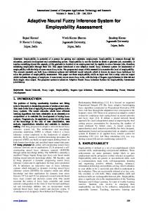
Inference System as input data set for training. The digital input of patient with seizure has ‘1’ as its output while the patient without Seizure has ‘0’ as its data output. The sufficiency in the functioning of ANFIS depends on the size of the training set and test set. In this study, we used 258 * 15 features in training and 129 *15 features were used for testing in order to verify the accuracy and the effectiveness of the trained ANFIS for the detection of epileptic seizure in patient with generalized tonic-clonic seizure. And the training data set is different from the test data set in order to improve the general capabilities of our ANFIS. 2.2 Adapting ANFIS to Seizure phenomena The ANFIS is a fuzzy Sugeno model put in the framework of adaptive systems to facilitate learning and adaptation [38]. In 1993, Jang introduced ANFIS as a model that maps inputs through input membership functions (MF) and associated parameters, and then through output MF to crisp output [39]. However, the initial membership functions and the rule base for fuzzy inference system can be designed by employing human expertise about the target system to be modelled. Such framework makes the ANFIS modelling more systematic and less reliant on expert knowledge. Thus, to present the ANFIS architecture, two fuzzy if– then rules based on a first order Sugeno model are considered: Rule 1: If
and
then
Rule 2: If
and
then
f1 p1 x q1 y r1
f 2 p2 x q2 y r2
where x and y are the inputs; sets;
f1
A1 and B1 are the fuzzy
are the outputs within the fuzzy region
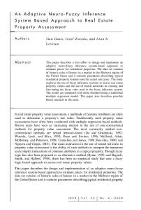
specified by the fuzzy rule; p1, qi and r1 are the design parameters that are determined during the training process. Moreover, the ANFIS architecture to implement these two rules is shown in Figure 4, in which a circle indicates a fixed node, whereas a square indicates an adaptive node. In the first layer, all the nodes are adaptive nodes. Thus, the outputs of layer 1 are the fuzzy membership grade of the inputs, which are given by equations (1) and (2) respectively: ( ) 1, (1) ( ) , (2) where
Ai ( x), Bi2 ( y)
can
adopt
any
fuzzy
membership function. Vol. 37, No. 2, April 2018
528
AN ADAPTIVE NEURO-FUZZY INFERENCE SYSTEM FOR THE PHYSIOLOGICAL PRESENTATION OF SEIZURE DISORDER, O. O. E. Ajibola & A. N. Aladefa
the output of each node in this layer is simply the product of the normalized firing strength and a first order polynomial (for a first order Sugeno model). The outputs of this layer are given by: ) 1, ̅ ̅̅̅( (6) In the fifth layer, there is only one single fixed node labeled with ɛ. Thus, this node performs the summation of all incoming signals. Hence, the overall output of the model is given by:
Source: Xiaomi · Press & Media
2 wifi 2 Oi5 w ifi i 1 w w i 1 1 2
Figure 3: First Order Sugeno Fuzzy Model
(7)
However, it can be observed that there are two adaptive layers in this ANFIS architecture namely; the first layer and the fourth layer. In the first layer, there are three modifiable parameters {ai; bi; ci}; which are related to the input membership functions. These parameters are the premise parameters. In the fourth layer, there are also three modifiable parameters {pi; qi; ri}; pertaining to the first order polynomial which represent the consequent parameters.
Figure 4: ANFIS Architecture For example, if the bell-shaped membership function is employed,
Ai ( x)
Ai (x)
is given by:
1 x c i 1 a i
2
bi
(3)
where ai, bi and ci are the parameters of the membership function, governing the bell-shaped functions accordingly. However, each node in this layer generates a membership grade of a linguistic label, where ‘x’ is the input to node. In the second layer, the nodes are fixed nodes. Thus, they are labeled with Π indicating that they perform as simple multipliers. The outputs of this layer can be represented as: ( ) ( ) 1, ( ) They are the so-called firing strengths of the rules. However, each node in this layer calculates the strength of the rule via multiplication. In the third layer, the nodes are also fixed nodes. Thus, they are labeled with N indicating that they play a normalization role to the firing strengths from the previous layer. However, the outputs of this layer can be represented as: w ̅ 1, ( ) They are the so-called normalized firing strengths. And in the fourth layer, the nodes are adaptive nodes. Thus, Nigerian Journal of Technology,
2.3 Learning algorithm of ANFIS The task of the learning algorithm for this architecture is to tune all the modifiable parameters, namely {a i; bi; ci} and {pi; qi; ri}; to make the ANFIS output match the training data. However, for any premise parameters ; and of the membership function, the output of the ANFIS model can be written as:
f
w1 w1 f1 f2 w1 w2 w1 w2
(8)
Substituting Eq. (6) into Eq. (8) yields:
f wifi w 2 f 2
(9)
Insert the fuzzy if–then rules into Eq. (9), it becomes:
f w1 ( p1 x q1 y r1 ) w2 ( p2 x q2 y r2 )
(10)
After rearrangement, the output can be expressed as:
f ( w1 x) p1 ( w1 y )q1 ( w1 y )r1 ( w2 x) p2 ( w2 y )q2 ( w2 y )r2
(11)
Equation (11) is a linear combination of the modifiable consequent parameters p1, q1, r1, p2, q2 and r 2. However, the least squares method can be used to identify the optimal values of these parameters easily. When the premise parameters are not fixed, the search space becomes larger and the convergence of the training becomes slower. Thus, a hybrid algorithm combining the least squares method and the gradient descent method is adopted to solve this problem. Furthermore, the hybrid algorithm is composed of a Vol. 37, No. 2, April 2018
529
AN ADAPTIVE NEURO-FUZZY INFERENCE SYSTEM FOR THE PHYSIOLOGICAL PRESENTATION OF SEIZURE DISORDER, O. O. E. Ajibola & A. N. Aladefa
forward pass and a backward pass, the least squares method (forward pass) is used to optimize the consequent parameters with the premise parameters fixed. Once the optimal consequent parameters are found, the backward pass starts immediately, the gradient descent method (backward pass) is used to adjust optimally the premise parameters corresponding to the fuzzy sets in the input domain. And the output of the ANFIS is calculated by employing the consequent parameters found in the forward pass. Nevertheless, the output error is used to adapt the premise parameters by means of a standard backpropagation algorithm. It has been proven that this hybrid algorithm is highly efficient in training the ANFIS [38 & 39]. Therefore, in the present study the proposed ANFIS model was trained with the backpropagation gradient descent method in combination with the least squares method when 15 channels of EEG waveforms were used as inputs. Finally, the coding of this algorithm is carried out in the MATLAB (R2013a) environment. The program written in the MATLAB editor was based on the source data from the digital extraction of the EEG recordings of patient with seizure and the non-seizure. 3. RESULTS AND DISCUSSION In this study, the ANFIS architecture consists of five layers, the first layer which is an adaptive node generates the membership grade of a linguistic label, where ‘x’ in Equation (3) is the input parameter.
However, the Linguistic label (Ai) is classified based on the range of the amplitude namely Low, Medium and High. Thus, we have three input membership variables described by (Ai) viz: low, medium and high for each input channel with the range between 0 and 1. The following graphs were obtained from the data used in training the ANFIS model. Figure 5 shows the outputs of all the 15 inputs in layer 1 of the ANFIS architecture (i.e. the fuzzy membership grade of all the fifteen inputs), this shows their strength and spread. It maps non-fuzzy input values to fuzzy linguistic terms and vice versa. In Equation (3), a i, bi, ci are the parameter set that determine the shape of the membership function (MF) with maximum of 1 and minimum of 0. However, the red color is for the low amplitude, the green color is for the medium amplitude, while the blue color is for the high amplitude. Figure 6 represents the ANFIS test result after the first training for a seizure patient. However, the data sets are still far away from the expected output of 1. In Figure 7 shows the ANFIS test result after the first training for a non-seizure patient. However, the data sets were far away from the expected output of 0. Figure 8 is the ANFIS test result after the second training for a seizure patient. The figure reveals that there is converging trend towards the expected output of 1.
Figure 5: Membership Function for all the Fifteen Inputs Nigerian Journal of Technology,
Vol. 37, No. 2, April 2018
530
AN ADAPTIVE NEURO-FUZZY INFERENCE SYSTEM FOR THE PHYSIOLOGICAL PRESENTATION OF SEIZURE DISORDER, O. O. E. Ajibola & A. N. Aladefa
Figure 6: Training test results after the first ANFIS for a Seizure Patient
Figure 7: Training test results after the first ANFIS for a Non- Seizure Patient
Figure 8: Training test results after the second ANFIS for a Seizure Patient
Figure 9: Training test results after the second ANFIS for a Non - Seizure Patient
Figure 9 shows the ANFIS test result after the second training for a Non-seizure patient. The figure reveals that there is converging trend towards the expected output of 0. Figure 10 shows the comparison between the training data and the test data for a Non-seizure patient. Figure 11 shows the comparison between the training data and the test data for a seizure patient. Figure 12 shows the graphical representation of the RMSE for the ANFIS model against the Epoch. The test performance of the ANFIS model was determined by the computation of the statistical parameters such as sensitivity, specificity and total classification accuracy. The sensitivity, specificity and total classification accuracy are defined as follows: Sensitivity: The ratio of the number of True Positives (TP) to the sum of true positives and False Negatives (FN). Specificity: The ratio of the number of True Negatives (TN) to the sum of true negatives and False Positives (FP) Accuracy: It is the number of patterns detected by the total number of patterns. Out of the 129 feature vectors of seizure 129 patterns were detected (TP) leaving 0 FN. Also, out of 108 feature vectors of non-seizure 86 patterns were Nigerian Journal of Technology,
detected (TN) implying that 22 were not detected. Therefore, the performance of the classifier for normal and seizure conditions are shown in Table 1 and success rate in Table 2 respectively. Diagnosing epileptic seizure on EEG rely on the detection of a particular signal which requires the observation of the patient, EEG recordings, and gathering of additional clinical information.
Table 1: The performance of the classifier for Normal and Seizure Conditions. Class Normal Seizure
Total Feature 108 129
Correctly Detected 86 129
Incorrectly Detected 22 0
Table 2: Accuracy of the Network. Test Outcome Specificity Sensitivity Accuracy
Total Feature 129 108 237
Correctly Detected 129 86 215
Accuracy (%) 100 79.6 90.7
Vol. 37, No. 2, April 2018
531
AN ADAPTIVE NEURO-FUZZY INFERENCE SYSTEM FOR THE PHYSIOLOGICAL PRESENTATION OF SEIZURE DISORDER, O. O. E. Ajibola & A. N. Aladefa
Figure 10: Training Vs. Test data for a Non-Seizure Patient
Figure 11: Training Vs. Test data for a Seizure Patient
Figure 12: Root Mean Squared Error (RMSE) Vs. Epoch However, conventional analysis of EEG recordings by a trained professional are very tedious and time consuming especially a long term recordings obtained from an ambulatory recording systems of a patient which includes EEG recordings of up to 1 week period and this can cause errors which may frustrate the outcomes of clinical analyses. Therefore, the advantages of using an automatic detection system cannot be overemphasized. In this study, we described the detection of epileptic seizures from EEG signals using statistical features in amplitude extracted directly from the EEG machine and a diagnostic ANFIS to assist an expert to draw inference from EEG without going through a process of analyzing cumbersome Nigerian Journal of Technology,
recordings in real time. This will provide a valuable diagnostic decision support tool for physician treating epileptic seizure. The ANFIS used 258 *15 training data set in 300 training periods and the step size for parameter adaptation had an initial value of 0.0075 with the Root Mean Square Error (RMSE) of 0.0122, the curve of network error convergence of the ANFIS as shown in Figure 12 is 9.1645 × 10¯6. However, the error decreases exponentially as the system epoch increases. The ANFIS performance as displayed in Table 2 shows the classification of normal subject and seizure patient using ANFIS model which were done with the accuracy of 79.6 and 100% respectively while total accuracy of 90.7% is achieved. The classification results, the values of statistical parameters and performance evaluation parameters indicated that testing of the ANFIS was successful. The phenomenon captured in Figure 11 is the EEG manifestation of the aberrant hyper-excitable state and these delineates the clinical syndrome of Seizure disorder (i.e. paroxysmal depolarization shifts, or interictal spikes) which is an abrupt, all-or-none depolarization which occurred during interictal discharges lasting a few hundred milliseconds [40]. However, it consist of giant Excitatory Postsynaptic Potentials (EPSPs) and these potentials are the excitation and the manifestation of synchronous burst firing of many neurons, and the EPSP builds up from the resting potential to the point of threshold [41]. According to literature, the all-or-none depolarization (i.e. an action potential) propagates down to induce neurotransmitter release at the axon terminal [42 – 44]. However, there exists no such thing as a weak or partial action potential. Either the threshold potential is reached and an action potential occurs, or action potential does not occur [45 – 47]. Therefore, if IPSP fails to match the EPSP, the excessive precipitation of excitatory physiological information (EPI) that could attain threshold level occurs, and this pathologically increases the likelihood of depolarization and synchronized firing of groups of neurons, then a frank seizure activity is initiated. 4. CONCLUSIONS In this work, a major headway has been made in medical practice by formulating an ANFIS that captures the physiological presentation of seizure disorder for the purpose of analysing its clinical symptoms. Efforts targeted at management of seizure through Electroconvulsive Therapy (ECT) has not yielded any encouraging result due to the fact that the point of trigger of a seizure is not necessarily the same for successive presentation of seizure even for the same Vol. 37, No. 2, April 2018
532
AN ADAPTIVE NEURO-FUZZY INFERENCE SYSTEM FOR THE PHYSIOLOGICAL PRESENTATION OF SEIZURE DISORDER, O. O. E. Ajibola & A. N. Aladefa
patient. However, it should be possible to deploy an expert system that would be able to manage the Clinical Symptoms of Seizure. To achieve this aim, we have made an efforts to analyze the physiological attributes of Seizure disorder by using an Adaptive Neuro-Fuzzy Inference system so that we could proffer a workable model for the Clinical Symptoms of Seizure. In the nearest future, we hope to develop a management model based on the same paradigm as a pedestal for the development of an Expert system for the management of seizure disorder. 5. ACKNOWLEDGMENT The authors which to acknowledge the contributions of Clinix Healthcare, Ilupeju, Ikeja, Lagos, Nigeria to the success of this study. 6. REFERENCES [1] Adeli, H., Zhou Z., Dadmehr, N. “Analysis of EEG records in an epileptic patient using wavelet transform”. J Neurosci Meth, Vol. 123, pp. 69–87. 2003. [2]
[3]
lhm, S.Y. "Epilepsy Foundation (formerly Epilepsy Foundation of America)", Electronic Resources Review, Vol. 3, No. 7, pp. 73-75, 1999. World Health Organization, Epilepsy. http://www.who.int/mental_health/ neurology/epilepsy/en/index.html. 2012.
[4] Ramsay, R.E., Rowan, A.J., Pryor, F.M. Special considerations in treating the elderly patient with epilepsy. Neurology. Vol. 62, No. 5, pp. S24-529. 00 .” [5] Shah, S,V. “Diseases of the brain and nervous system”. Mudresh Jitendra Purohit, Surya Offset. India. 2008. [6] Lopez-Munoz, F., Boya, J., and Alamo, C. “Neuron theory, the cornerstone of neuroscience, on the centenary of the Nobel Prize award to Santiago Ramon Y Cajal”. 18 : Brain Res Bull. Vol. 70: pp. 391-405. 2006. [7] Hodgkin, A. L. “Evidence for electrical transmission in nerve: Part I”. J. Physiol. Vol. 90: pp. 183-210. 1937a. [8] Hodgkin, A. L. “Evidence for electrical transmission in nerve: Part II”. J Physiol .Vol. 90, pp. 211-232. 1937b. [9] Ajibola, O. O. E., Ibidapo-Obe, O. and OIunloyo, V. O. S. “A Model for the Management of Gait Syndrome in Huntington’s Disease Patient”. Journal of Bioinformatics and Intelligent Control. Vol. 3, No. 1, pp. 15 – 22.. 2014. [10] Greenamyre, J. T., Porter, R. H. “Anatomy and physiology of glutamate in the CNS”. Neurology. Vol. 44, pp. S7–S13. 1994. Nigerian Journal of Technology,
[11]
Rogawski, M.A., “Excitatory amino acids and seizures". In: Stone TW (ed.), CNS. 1995. [12] Nardone, R., Brigo, F. and Trinka, E. “Acute Symptomatic Seizures Caused by Electrolyte Disturbances”. J Clin Neurol. Vol. 12, No. 1, pp. 2133. 2016. [13] Jefferys, J. G., Haas, H. L. “Synchronized bursting of CA1 hippocampal pyramidal cells in the absence of synaptic transmission”. Nature, Vol. 300, pp. 448–450. 1982. [14] Liu, Y-C, Lin, C-C.K., Tsai, J-J. and Sun, Y-N “Model-Based Spike Detection of Epileptic EEG Data”. Sensors. Vol. 13, pp.12536-12547. 2013. [15] Ajibola, O.O.E., Olunloyo, V. O. S. and IbidapoObe, O. “Artificial neural network simulation of arm gait of Huntington disease patient”. Int. J. Biomechatronics and Biomedical Robotics, Vol. 1, No. 3, pp. 133 – 140. 2011. [16] Shi, Z., He, L., Nakamura, T., Suzuki, K. and Itoh, H. “Survey on neural networks used for medical image processing”. Inter. J. Computational Science. Vol. 3, No. 1, 86-100. 2009, [17] Shi, Z. and He, L. “Application of Neural Networks in Medical Image Processing”.
Proceedings of the Second International Symposium on Networking and Network Security. Jinggangshan, P. R. China, pp. 023-026. 2010. [18]
Guler, I., Ubeyli, E.D. “Detection of ophthalmic artery stenosis by leastmean squares backpropagation neural network”. Comput Biol Med, Vol. 33, No. 4, pp. 333–43. 2003. [19] Ubeyli, E.D., Guler, I. “Neural network analysis of internal carotid arterial Doppler signals: Predictions of stenosis and occlusion". Expert System Appl., Vol. 25, No. 1, pp. 1–13. 2003. [20] Wilson, S.B. and Emerson, R. “Spike detection: a review and comparison of algorithms”. Clinical Neurophysiology. Vol. 113, pp.1873–1881. 2002. [21] Xu, G., Wang, J., Zhang, Q. and Zhu, J. “An Automatic EEG Spike Detection Algorithm Using Morphological Filter”. IEEE International
Conference: Automation Science and Engineering, [22]
CASE '06. 2006. Baikun ,WAN., Bikash, Dhakal., Hongzhi QI., Xin zhu. “Multi-method synthesizing to detect and classify epileptic waves in EEG”. Proceedings of the
Fourth International Conference on Computer and InformationTechnology, Vol. 10, pp.922 -926. 2004. [23]
Tulga, K., Ozcan, O., Nurgun E. “The use of wavelet transform as a preprocessor for the neural network detection of EEG spikes”. IEEE Engineering in Medicine and Biology: pp.1 – 3. 1994. [24] Guler, I., Ubeyli, E. D. “Adaptive neuro-fuzzy inference system for classification of EEG signals using wavelet coefficients”. Journal of Neuroscience Methods: pp.1-9. 2005. [25] Yaunanghi, N., Najumnissa, D. and Shenbagadevi, S. “Feature extraction and detection of epileptic seizure”. Proceedings of the Vol. 37, No. 2, April 2018
533
AN ADAPTIVE NEURO-FUZZY INFERENCE SYSTEM FOR THE PHYSIOLOGICAL PRESENTATION OF SEIZURE DISORDER, O. O. E. Ajibola & A. N. Aladefa
National Conference on Signals, Systems and Communication, NCSSC-2006, Chennai, India: pp. 8–12. 2006. Zadeh, L.A. “Fuzzy sets”. Inf. Control. Vol. 8, No. 3, pp. 338–353. 1965. [27] Sugeno, M., Yasukawa, T. ”A fuzzy-logic based approach to qualitative modeling”. IEEE Trans. Fuzzy Syst. Vol. 1, No. 1, pp. 7–31. 1993. [28] Aminifar, S, and Yosefi, G. “Application of adaptive neuro fuzzy inference system (ANFIS) in implementing of new CMOS fuzzy logic controller (FLC) chip”. In: AIP Conference Proceedings, pp 49–53. 2007. [29] Nauck, D., Kruse, R. “Obtaining interpretable fuzzy classification rules from medical data”. Artif. Intell. Med. Vol. 16, pp. 149–169. 1999. [30] Subasi, A. “Application of adaptive neuro-fuzzy inference system for epileptic seizure detection using wavelet feature extraction”. Computers in Biology and Medicine. Vol. 37, pp. 227 – 244. 2007. [31] Najumnissa, D., Rangaswamy, T. R. “Detection and Classification of Epileptic Seizures using Wavelet feature extraction and Adaptive NeuroFuzzy Inference System”. International Journal Of Computational Engineering Research, Vol. 2, No. 3, pp.755-761. 2012. [32] Guler, N. F., Ubeyli, E. D., “Gulaer, I. “Recurrent neural networks employing Lyapunov exponents for EEG signals classification”. Expert Systems with Applications, Vol. 29, pp.506–514. 2005. [33] Vieira, J., Dias, F.M., Mota, A. “Artificial neural networks and neuro-fuzzy systems for modelling and controlling real systems: a comparative study”. Eng. Appl. Artif. Intell. Vol. 17, pp. 265–273. 2007. [34] Virant-Klun, I., Virant, J. “Fuzzy logic alternative for analysis in the biomedical sciences”, Comput. Biomed. Res. Vol. 32, pp. 305–321. 1999. [35] Patra, K.P. and Lewis, D.W. “Electroencephalography: a primer”. Pediatr. Rev. Vol. 33, No. 5, pp. 226-31. 2012. [36] Almási A-D, Woźniak S, Cristea V, Leblebici Y, Engbersen T. Review of advances in neural networks: neural design technology stack. Neurocomputing Vol. 174, pp.31–41. 2016. [26]
Nigerian Journal of Technology,
[37]
Koessler, L., Maillard, L., Benhadid, A., Vignal, J.-P., Braun, M. and Vespignani, H. 2007. Spatial localization of EEG electrodes Localisation spatiale des ´electrodes EEG. Clinical Neurophysiology. Vol. 37, pp.97—102. 2007. [38] Desouky, S.F and Schwartz, H.M. “Self-learning fuzzy logic controllers for pursuit–evasion differential games”. Robotics and Autonomous Systems. Vol 59, No. 1, pp. 22-33. 2011. [39] Karaboga, D. and Kaya, E. “Adaptive network based fuzzy inference system (ANFIS) training approaches: a comprehensive survey”. Artificial Intelligence Review. Vol. 23, No. 3, pp. 1–31. 2018. [40] Matsumoto, H., Ajmone-Marsan, C. “Cortical cellular phenomena in experimental epilepsy: interictal manifestations”. Experimental Neurology. Vol. 9, pp. 286–304. 1964. [41] Ajibola, O.O.E., Olunloyo, V. O. S. and IbidapoObe, O. “Fuzzy modelling of arm gait in Huntington’s disease patient”. J. Bioinformatics and Intelligent Control. Vol. 1, No.5, pp. 71 – 75. 2012. [42] Ibidapo-Obe, O., Ajibola, O.O.E. and Olunloyo, V.O.S. “On equation of arm gait of Huntington Disease patient”. Int. J. Biometrics, Vol. 2, No. 1, pp. 53 – 70. 2010. [43] Jasper, H. H. “Report of the Committee on Methods of Clinical Examination in Electroencephalography”. Electroenceph. Clin. Neurophysiol. Vol. 10, pp. 370-371. 1958. [44] Jasper, R. Daube, and Squire, M. Stead, “Basic for Neurophysiology”. Clinical Physiology, Third Edition, Contemporary Neurological Series, Oxford University Press, Inc., New York. 2009. [45] Cooper, R., Osselton, J. W., Shaw, J. C., “EEG Technology”, 2nd ed., Butterworths, London, pp. 275. 1969. [46] Park, H.S., Lee, Y.H., Lee, D.O., Kim, S.I. “Detection of Epileptiform activity using wavelet and neural network”, Proceedings of the 19th
Annual International Conference of the IEEE / [47]
EMBS. Vol. 3, pp. 1194 – 1197. 1997. Reeves, A. G. “Epilepsy and the Corpus Callosum”. New York, Plenum Press. 1995.
Vol. 37, No. 2, April 2018
534
AN ADAPTIVE NEURO-FUZZY INFERENCE SYSTEM FOR THE PHYSIOLOGICAL PRESENTATION OF SEIZURE DISORDER, O. O. E. Ajibola & A. N. Aladefa
7. APPENDIX
Figure 13: EEG recording of a Seizure patient
Figure 14: EEG recording of a Non-seizure patient
Nigerian Journal of Technology,
Vol. 37, No. 2, April 2018
535
AN ADAPTIVE NEURO-FUZZY INFERENCE SYSTEM FOR THE PHYSIOLOGICAL PRESENTATION OF SEIZURE DISORDER, O. O. E. Ajibola & A. N. Aladefa
Figure 15: Data Extraction from the corresponding EEG recordings of a Seizure patient
Figure 16: Data Extraction from the corresponding EEG recordings of a Non-seizure Patient
Nigerian Journal of Technology,
Vol. 37, No. 2, April 2018
536