An experimental study on neural crest migration in Barbus conchonius. (Cyprinidae, Teleostei), with special reference to the origin of the enteroendocrine cells.
J. Embryol. exp. Morph. Vol. 62, 309-323 pp. 1981 Printed in Great Britain © Company of Biologists Limited 1981
3Q9
An experimental study on neural crest migration in Barbus conchonius (Cyprinidae, Teleostei), with special reference to the origin of the enteroendocrine cells By C. H. J. LAMERS, J. W. H. M. ROMBOUT AND L. P. M. TIMMERMANS From the Agricultural University, Department of Experimental Animal Morphology and Cell Biology, Wageningen, The Netherlands
SUMMARY A neural crest transplantation technique is described for fish. As in other classes of vertebrates, two pathways of neural crest migration can be distinguished: a lateroventral pathway between somites and ectoderm, and a medioventral pathway between somites and neural tube/notochord. In this paper evidence is presented for a neural crest origin of spinal ganglion cells and pigment cells, and indication for such an origin is obtained for sympathetic and enteric ganglion cells and for cells that are probably homologues to adrenomedullary and paraganglion cells in the future kidney area. The destiny of neural crest cells near the developing lateral-line sense organs is discussed. When grafted into the yolk, neural crest cells or neural tube cells appear to differentiate into 'periblast cells'; this suggests a highly activating influence of the yolk. Many neural crest cells are found around the urinary ducts and, when grafted below the notochord, even within the urinary duct epithelium. These neural crest cells do not invade the gut epithelium, even when grafted adjacent to the developing gut. Consequently enteroendocrine cells in fish are not likely to have a trunkor rhombencephalic neural crest origin. Another possible origin of these cells will be proposed.
INTRODUCTION In all vertebrate embryos the neural crest appears directly after neurulation in an anterior-posterior wave-like movement along the embryonic axis. Almost immediately after its development the neural crest cells begin to disperse throughout the embryo. However, most of the early intra-embryonic cell movements escape observation as the cells are undifferentiated and cannot be distinguished from the tissues in which they move. Therefore, several experimental techniques have been developed, mainly based on extirpation, explantation and transplantation (c.f.: Weston, 1970; Le Douarin, 1976) to study the 1
Author's address: Agricultural University, Department of Experimental Animal Morphology and Cell Biology, Zodiac, Marijkeweg 40, 6709 PG, Wageningen, The Netherlands.
310
C. H. J. LAMERS AND OTHERS
destiny and the pathways of migration of neural crest cells. In fish, migration pathways of pigment cells have been extensively studied, for pigment formation takes place in an early stage of migration (Orton, 1953; Shepard, 1961). The relatively few experimental studies on other neural crest cells in fishes were restricted to extirpation and explantation experiments (Lopashov, 1944; Damas, 1951; Newth, 1951, 1956). Except the pigment cells and cells of the spinal ganglia, no other cells or structures are known to be of neural crest origin, but this origin has been assumed for ganglia and arteries in the head region and for some parts of the visceral and head skeleton. The scarce information on the destiny of the neural crest has been the main motive for developing a technique to study migration of neural crest cells in fishes. Recently, enteroendocrine cells and their relation to the APUD-system (acronym stands for Amine Precursor Uptake and Decarboxylation) have been described for B. conchonius (Rombout, 1977; Rombout, Lamers & Hanstede, 1978). The cells of the gastro-entero-pancreatic (GEP) endocrine system, together with many other endocrine cells, belong to the APUD-series, which is presumed to be derived from a common neural ancestor (Pearse, 1969; Pearse & Takor Takor, 1979); such an origin was proved for some cell types of this series (Le Douarin, 1978; Pearse & Polak, 1978) but not for cells of the GEP endocrine system (Pearse, 1979). Therefore, special attention will be paid to a possible contribution of the neural crest cells to the gut. Our experiments were restricted to the trunk and the late rhombencephalic region. A stable cell-labelling technique as applied in birds (Le Douarin, 1973) was not available for fish. So [3H]thymidine, which was also a suitable marker for studying migration of neural crest cells in avian and amphibian embryos (Weston, 1963; Chibon, 1966; Johnston, 1966) was used.
MATERIALS AND METHODS
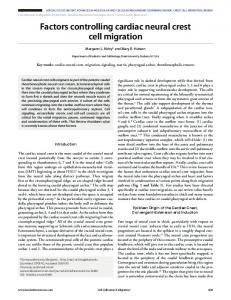
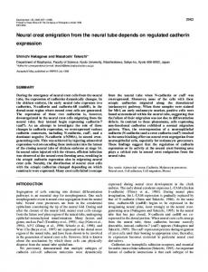
Embryos of Barbus conchonius have been used for this investigation. Early developmental stages were determined by counting the somites, and later stages were determined by normal developmental stages at 25 °C. The small size of the embryo ( 0 ±0-8 mm), the convex shape and the pressure in the yolk prevented the transplantation of parts of the neural tube. Therefore, the transplantation technique was modified as will be described. [H3]thymidine was used as nuclear marker (spec. act. 24 Ci/mmol: Radio-chemical Centre, Amersham, England). Experimental procedures (Fig. la-g): After fertilization the chorion of the eggs of B. conchonius was removed (Fig. la) with 0-5% protease (Sigma, chem. comp. P5130). The eggs were then rinsed and cultured in 10 % Holfreter solution at 19-20 °C. [H3]thymidine (10—30 nl: 1 mCi/ml) was injected into the yolk of the eggs of the donor
Neural crest migration in fish
311
•••
• ••
Fig. 1. (a-g) steps in experimental procedure.
embryos, just below the cells at the 2- to 16-cell stage (Fig. \b). At that stage the cells are still not separated from the yolk by a basal membrane. Transplantations were carried out 24-30 h after fertilization between the 1st and the 16th somite stage (fig. 1 c). At each stage the neural primordium at the place of the last formed five to six somites was mechanically excised together with the surrounding tissue, and isolated by incubation in 0-03 % trypsin solution in a NRHC medium (Sigma, chem. comp. T8253) (fig. 1 c, d, e). In most cases the notochord remained attached to the neural primordium after trypsinization, permitting an exact orientation. The dorsal part of the neural primordium was excised mechanically to obtain small pieces of tissue that contained mainly neural crest cells among the cells of the dorsal part of the neural tube (Fig. le). This cell material was injected into host embryos by microinjection with glass capillaries, preferably dorsally between neural tube and somites, the place whence normally the migration of the neural crest cells has its origin (Fig. 1/, / ' ) . The host embryos (between the first and the sixteenth somite stage) received grafts in the rhombencephalic and trunk region; the anus is at the level of somite 17/18. The migratory capacity of neural tube cells was checked by injecting cells of labelled ventral neural tube. After transplantation the host embryos were kept for 1-5 days at 25 °C. Embryos were fixed in Bouin solution, and cross sectioned at 5/*m after
312
C. H. J. LAMERS AND OTHERS
embedding in Paraplast Plus (Sherwood). Sections were prepared for autoradiography with Kodak NTB-2 emulsion. After 1-6 weeks exposure the slides were developed with Kodak D-19 developer and stained with haemalum and eosin. RESULTS
(1) Marker After injection of [3H]thymidine, the embryos developed normally without any noticeable retardation in comparison to untreated control animals and no radiation damage has been observed. The cell nuclei proved to be heavily labelled (Fig. 4), at least until the larval stages (6 days). This method was suitable for our experiments, as after transplantation the cells can be easily recognized with radioautography. Of the 410 host embryos that received grafts, 243 survived and in 120 of these labeled cells could be recognized. In most cases, clusters of probable neural tube cells remained at the place of injection; this served as a point of reference for the exact spot of injection. In 41 embryos this point of reference was located at the intended place, dorsally between neural tube and somites; these grafts can be considered as isotopic ones. Only this group has been used for studying migration. In another 40 embryos the grafts were traced in more ventral places, and can be considered as ectopic grafts; the periblast of 21 of these embryos contained labelled cells. The remaining group of embryos with labelled cells was not used fordifferent reasons, e.g. no distinctpoint of reference, high background or inadequate fixation. (2) Early development and neurulation Gastrulation takes place between 7 and 10 h (25 °C) after fertilization. Within 1 h after closure of the blastopore the notochord becomes visible in the midline of the embryo (Figs 2, 3d). Neurulation begins at the same time in the head region and continues in an anterior-posterior wave along the embryonic axis; cells of the inner layer of the dorsal ectoderm move towards the midline and form a wedge-shaped mass of cells, the neural keel (Figs 2b, 3 b, c, f). Between 12 and 13 h the first somites are formed and the future optic vesicles appear as lateral enlargements of the forebrain. At 19-20 h, when all somites have developed (about 33), neurulation is completed in the caudal part of the embryo. The end of neurulation at a certain place is marked by the appearance of a basement membrane at the dorsal side of the neural tube; at that time neural crest cells can be recognized as a distinct group of cells between ectoderm and neural tube (Figs 2 c, 3d). The cells within the neural cord become rearranged into right and left groups facing the midline. At the same time the neural crest cells become separated and begin to migrate (Figs 2d, 3e). The start of this cell migration is gradually transmitted in the caudal direction and follows the formation of the somites (at a distance of about nine
Neural crest migration in
fish
313
somites). The duration of migration could not be determined; the beginning is noticed in the head region 14 h after fertilization, whereas some pigment cells still seem to be moving away from the neural tube 32 h after fertilization. By cavitation a lumen is formed within the neural anlage. This phenomenon comes about first in the head region in a 12-somite embryo and can be seen in the trunk region after a short time (15 somites). In 24-somite stage the whole neural anlage is provided of a clear lumen. (3) Migration of neural-crest cells in isotopic grafts After 2-6 h the injected cells were well incorporated in the host tissue (Fig. 5) and 6 h after transplantation a dispersion of these cells can be noticed. After 17 h the cells were found in different places, from neural tube to ventral region (Fig. 6). At that time migration seems to be completed, and only growth and differentiation of the tissues takes place in the following days of development. No principal differences have been observed in the location of the migrated cells at various developmental stages (1-6 days), and the results have been combined. Only the cells that had distinctly moved away from the grafts are considered as migrated cells. The distribution of the migrated cells from isotopic grafts is represented in Fig. 7. Neural crest cells migrate from their initial place along two ways (Figs 6, 7): one pathway in the ventral direction passing the area between somite and neural tube/notochord, the other pathway in the ventrolateral direction between somites and ectoderm. Cells migrating via the first pathway have been found: (i) In the area of the developing spinal ganglia and within the ganglia, (ii) At the periphery of the notochord; nuclei of these cells are oblong. Some labelled oblong nuclei are also found in the septa between the myotomes. (iii) In the area between notochord and intestine where aorta, cardinal vein, and primary urinary ducts are located, and where sympathetic ganglia are formed (Fig. 8); the cells are found ventrolaterally of the aorta, near the cardinal vein, in the walls of the cardinal vein and aorta, and near the primary urinary ducts, (iv) In the area around the gut; labelled cells were not observed, however, in the intestinal epithelium (Fig. 9). Cells migrating via the second pathway have been found: (i) In the skin, where they are mainly differentiated as pigment cells; melanin formation reduces the possibility of recognizing the [3H]thymidine label, and only some of the pigment cells could be traced, (ii) Near the lateral line organ and in the septa at the place of this organ. In several embryos labelled cells appear to be located in the neural tube, and some were noticed in the myotomes. The latter are probably myoblasts or neural tube cells which had become intermingled with the graft.
314
C. H. J. LAMERS AND OTHERS oe
nc
oe
•
-
*.
J'
Neural crest migration in
fish
315
(4) Neural crest cells in ectopic grafts In embryos in which the grafts had been injected at the ventral side of the embryos or even in the yolk, the following observations were made: when the graft had been injected near the future primary urinary ducts and intestine, distinctly labelled cells were noticed in the epithelium of the primary urinary duct but not in the intestinal epithelium, whereas several labelled cells are present in the gut wall (Fig. 10). It was noticed that the wall of the gut in ectopic grafts contained a much greater number of cells (n == 34) than in the isotopic grafts (n = 9). Participation of neural crest cells in the development of the primary urinary duct epithelium is not accompanied by abnormal shape or malformation. When neural crest or neural tube cells were accidentally injected into the yolk, some remained there but others appeared to have fused with the periblast. The labelled nuclei of these cells were just as large as other nuclei in the periblast (Fig. 11). (5) Grafts of fragments from the ventral part of the neural tube It is not yet clear whether the labelled cells in the neural tube are neural crest cells or cells of the dorsal part of the neural tube, which had been injected together with the neural crest cells. As a control on the migratory capacity of the neural tube cells, fragments of ventral neural tube were injected (± 40 grafts) to prevent any intermingling with neural crest cells. Migration of cells from the graft has not been noticed. Moreover, ventral neural tube cells do not fuse with the host neural tube.
F I G U R E S 2 AND 3
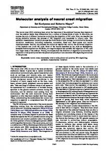
Fig. la-d. Scheme of the process of neurulation in the trunk region. Fig. 3 a-/ Cross of sections embryos during several stages of neurulation. (bar = 20/*m). (a) Notochord just formed and not yet separated from the endodermal layer. The inner layer of the endoderm becoming thicker in the midline. Note the large periblast nuclei (embryo 6 somites; section at the level of the future s. 14). (b)The inner ectodermal layer becoming more wedge shaped in the midline, forming the neural keel (embryo 6 s.; section at the future s. 8). (c) The process of neurulation more advanced than in b (embryo 12 s.; section at s. 11). (d) Neurulation completed and the basement membrane is formed at the dorsal side. Neural crest becomes visible above the neural tube (arrow) (embryo 16s.; section at s. 12). (e) Neural crest cells start to migrate (arrows) and the notochord becomes vacuolized (embryo 16 s.; section at s. 7). (/) Section in the region of the first somite (rhombencephalic) (embryo 6 s.). The neural anlage more pronounced than in the trunk region. The entodermal layer clearly visible in this region, ch, notochord; e, endoderm; ie, inner ectodermal layer; w, mesoderm; nc, neural crest; nk, neural keel; nt, neural tube; oe, outer ectodermal layer; p, periblast.
316
C. H. J. LAMERS AND OTHERS
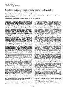
6(c) 8
Fig. 4. Autoradiograph 23 h after [ H]thymidine injection. All nuclei of the embryo heavily labeled (cross section, bar = 20 /tm). Fig. 5. Autoradiograph of a section at the level of the first somite 2 h after grafting. The graft (arrow) well incorporated in the host tissue (bar = 20 fim). Fig. 6. Autoradiographs of sections at different levels of the same embryo 17 h after grafting (bar = 20 /im). (a) a group of labelled cells located at the dorsolateral side of the neural tube (arrow); these cells have not migrated, indicating the place of injection, (b, c) sections at 35 fim and 90/tm caudally from (a) respectively. A few cells are still located near the neural tube (arrow). Most cells have migrated from the graft in two directions, one ventral and one lateroventral direction, ch, notochord n, neural tube; y, yolk.
Neural crest migration in fish
317
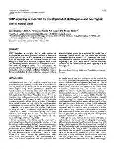
Fig. 7. Diagram of a cross section through the trunk region of a 3- to 4-day-old embryo showing the total number of migrated cells in different areas, summarized for 41 embryos. Each point represents four labelled cells. 0 ; four labelled pigment cells. DISCUSSION 3
In the present study [ H]thymidine was used as nuclear marker. This is the first reliable method for studying the migration behaviour of neural crest cells in fish, for no other cell marker is known in this class of vertebrates. Two migratory pathways of neural crest cells have been recognized, which lead to the destination of these cells near the ectoderm, lateral line organ, aorta, cardinal vein, and urinary ducts and in the spinal ganglia and gut wall. Although in our experiments the neural crest cells were added to those normally present in the embryo, abnormalities have not been observed in development, and migration pathways of the neural crest cells seemed to be normal. The preparation of the neural crest by trypsinization in fish is more complicated than in birds because of the massiveness of the structures; in a number of cases the neural crest was not totally detached from the adjacent tissues (e.g. myoblasts and neural tube cells). Contamination of the graft with II
EMB
62
C. H. J. LAMERS AND OTHERS
>; ^^^^^^^^__
Neural crest migration in
fish
319
myoblasts may explain the presence of labelled cells in the myotomes. Neural tube cells possibly do not migrate, as can be concluded from the experiments with ventral neural tube cells and from the clustering of dorsal neural tube cells at the place of injection. So it may be concluded that the labelled cells observed in the host neural tube are neural crest cells. However, the experiments are not fully conclusive as to the (migratory) capacities of dorsal neural tube cells. With the applied transplantation method it is possible to study the migration and fate of the neural crest cells of fish; until now only suggestions were available from experiments in birds and amphibia and from scarce extirpation and explantation experiments in fish. Our results confirm the findings of Newth (1951, 1956) and Lopashov (1944) who found in the trunk region a contribution of neural crest cells to the spinal ganglia, as already generally known for amphibia and birds (c.f. Weston, 1970). The results obtained for pigment cells are in agreement with theories on their origin in fish by Orton (1953), Newth (1956) and Shepard (1961) and with the generally accepted theories on their origin in the neural crest (c.f. Weston, 1970). Newth (1951, 1956), Lopashov (1944) and Damas (1951) suggested that the neural crest of the head region participates in the formation of the head ganglia, the arteries and the cranial and visceral skeleton. As our experiments were restricted to the trunk region their view could not be confirmed. It was first proved in chick embryos that the migration of the neural crest cells takes place in two well-defined pathways (Weston, 1963): a dorsolateral stream of cells towards and in some places below the ectoderm and a ventral stream towards the mesenchyme between the neural tube and the myotome and penetrating the somitic mesenchyme. The present study indicates a similar process in fish as observed in birds, but there is one difference, i.e. the ventral stream does not penetrate the mesoderm but migrates, within the space between FIGURES
8-11
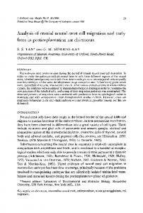
Fig. 8. Autoradiographs of a host embryo, 5 days after grafting (bar = 10 /*m). (a) two labelled cells located near the aorta (arrows). In this region the sympathetic ganglion will be formed, (b) one labelled cell between aorta and cardinal vein (arrow). Fig. 9. Autoradiograph of a host embryo 4 days after grafting; one labelled cell near the basement membrane of the gut epithelium, and two labelled cells between aorta and cardinal vein (arrows) (bar = 5 /tm). Fig. 10. Autoradiograph of a host embryo, 3 days after grafting. The graft was injected near the developing primary urinary duct; four labelled cells in the epithelium of the primary urinary duct, and two cells nearby. Just below the basement membrane of the gut epithelium are three labelled cells (bar == 5 /tin). Fig. 11. Autoradiograph of a host embryo 6 days after grafting labelled cells into the yolk. Labelled cells with the characteristic large nuclei of the periblast. The section was made at a level where the yolk is completely absorbed and only the periblast is present underneath the gut (bar = 10/mi), a, aorta; c, cardinal vein; ch, notochord; g, gut; p, pigment cell; u, urinary duct.
320
C. H. J. LAMERS AND OTHERS
the neural tube/notochord and the somite; as a result the spinal ganglia develop in the space between the somites and the neural tube, and not in the somites as they do in birds and amphibians (Weston, 1970). The explanation may be that contrary to the developmental rate of the other organs, in fish the somites have an explosive growth and differentiation in the early developmental stages and become functional within a few hours after their formation. The present study clearly indicates a contribution of the neural crest to the spinal ganglia and to the region of aorta and postcardinal vein. It is known that the sympathetic ganglia appear near the aorta and that the nephritic tissue is formed around the post-cardinal vein. Chromaffin elements are scattered in the nephritic mesenchyme, always near or just below the endothelium of the postcardinal vein. This tissue is analogous to the adrenal medulla and paraganglia of higher vertebrates (Grasse, 1958; Bolk, Goppert, Kallius & Lubosch, 1934). This indicates participation of neural crest cells in the formation of the sympathetic ganglia and chromaffin tissue. Confirmation requires information on the differentiation of these cells during later developmental stages. A distinct class of cells with an oblong nucleus has been observed near the notochord and in the septa between the myotomes. These cells probably correspond to Schwann sheath cells; the origin of these cells from the neural crest has been established in birds and amphibia (c.f. Weston, 1970). Regularly, cells were found near the developing lateral line organ. Grafting experiments in amphibia indicated that neuromasts of the lateral line organ are derived from the auditory placode by backward migration of cells out of this region (Harrison, 1903). Several authors assumed that neuromasts in fish develop from epithelial cells, possibly induced by the lateralis nerve (c.f. Grasse, 1958). In the present study, neural crest cells were found near the future lateral line organ before nerves can be detected; therefore, these crest cells might be involved in the differentiation of ectoderm cells into neuromasts. The neural crest cells were found to penetrate the urinary duct epithelium only when the graft is placed in the vicinity of this future organ. In birds, neural crest cells contribute to the metanephritic mesenchyme (Fontaine, 1974), but a contribution to the primary urinary duct or future urethra epithelium has never been reported. The affinity of the neural crest cells to the urinary duct epithelium may be due to the ectopic location of the neural crest. Several neural crest cells are found in the wall of the gut, and their number increases strongly when neural crest was grafted near the future gut; however, in the stages investigated (up to 6 days), none of these cells penetrated the future intestinal epithelium. This is in agreement with the results obtained in birds by Le Douarin & Teillet (1973) who transplanted neural tubes including neural crest from quail to chick, and of Le Douarin & Teillet (1974), Smith, Cochard & Le Douarin (1977) and Teillet, Cochard & Le Douarin (1978), who cultured quail neural primordia together with chick aneural hindgut fragments. The latter experiments are comparable to our ectopic neural crest
Neural crest migration in
fish
321
grafts and indicate that neural crest cells grafted in this way maintain their migratory capacity and their ability to differentiate in a normal way. It may be concluded that neural crest cells of B. conchonius, just as in birds, do not have any affinity to the future intestinal epithelium (endoderm); such a contribution was clearly shown to be possible for the epithelium of the primary urinary duct (mesoderm). The neural crest cells in the gut wall probably contribute to enteric ganglia, as described by Le Douarin and her group. In birds these structures originate in distinct levels of the neural axis (the region between somite 1-7, and caudally of 28 S; Le Douarin & Teillet, 1973). Our method does not establish the exact origin. The present study indicated a trunk or rhombencephalic neural crest origin as unlikely for enteroendocrine cells of B. conchonius, contrary to Pearse (1969, 1973) and Pearse & Polak (1971). This confirms the results obtained in birds by Andrew (1963, 1974) and Le Douarin & Teillet (1973), but the conclusion of an endodermal origin of the enteroendocrine cells, presented in the Unitarian hypothesis of Cheng & Leblond (1974) and suggested by Rawdon, Andrew & Kramer (1980), seems to be premature. A neural origin of the enteroendocrine cells cannot be rejected because of the following arguments: — the permanent or ephemeral presence of the APUD characteristics and, consequently, their participation in the APUD series (c.f. Pearse & Takor Takor, 1979; Rombout et al., 1978). — the presence of several peptides common to gut endocrine cells and nervous system (Pearse, 1977, Pearse & Takor Takor, 1979). — the presence of a neurone-specific enolase in cells of the APUD series, and in some GEP endocrine cells (Schmechel, Marangas & Brightman, 1978). — the neuron-like processes in some gut endocrine cells (Larsson et al., 1979). — the penetration of endocrine cells from the lamina propria into the gut epithelium of 4-5 months old human fetuses (Osaka & Kobayashi, 1976). When the neural crest, and even the whole neurectoderm (Fontaine & Le Douarin, 1977) was excluded as a possible origin of the gut endocrine cells in birds, the hypothesis on the origin of the GEP endocrine cells was modified (Le Douarin, 1978; Pearse, 1977, 1979), and they are now considered to be derived from neuroendocrine-programmed ectoblasts. This hypothesis is supported by recent views on the gastrulation in birds and mammals (Levak-Svajger, 1974; Pearse & Takor Takor, 1979), that formation of the definite endoderm (the secondary hypoblast) begins at the primitive-streak stage. Possibly cells " retaining a neuroendocrine program " may accompany the endodermal cells during migration from the ectoblast through the primitive streak. The classical view on the gastrulation process in fish has recently also been disputed by Ballard (1973). He concluded from his experiments that future endoderm cells are located at the most ventral side of the blastodisc and in contact with the
322
C. H. J. LAMERS AND OTHERS
periblast, which eliminates the need for dorsoventral migration of future endodermal cells. Therefore, the presumptive gut must be formed by a rearrangement of the deep-lying cells of the blastodisc. As the future endoderm and ectoderm are located close to each other in the blastodisc,' neuroendocrineprogrammed - ectoblasts' might be intermingled with future endoderm. The phenomenon that neuroectodermal cells may lose their neural properties and (de)differentiate into cells of another germ layer, the periblast, might be attributed to a strong inducing influence of the yolk or periblast. To our knowledge, this phenomenon has never been mentioned. The authors are grateful to Prof. Dr J. W. M. Osse for his critical comments; to Mr W. Valen for preparing the illustrations; to Mrs D. Nijenhuis and Mrs P. van Capelle for typing the manuscript; to Dr L. Boomgaart for correction of linguistic errors. REFERENCES ANDREW, A. (1963). A study of developmental relationship between enterochromaffin cells and the neural crest. / . Embryol. exp. Morph. 11, 307-324. ANDREW, A. (1974). Further evidence that enterochromaffin cells are not derived from the neural crest. / . Embryol. exp. Morph. 31, 589-598. BALLARD, W. W. (1973). A new fate map for Salmo gairdneri. J. exp. Zool. 184, 49-73. BOLK, L. GOPPERT, E., KALLIUS, E. & LUBOSCH, W. (1934). Handbuch der vergleichende Anatomie der Wirbeltiere, vol. n, 1, pp. 777-804. Amsterdam: Asher & Co. CHENG, H. & LEBLOND, C. (1974). Origin, renewal and differentiation of four main epithelial cell types in the mouse small intestine. V. Unitarian theory of the origin of the four epithelial cell types. Am. J. Anat. 141, 537-561. CHIBON, P. (1966). Marquage nucleair par la thymidine tritiee des derives de la crete neurale chez 1'amphibien Urodele. Pleurodeles waltlii Michah. /. Embryol. exp. Morp. 18, 343358. DAMAS, H. (1951). Observations sur le developpement des ganglions craniens chez Lampreta fluviatilis (1). Archs Biol. Liege 62, 55-95. Fontaine, J. (1974). Presence de cellules a catecholamines dans le mesenchyme metanephritique de l'embryon de poulet. Annls Embryol. Morph. 7, 199-204. FONTAINE, J. & LE DOUARIN, N. M. (1977). Analysis of endoderm formation in the avian blastoderm by the use of quail-chick chimaeras. The problem of the neurectodermal origin of the cells of the APUD series. / . Embryol. exp. Morph. 41, 209-222. GRASS£, P. P. (1958). Traite deZoologie; Tome XIII, 2, Agnathes etPoissons. Paris: Masson. HARRISON, R. G. (1903). Experimented Untersuchungen iiber die Entwicklung der Sinnenorgane der Seitenlinie bei den Amphibien. Arch. mikr. Anat. 63, 35-149. JOHNSTON, M. (1966). A radioautographic study of the migration and fate of cranial neural crest cells in the chick embryo. Anat. Rec. 156, 143-155. LARSSON, L. I., GOLTERMANN, M., DE MAGISTRIS, L., REHFELD, J. F., SCHWARTZ, T. W. (1979). Somatostatin cell processes as pathways for paracrine secretion. Science 20 (4413), 13931395. LE DOUARIN, N. M. (1973). A biological cell labeling technique and its use in experimental embryology. Devi Biol. 30, 217-222. LE DOUARIN, N. M. (1976). Cell migration in early vertebrate development; studies in interspecfiic chimaeras. In Embryogenesis in Mammals. Ciba Foundation Symposium 40, p. 71. Amsterdam-Oxford-New York; K. Elliot & M. O'Connor; Elsevier, NorthHolland : Excerpta Medica. LE DOUARIN, N. M. (1978). The embryological origin of the endocrine cells associated with the digestive tract. In Gut Hormones (ed. S. R. Bloom), pp. 49-56. London: Churchill Livingstone.
Neural crest migration in
fish
323
N. M. & TEILLET, M. A. (1973). The migration of neural crest cells to the wall of the digestive tract in avian embryo. /. Embryol. exp. Morph. 30, 31-48. LE DOUARIN, N. M. & TEILLET, M. A. (1974). Experimental analysis of the migration and differentiation of neuroblasts of the autonomic nervous system and of neuroectodermal mesenchymal derivatives using biological cell marking technique. Devi Biol. 41, 162-184. LEVAK-SVAJGER, B. & SVAJGER, A. (1974). Investigation on the origin of the definitive endoderm in the rat embryo. /. Embryol. exp. Morph. 32, 445-459. LOPASHOV, G. V. (1944). Origin of pigment cells and visceral cartilage in teleosts. C.r. Acad. Sci. URSS, 4, 169-172. NEWTH, D. R. (1951). Experiments on the neural crest of the lamprey embryo. J. exp. biol. 28, 247-260. NEWTH, D. R. (1956). On the neural crest of the lamprey embryo. /. Embryol. exp. Morph. 4, 358-375. ORTON, G. L. (1953). Development and migration of pigment cells in some teleost fishes. J. Morph. 93, 69-99. OSAKA, M., KOBAYASHI, S. (1976). Duodenal basal-granulated cells in the human fetus with special reference to their relationship to nervous elements. In Endocrine gut and pancreas (ed. T. Fujita), pp. 145-158. Amsterdam: Elsevier. PEARSE, A. G. E. (1969). The cytochemistry and ultrastructure of polypeptide hormoneproducing cells (the APUD series) and the embryologic, physiologic and pathologic implications of the concept. /. Histochem. Cytochem. 17, 303-313. PEARSE, A. G. E. (1973). Cell migration and the alimentary system: Endocrine contributions of the neural crest to the gut and its derivatives. General Review Digestion 8, 372-385. PEARSE, A. G. E. (1977). The diffuse neuroendocrine system and the 'common peptides'. In Molecular Endocrinology (ed. Mclntyre & Szelke), pp. 309-323. Amsterdam: Elsevier. Pearse, A. G. E. (1979). The endocrine division of the nervous system. A concept and its verification. In: Molecular Endocrinology, (ed Mclntyre & Szelke), pp. 3-18. Amsterdam: Elsevier. PEARSE, A. G. E. & POLAK, J. (1971). Cytochemical evidence for the neural crest origin of mammalian ultimobranchial C-cells. Histochemie, 27, 96-102. PEARSE, A. G. E. & POLAK, J. M. (1978). The diffuse neuroendocrine system and the APUD concept. In Gut Hormones (ed. S. R. Bloom), pp. 33-39. London: Churchill Livingstone. PEARSE, A. G. E. & TAKOR TAKOR, T. (1979). Embryology of the diffuse neuroendocrine system and its relationship to the common peptides. Fed. Proc. 38, 2288-2294. RAWDON, B. B., ANDREW, A. & KRAMER, B. (1980). The embryonic origin of intestinal endocrine cells in the chick: a preliminary report. Proc. 10th. Conf. Europ. Comp. Endocrinologists. Gen. Comp. Endocrinol. 40, 351. ROMBOUT, J. H. W. M. (1977). Enteroendocrine cells in the digestive tract of Barbus conchonius (Teleostei, Cyprinidae). Cell. Tiss. Res. 185, 435^450. ROMBOUT, J. H. W. M., LAMERS, C. H. J. & HANSTEDE, J. G. (1978). Enteroendocrine APUD cells in the digestive tract of larval Barbus conchonius. (Teleostei, Cyprinidae). /. Embryol. exp. Morph. 47, 121-135. SCHMECHEL, D., MARANGAS, P. J. & BRIGHTMAN, M. (1978). Neurone-specific enolase is a molecular marker for peripheral and central neuroendocrine cells. Nature, Lond. 276, 834-836. SHEPARD, D. C. (1961). A cytological study of the origin of melanophores in the teleosts. Biol. Bull. 120, 206-220. SMITH, J., COCHARD, P., LE DOUARIN, N. M. (1977). Development of choline acetyltransferase and cholinesterase activities in enteric ganglia derived from presumptive adrenergic and cholinergic levels of the neural crest. Cell. Diff. 6, 199-216. TEILLET, M. A., COCHARD, P. & LE DOUARIN, N. M. (1978). .Relative roles of the mesenchymal tissues and of the complex neural tube-notochord on the expression of adrenergic metabolism in neural crest cells. Zoon 6, 115-122. WESTON, J. A. (1963). A radioautographic analysis of the migration and localization of trunk neural crest cells in the chick. Devi Biol. 6, 279-310. WESTON, J. A. (1970). The migration and differentiation of the neural crest cells. Adv. Morphogenesis 8, 41-114. LE DOUARIN,
{Received 1 August 1980, revised 20 October 1980)