2181
Development 126, 2181-2189 (1999) Printed in Great Britain © The Company of Biologists Limited 1999 DEV8596
Evidence for collapsin-1 functioning in the control of neural crest migration in both trunk and hindbrain regions Britta J. Eickholt1, Sarah L. Mackenzie1, Anthony Graham1, Frank S. Walsh2 and Patrick Doherty1,* 1Molecular Neurobiology Group, GKT School 2SmithKline Beecham Pharmaceuticals, New
of Medicine, King’s College, London Bridge, London SE1 9RT, UK Frontiers Science Park, Third avenue, Harlow, Essex CM19 5AW, UK
*Author for correspondence (e-mail:
[email protected])
Accepted 25 February; published on WWW 19 April 1999
SUMMARY Collapsin-1 belongs to the Semaphorin family of molecules, several members of which have been implicated in the coordination of axon growth and guidance. Collapsin-1 can function as a selective chemorepellent for sensory neurons, however, its early expression within the somites and the cranial neural tube (Shepherd, I., Luo, Y., Raper, J. A. and Chang, S. (1996) Dev. Biol. 173, 185-199) suggest that it might contribute to the control of additional developmental processes in the chick. We now report a detailed study on the expression of collapsin-1 as well as on the distribution of collapsin-1-binding sites in regions where neural crest cell migration occurs. collapsin-1 expression is detected in regions bordering neural crest migration pathways in both the trunk and hindbrain regions and a receptor for collapsin-1, neuropilin-1, is expressed by migrating crest
cells derived from both regions. When added to crest cells in vitro, a collapsin-1-Fc chimeric protein induces morphological changes similar to those seen in neuronal growth cones. In order to test the function of collapsin-1 on the migration of neural crest cells, an in vitro assay was used in which collapsin-1-Fc was immobilised in alternating stripes consisting of collapsin-Fc/fibronectin versus fibronectin alone. Explanted neural crest cells derived from both trunk and hindbrain regions avoided the collapsin-Fc-containing substratum. These results suggest that collapsin-1 signalling can contribute to the patterning of neural crest cell migration in the developing chick.
INTRODUCTION
(Kalcheim and Teillet, 1989; Stern and Keynes, 1987). A number of molecules have been shown to be selectively expressed in the caudal somite, several of which exert dual activity, modulating both axonal outgrowth and neural crest migration. For example, chondroitin sulfate proteoglycan, collagen IX and peanut agglutinin (PNA)-binding molecules inhibit axonal growth and neural crest cell migration in vitro (Davies et al., 1990; Krull et al., 1995; Newgreen et al., 1986; Ring et al., 1996). Versican, a large aggregating proteoglycan, is selectively expressed in the regions that act as barriers to both neural crest cell migration and axon outgrowth (Landolt et al., 1995). Since versican inhibits cellular interactions with fibronectin (FN), laminin and collagen I, it is believed to function by inhibiting the migratory activity of these molecules. The restricted expression of transmembrane ligands of the Eph family of tyrosine kinases, ephrin-B1 in chick and ephrin-B2 in rat, to the caudal somite halves also points to a function in segmental patterning (Krull et al., 1997; Wang and Anderson, 1997). In vitro, ephrin-B2 can inhibit both crest migration and motor axon outgrowth (Wang and Anderson, 1997), and application of soluble ephrin-B1 molecules to whole chick trunk sections in vitro disrupts the metameric pattern of neural crest cell migration (Krull et al., 1997). Therefore, members of the Eph family appear to play a prominent role in the process of segmental patterning and
The migration of neural crest cells is a co-ordinated process controlled by various signals present in the immediate environment of the migratory route. Crest cells move along specific pathways that are highly segmented (Bronner-Fraser, 1993). In the trunk, crest cells emerging from the neural tube move selectively through the rostral half somite (BronnerFraser, 1986; Rickmann et al., 1985), allowing for the formation of the repeated pattern of paired dorsal root and sympathetic ganglia on either side of the neural tube in trunk regions (Goldstein and Kalcheim, 1991; Kalcheim and Teillet, 1989; Teillet et al., 1987). In addition, motor axons that exit the spinal cord extend into the periphery through the rostral half sclerotome (Keynes and Stern, 1984, 1988). This segmental pattern of crest migration and axon extension has been shown to be controlled by the intrinsic rostrocaudal polarity of the somites. Ablations of entire somites result in the disruption of the patterned outgrowth of motor axons (Tosney, 1988), and 180° rotations of the neural tube or presumptive somites result in both neural crest and motor axon migration through the caudal somite (originally the rostral somite) (Bronner-Fraser and Stern, 1991; Keynes and Stern, 1984). Moreover, motor axon outgrowth through somites consisting of multiple rostral halves is unsegmented
Key words: Neural crest cell, Migration, Collapsin-1, Neuropilin-1, Chick
2182 B. J. Eickholt and others this role is evident in both trunk and hindbrain regions (Smith et al., 1997). The dual activity of molecules in motor axon guidance and neural crest cell migration prompted us to examine the potential for collapsin-1 to function in the establishment of the segmented body. Collapsin-1 (Sema III/SemD) belongs to the Semaphorin family of proteins, members of which have been shown to function in the control of growth cones guidance (Mark et al., 1997). Collapsin-1/Sema III functions as a soluble molecule that can induce growth cone collapse (Luo et al., 1993) and neurons extending from a DRG explant placed in proximity to COS cells expressing collapsin-1/Sema III in a collagen gel avoid the collapsin-1 source (Messersmith et al., 1995; Shepherd et al., 1997). It has also been demonstrated that growth cones of spinal and several cranial motoneurons (MNs) are sensitive to collapsin-1 in vitro (Shepherd et al., 1996; Varela-Echavarria et al., 1997). Within the chick trunk, collapsin-1/Sema III is expressed in the dermamyotome and overlying ectoderm (Shepherd et al., 1996; Giger et al, 1996; Wright et al., 1995); tissues that have been shown to secrete a factor that repulsively guides DRG neurons in vitro (Keynes et al., 1997). Collapsin-1 is also selectively expressed in the caudal half of the somite in the rat (Giger et al., 1996; Wright et al., 1995). In the present study, we report on the distribution of collapsin-1 receptors in chick embryos with respect to neural crest migration using a collapsin-Fc chimera as a receptor probe (Eickholt et al., 1997). We compared this with the expression of an established neuronal receptor for collapsin-1, neuropilin-1 (He and Tessier-Lavigne, 1997; Kolodkin et al., 1997). Our results demonstrate that migrating crest cells specifically bind collapsin-1 and express neuropilin-1. Furthermore, we show that the mRNA encoding collapsin-1 is highly expressed in regions that crest cells avoid. An established in vitro test system has been used to demonstrate that migrating crest cells are responsive to collapsin-1: when offered a choice between narrow stripes consisting of collapsin-Fc/FN (FN) versus FN alone, both hindbrain and trunk crest avoid the collapsin-Fc-coated substratum. Furthermore, we show that collapsin-1 induces rapid changes in the morphology of crest cells that is not unlike a neuronal growth cone collapse response. We conclude that collapsin-1 might contribute to the molecular mechanisms that determine the segmental pattern of both trunk and hindbrain crest cell migration, and identify neuropilin-1 as a candidate receptor that might mediate this response. MATERIALS AND METHODS Animals Embryos from Rhode Island Red hens’ eggs were obtained from a local flock (Needle farm, Enfield) and incubated at 37°C to required developmental stages according to Hamburger and Hamilton (1951). In situ hybridisation The collapsin-1 cDNA fragment was isolated as previously described (Eickholt et al., 1997) and subcloned into the Bluescript vector. The Neuropilin-1 Bluescript vector was kindly provided by Professor Fujisawa. Antisense digoxigenin (DIG) riboprobes were synthesised by the incorporation of DIG-labelled UTP (Boehringer) from the linearised collapsin-1 and neuropilin-1 template using T3 and T7
RNA polymerase (Progema) respectively. Whole-mount in situ hybridisations were modified from Henrique et al. (1995). Embryos were fixed in 4% PFA and washed in PTW (PBS/0.1% Tween). Embryos were treated with 50% methanol/PTW and then with 100% methanol, before they were rehydrated through a series of 75%, 50%, 25% MetOH/PTW. After several washes with PTW, embryos were treated for 14 minutes with 10 µg/ml proteinase K/PTW and postfixed in 4% PFA/0.1% glutaraldehyde in PTW. Following several washes with PTW, embryos were rinsed in 1:1 PTW/hybridisation mix (50% Formamide, 1.3× SSC, 5 mM. EDTA, 50 µg/ml yeast RNA, 0.2% Tween, 0.5% CHAPS, 100 µg/ml Heparin) and then with pure hybridisation mix, before the hybridisation mix containing 1 µg/ml DIG-labelled probe was added (overnight at 70°C). Embryos were washed with hybridisation mix and 1:1 hybridisation mix/MABT (100 mM maleic acid, 150 mM NaCl, 1% Tween, pH 7.5) at 70°C. Embryos were then rinsed and washed with MABT, before they were treated with MABT containing 2% Boehringer Blocking Reagent (Boehringer) and 20% goat serum for 1 hour. Anti-DIG-AP antibody (Boehringer, 1:2000 in blocking solution) was applied and embryos were incubated overnight at 4°C. After extensive washes in MABT the embryos were washed with NTMT (100 mM NaCl, 100 mM TrisHCl, 50 mM MgCl2, 1% Tween, pH 9.5). To activate the alkaline phosphates, NTMT containing NBT/BCIP (1:100, Boehringer) was added. When the colour had developed to the desired extent, the embryos were washed 3× with PTW. In situ hybridisations of cultured neural crest cells were performed the same way excluding the proteinase K treatment. Whole-mount immunohistochemistry Embryos were collected in Howard’s Ringer solution and before fixation in 4% PFA, forebrains were cut to prevent trapping of the antibody. Endogenous peroxidases were inactivated by incubating the embryos with 0.05% hydrogen peroxide in blocking buffer (PBS/1% Triton/2% BSA) overnight at 4°C. After several washes in blocking buffer, HNK-1 antibody was applied (Zymed, 1:100 in blocking buffer) and incubated for 1 day at 4°C with rocking. After extensive washes in blocking buffer, the secondary antibody was added (diluted in blocking buffer) and incubated overnight at 4°C. Embryos were then washed several times in PBS/1% Triton and preincubated for an hour with DAB developing buffer (0.5 mg/ml in 0.1 M Tris/HCl pH 7.4). The buffer was then replaced by an active DAB developing buffer containing 0.03% hydrogen peroxide. Embryos were developed until the colour had gained satisfactory intensity and vibrotomed into 50 µm sections. Collapsin-1 Fc-chimeric proteins Fc-chimeras of chick collapsin-1 (Sema III/SemD), containing essentially the full coding sequence (aa 1-748) were designed as previously described in Eickholt et al. (1997). Briefly, chick collapsin1 cDNA was subcloned into the pIG I expression vector, which encodes the Fc-region of human IgG1 (Fawcett et al., 1994). Plasmid DNA was transiently transfected into COS-7 cells and the Fc-chimera was then purified from conditioned medium by protein-A Sepharose affinity chromatography. Binding distribution of collapsin-1-Fc in vitro and in situ Cultured neural crest cells (see below) were washed with F12/10% FCS and incubated with collapsin-Fc at 10 µg/ml in F12/10% FCS for 60 minutes at RT. After four washes with F12, the cultures were fixed with 4% PFA and washed with blocking buffer (2% BSA, 0.1% Triton in PBS). Bound collapsin-Fc was detected using goat anti-Fc antibody (Sigma, 1:200), followed by a TRITC-conjugated anti-goat antibody (Sigma, 1:200). Cells were mounted in Mowiol (15% in glycerin/PBS) and examined by fluorescence microscopy. For the localisation of collapsin-1-binding sites in situ, unfixed chick embryos were snap frozen in liquid nitrogen cooled isopentene. Serial sections (10 µm) were cut, air dried and fixed with 1%
Collapsin-1 and neural crest migration in chick 2183 PFA/PBS followed by a methanol treatment (20 minutes each at 4°C). Sections were blocked with 5% milk/0.1% Triton in PBS. Then collapsin-Fc (20 µg/ml) or human IgG (20 µg/ml) was applied and incubated for 1 hour at room temperature. After several washes, collapsin-Fc binding was detected as described above. Cell nuclei were visualised with Hoechst dye (1:10000 in PBS/0.1% Tween). Sections were mounted in Mowiol and examined by fluorescence microscopy. Neural crest explant cultures Hindbrain and trunk regions were dissected out in Howard’s Ringer solution, collected according to their somite stage (for the study of hindbrain and trunk crest, stage 10 and stages 12-13 chick embryos were used, respectively) and incubated for 5 minutes in dispase (1 mg/ml in L-15, Boehringer). Tissues were rinsed in L-15 medium and the mesenchyme and the notochord were removed. For trunk neural crest, pieces of neural tube extending from somite 4 to the 10th somite region were cut out (leaving some ectoderm attached to the neural tube). Alternatively, single rhombomeres were cut from the neural tube at the hindbrain level. The explants were placed on FN-coated chamber slides (10 µg/ml) and cultured for 20 hours in F12/SATO. In the collapse assay, human IgG or collapsin-Fc (both at 1µg/ml) was added and cultures were incubated for 30 minutes before they were carefully fixed with 4% PFA/PBS. Cultures were stained with the HNK-1 antibody and phalloidin-TRITC (1.6 µM, Sigma). The cell area of individual cells was determined automatically with a standard image analysis program. Neural tube explants were also placed onto coverslips, which had been coated with alternating substratum stripes as described in Vielmetter et al. (1990). Briefly, silicone matrices were sterilised and airdried, and airdried poly-L-lysine-mounted coverslips (20 µg/ml) were placed on the matrices and pressed on tightly. A mix of the purified collapsin-Fc (80 µg/ml) or control IgG (Pierce, 80 µg/ml) plus FN (1 µg/ml) in PBS was injected into the canals and incubated for 1 hour at 37°C. PBS was injected into the canals to remove unbound protein. Coverslips were then removed from the matrices, washed with PBS and incubated for 1 hour with FN (10 µg/ml). After 20 hours in F12/SATO, the explant cultures were fixed in 4% PFA. Neural tube explants cultured on striped substrata were examined using the HNK-1 antibody (Zymed, 1:100). The striped substratum pattern was visualised with a FITC-conjugated antihuman Fc- antibody (Sigma, 1:100). In addition, nuclear staining was performed (Hoechst, 1:10 000 for 5 minutes). After several washes in PBS, the explants were mounted in Mowiol and evaluated by fluorescence microscopy. In control experiments, the homogeneity of FN was tested using an anti-FN antibody (Sigma, 1:200), followed by Texas Red-conjugated secondary antibody (DAKO).
RESULTS Neural crest migration correlates with collapsin-1 expression Neural crest cells arise from the dorsal region of the neural tube of vertebrate embryos and migrate in a segmented fashion at both trunk and hindbrain levels. In situ hybridisations of whole-mount embryos were performed using a collapsin-1 probe in order to correlate its expression at specific developmental time points in relation to active neural crest migration. Within the hindbrain collapsin-1 expression follows the segmental ground plan into eight rhombomeres. Expression is conspicuous at stage 10 in rhombomere (r) 5 (not shown) that coincides with the pattern of neural crest cells emerging from
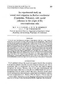
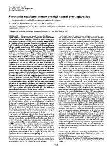
Fig. 1. collapsin-1 expression pattern in hindbrain and trunk regions coincides with the migration of neural crest cells. (A) Coronal section through the hindbrain of a stage 12 whole-mount embryo probed with collapsin-1. Rostral is at the top. Expression is seen in rhombomere (r) 3 and r5, with particularly strong levels in r5. Within the otic vesicle collapsin-1 signal is limited to the region laterally adjacent to r5 (arrowhead). (B) Cross sections through the trunk at stage 12− demonstrate strong expression within the dorsal and lateral portion of the immature somite. In addition, collapsin-1 is strongly expressed within the dorsal ectoderm and lateral plate mesoderm. (C) Longitudinal section of a stage 18 embryo labelled with the HNK-1 antibody. Neural crest migration is limited to the rostral (R) half somite. (D) Longitudinal section taken from an embryo at stage 18 demonstrating that the collapsin-1 mRNA is associated with the caudal (C) sclerotome and the dermatome with elevated levels of expression seen in the caudal half. Scale bars, 50 µm (A), 25 µm (B), 50 µm (D).
the neural tube (Lumsden et al., 1991; Noden, 1975). At stage 12, the collapsin-1 transcript is localised in r1, r3 and r5 with weak expression in r1 and particularly strong expression in r5 (A). In addition, collapsin-1 signal is detected within the otic vesicle with elevated levels seen in the region flanking r5 (Fig. 1A, arrowhead). At the onset of neural crest migration within the trunk region, strong expression of collapsin-1 mRNA is seen in the dorsal and lateral portion of the immature somite (Fig. 1B) with slightly elevated levels in the caudal somite in comparison to the rostral somite (not shown). In addition, the dorsal ectoderm and the lateral plate mesoderm show strong expression of collapsin-1. Neural crest cells that arise from the neural tube in the trunk initially migrate in a non-segmental fashion (Bronner-Fraser, 1986). Crest cells invading the sclerotome are
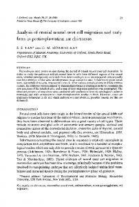
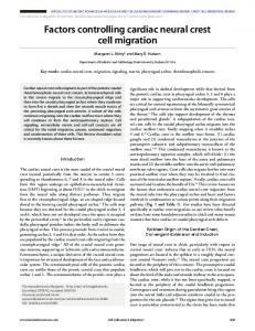
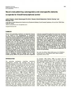
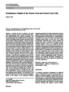
2184 B. J. Eickholt and others found in the rostral halves of the somites (Rickmann et al., 1985; Tucker et al., 1984; Vincent and Thiery, 1984; Vincent et al., 1983). Crest cells, visualised with the HNK-1 antibody, are exclusively seen in the rostral sclerotome, defining sharp borders between the rostral and caudal halves (Fig. 1C). The corresponding collapsin-1 in situ pattern at stage 18 (Fig. 1D) reveals that expression is clearly associated with the caudal sclerotome, whereas expression is absent in the rostral sclerotome. Sagittal sections revealed that this restriction is also seen throughout the dorsoventral axis of the sclerotome (not shown). However, even in the most rostral somite, collapsin-1 expression in the sclerotome is not established before stage 15. In addition, collapsin-1 message is detected within the whole dermatome, and it appears that elevated levels of expression are present in each caudal half in comparison to the rostral half (Fig. 1D). At this stage, the neural tube does not express collapsin-1, which is consistent with recently reported collapsin-1/Sema III distributions (Giger et al., 1996; Shepherd et al., 1996; Wright et al., 1995). Collapsin-Fc binds to both hindbrain and trunk neural crest in situ and in vitro We used an Fc-chimeric version of the collapsin-1 molecule to localise the expression of collapsin receptors within the developing hindbrain and the trunk of stage 12-18 chick embryos (Eickholt et al., 1997). At stage 12, receptors specifically recognised by the collapsin-Fc in the hindbrain region are associated with a stream of cells emerging from the neural tube, shown at the level of r2 in Fig. 2A. Staining with an HNK-1 antibody identifies this population as neural crest cells (Fig. 2C). Binding within the branchial region was seen in crest cells emerging from the different levels of the hindbrain and the different regions along their pathways (not shown). Control experiments with human IgG testify to the specificity of the collapsin-Fc binding (Eickholt et al., 1997). To test whether collapsin-Fc also recognises trunk crest, a second series of stainings were performed. At stage 18, the collapsin-Fc also binds to trunk neural crest (Fig. 2B). In contrast to the binding seen on hindbrain crest, the collapsinFc appears to bind more strongly to crest in close proximity to the neural tube relative to crest cells that have invaded the sclerotome. This might reflect a lack of sensitivity of the method since neural crest emerges from the hindbrain neural tube in streams of cells. Alternatively, trunk crest cells might downregulate putative collapsin-1-binding sites. Experiments on neural crest cells grown in vitro demonstrate that crest cells emerging both from a rhombomere explant (Fig. 3B) and from a trunk neural tube explant (Fig. 3D) bind the collapsinFc to a similar extent.
Fig. 3. Collapsin-Fc binds to cultured neural crest cells. Neural tube explants derived from both hindbrain (A,B) and trunk regions (C,D) were cultured for 20 hours. (A,C) Neural crest cells, visualised with the HNK-1 antibody bind the collapsin-Fc (B,D). The two populations of crest cells show no obvious differences in the number of cells that bind the collapsin-Fc and/or the binding intensity. Scale bar, 50 µm.
Fig. 2. Collapsin-Fc binds to both hindbrain and trunk crest cells in situ. Cross section through a stage 12 hindbrain at the level of r2 (A,C,E) and the trunk at stage 18 (B,D,F) were stained with collapsin-Fc (A,B) and HNK-1 (C,D). The basic morphology of the sections visualised with Hoechst nucleus dye is demonstrated in (E,F). Within the hindbrain, binding of the collapsin-Fc chimera correlates with migrating neural crest cells, whilst binding within trunk regions correlates with crest cells in close proximity to the spinal cord. sc, Spinal cord. Scale bars, 50 µm (E), 50 µm (F).
Neuropilin-1 is expressed in migrating neural crest cells Neuropilin-1 has recently been identified as a neuronal receptor, or an essential part of a neuronal receptor complex, for collapsin-1. Collapsin-1/Sema III binds with high affinity to neuropilin-1 (Feiner et al., 1997; He and Tessier-Lavigne, 1997; Kolodkin et al., 1997) and antibodies generated against the extracellular domains of neuropilin-1 can inhibit the biological activity of collapsin-1/Sema III (He and TessierLavigne, 1997; Kolodkin et al., 1997). Within the hindbrain at stage 12, neuropilin-1 expression is found at the level of r1/2, r4 and r6 just beneath the ectoderm (Fig. 4A,C) with no or only a weak signal detected adjacent to r3 and r5 (Fig. 4B).
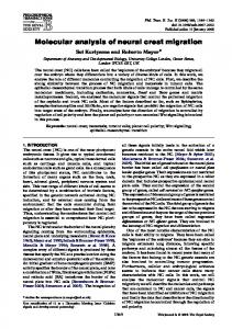
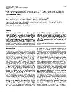
Collapsin-1 and neural crest migration in chick 2185 4D), whilst no expression was detected at stage 13. In longitudinal sections taken at stage 18, neuropilin-1 mRNA is expressed strongly within the rostral sclerotome (Fig. 4E). Neuropilin-1 expression in the motor column of the spinal cord (Fig. 4F) is consistent with previous studies (Takagi et al., 1995). In situ hybridisation was also used to confirm that HNK1-positive populations of cultured crest cells express neuropilin-1. As a control, the same cell population was negative for collapsin-1 expression (Fig. 5).
Fig. 4. Neuropilin-1 is expressed in regions of active neural crest migration. (A-C) Transverse sections through rhombomere (r) 4-r6 of a stage 12 embryo probed with neuropilin-1. Expression is seen in regions corresponding to pathways taken by neural crest cells emerging from the neural tube. (D) Lateral view of a whole embryo at stage 15 with rostral at the right. Neuropilin-1 expression is associated with the rostral half somites. (E) Longitudinal section of a wholemount embryo at stage 18. Neuropilin-1 signals are restricted to the rostral (R) somite halves. (F) Transverse section of a stage 18 embryo probed with neuropilin-1 reveals strong expression within the sclerotome and the motorcolumn (arrowhead). No expression is seen in the dermatome. Scale bars; 50 µm (C), 200 µm (D), 50 µm (E,F).
At this stage, no signal is detected within the neural tube. This restricted pattern of expression reflects the segmental pattern of late-migrating crest cells populating the branchial arches (Lumsden et al., 1991; Noden, 1975). The neuropilin1 transcript is not detected in the mesenchyme flanking the neural tube at the midbrain or more rostral levels (not shown). Within the trunk, neuropilin-1 expression is associated with regions of crest cells migrating through the rostral somite. This segmented expression pattern is clearly visible at stage 15 (Fig.
Fig. 5. Neuropilin-1 is expressed in cultured neural crest cells. Cultures of neural crest cells from the trunk region were probed for neuropilin-1 (A) and collapsin-1 (B) expression. (C) An overlap of HNK-1 immunoreactivity with neuropilin-1 expression characterises cells as neural crest. Scale bar, 25 µm.
Collapsin-1-Fc exhibits repulsive activities towards neural crest migration The restricted pattern of collapsin-1 expression within the somite and the fact that neural crest cells express collapsin receptor(s) suggest that neural crest may be responsive to collapsin-1 in a manner that might contribute to the definition of the migratory pathways. Two different in vitro assays were employed to test the capability of collapsin-1 to affect neural crest cell migration. Firstly, the collapsin-1-Fc was added for 30 minutes directly to cultures of neural crest cells (Fig. 6), at a concentration that is sufficient to induce the collapse of DRG growth cones (1 µg/ml, Eickholt et al., 1997). In comparison to control cultures that had been treated with 1 µg/ml human IgG, collapsin-Fc cause both trunk and hindbrain crest cells to round up with the loss of their outspread lammelipodia. Crest cells contain dense bundles of actin filaments that are disrupted by the collapsin-Fc treatment (Fig. 6). Changes in cell morphology could be correlated with the changes in cell area that were measured automatically. As shown in Fig. 7, collapsin-1-Fc reduces significantly the cell area in both crest cells from trunk and hindbrain regions. In contrast, no alterations in cell morphology were observed when the collapsin-Fc was added to cultures of NIH 3T3 cells (Fig. 7). This demonstrates the celltype specificity of the collapsin-1 effect. In a second set of experiments, collapsin-Fc was immobilised in alternating stripes consisting of the Fc-chimeric protein plus FN versus FN alone using a silicon matrix (Vielmetter et al., 1990). Isolated rhombomeres from stage 10 embryos, and segments of neural tube from stage 12 embryos, were placed on this substratum and, after 20 hours, the cultures were fixed and migrating neural crest cells visualised using HNK-1. Collapsin-Fc was localised with antibodies that recognise the Fc-portion of the chimeric molecule. When offered the choice between collapsin-Fc plus FN versus FN alone neural crest cells exhibit a preference to migrate on FN stripe, avoiding the stripes containing collapsin-1-Fc (Fig. 8A, see Table 1). Hindbrain crest cells show a similar migratory behaviour with crest cells migrating on FN in preference to the collapsin-Fc/FN substratum (Fig. 8B). In a series of control
2186 B. J. Eickholt and others (A) 600
area [mm2]
500
400
*
300
** 200
100
0
control
collapsin
Trunk crest cells
IgG
control
collapsin
IgG
Hindbrain crest cells
(B) 145000
Fig. 7. Measurement of the cell area of neural crest cells and 3T3 135000 cells. (A) Neural crest cells from 130000 both trunk and hindbrain regions 125000 were cultured for 20 hours. Cell 120000 areas of HNK-1-positive cells were determined in control 115000 cultures and cultures that have 110000 been treated for 30 minutes with 105000 collapsin-1 Fc-chimera or human 100000 control collapsin IgG (both at 1 µg/ml) using an automatic image analysis NIH 3T3 cells program. In both crest populations, a significant decrease in cell areas after collapsin-1-Fc addition was observed. (B) In contrast, NIH 3T3 cells do not respond to collapsin-1-Fc with any significant alterations in the cell area. Results show the mean value of the cell area pooled from 4 independent experiments, each value represents the mean ± s.e.m. Individual values in each experiment were determined by measuring the area of 30-40 isolated cells. *P