This article has been accepted for publication in a future issue of this journal, but has not been fully edited. Content may change prior to final publication.
An Implantable Closed-loop Asynchronous Drug Delivery System for the Treatment of Refractory Epilepsy Muhammad Tariqus Salam1, Marjan Mirzaei1, My Sandra Ly1, Dang Khoa Nguyen2 , and Mohamad Sawan1 1 Polystim Neurotechnologies Laboratory, École Polytechnique de Montréal, Montréal, Québec, Canada 2 Neurology Service, Department of Medicine, Centre hospitalier de l'Université de Montréal (CHUM) – Hôpital Notre-Dame, Montréal, Québec, Canada
[email protected]
Abstract – In this paper, we present an implantable device for intra-cerebral electroencephalography (icEEG) data acquisition and real-time epileptic seizure detection with simultaneous focal antiepileptic drug injection feedback. This implantable device includes a neural signal amplifier, an asynchronous seizure detector, a drug delivery system (DDS) including a micropump, and a hybrid subdural electrode (HSE). The asynchronous detection algorithm is based on data-dependent analysis and validated with Matlab tools. The detector and DDS have a power saving mode. The HSE contacts are made of Platinum (Pt) encapsulated with polydimethylsiloxane (PDMS). Given the heterogeneity of electrographic seizure signals and seizure suppression threshold, the implantable device provides tunable parameters facility through an external transmitter to adapt to each individual’s neurophysiology prior to clinical deployment. The proposed detector and DDS were assembled in Ø 50 mm and Ø 30 mm circular printed circuit boards, respectively. The detector was validated using icEEG recordings of 7 patients who had previously undergone an intracranial investigation for epilepsy surgery. The triggering of the DDS was tested and a predefined seizure suppression dose was delivered ~16 sec after electrographical seizure onsets. The device’s power consumption was reduced by 12% in active mode and 49% in power saving mode compared to similar seizure detection algorithms implemented with synchronous architecture. Index terms – Epilepsy, seizure detection, focal drug delivery.
I. INTRODUCTION Interest has grown recently in emerging prosthetic therapies for conventionally-untreatable neurological conditions, such as epilepsy [1], Parkinson’s disease [2], depression [3], and motor impairments [4]. Researchers are exploring the brain-machine interface to translate physiological activity and neuromodulation into machine language for therapeutic purposes. Brain signal monitoring, clinical state analysis and low-power dissipation in prosthetic devices for long-term treatment are key challenges for emerging prosthetic therapies. Epilepsy, which affects approximately 60 million people worldwide, is the second most common chronic neurological condition. It is characterized by a predisposition to unprovoked recurrent seizures. Despite the existence of a number of anticonvulsants, ~30% of patients continue to have disabling seizures. Half of them may benefit from epilepsy surgery if the epileptogenic zone (EZ) can be identified and resected without harm. Others, however, are not good surgical candidates. Due to the limited spatial or temporal resolution of currently available noninvasive localization techniques, accurate delineation of the EZ may sometimes be arduous, particularly in patients with nonlesional refractory epilepsy [5], [6]. Moreover, many patients
have an extensive area of epileptogenicity, multifocal epileptic foci, or an EZ overlying eloquent areas (language, primary motor or visual regions) that cannot be resected without permanent sequelae [6]. Hence, there is a need for alternative treatment modalities. Proof-of-principle experiments conducted in animals and humans with epilepsy have demonstrated the safety of successful chronic electrical stimulation [7], [8], focal pharmacological manipulations [9], [10], and thermal cooling [11] for the treatment of epilepsy. Much progress has been made with focal electrical stimulation therapy methods. Neuropace Inc. has developed a closed-loop-responsive neurostimulator (RNS) that records intra-cerebral electroencephalographic (icEEG) signals, analyzes them and triggers electrical stimulation only upon seizure detection. Preliminary results of electrical stimulation therapy are acceptable, but many patients do not respond to the stimulation and continue have disabling seizures [14], [15]. Focal drug delivery has shown promising seizure suppression results [9], [10]. Direct drug injection onto the epileptogenic zone could enhance the efficacy of medication while limiting side effects to a minimum. Table I shows drugs and doses used in pilot trials. Maximum safe tolerated dose was 150 µL per 20 min for a rat [10]. Two main approaches have been described for focal drug delivery: open-loop and closed-loop. The open-loop approach has been used to increase seizure threshold through continuous drug release while the closed-loop drug delivery system (DDS) is meant to release the drug only upon a seizure detection [10]. The advantages of closed-loop drug delivery over the open-loop method are (i) lower drug dose injection, (ii) potentially less adverse effects, and (iii) rapid focal drug injection. However, several issues of closed-loop systems remain to be addressed, such as the sensitivity and specificity of seizure detection and device power consumption. Good quality icEEG recording and effective automated seizure detection are obviously necessary for any closed-loop TABLE I PILOT STUDIES ON FOCAL DRUG DELIVERY THERAPY Drug Diazepam [10] pH-balenced vehicle [10] Adenosian [16] Lidocaine [17]
Therapy Closed-loop Closed-loop
Dose 35-50 µL 35-50 µL
Open-loop Open-loop
750 µL 5 µL
Gabapentine [18]
Open-loop
N/A
Result Seizure supression Seizure number reduction Seizure prevention Seizure severity reduction Seizure threshold increment
Copyright (c) 2011 IEEE. Personal use is permitted. For any other purposes, permission must be obtained from the IEEE by emailing
[email protected].
This article has been accepted for publication in a future issue of this journal, but has not been fully edited. Content may change prior to final publication.
system. Over the past few decades, several seizure detection mathematical models have been developed, with support vectors [19], artificial neural networks [20], wavelet transformation [21], wavelet decomposition [22], and nonlinear time series analysis [23]. These complex mathematical models are meant to be run on high-speed desktop computers for offline, long-term data processing to help clinicians find seizures but can hardly be employed in low-power implantable devices. Later, several implantable seizure detectors have been described in the literature on closed-loop epilepsy prostheses [24] - [32]. Power management is an important issue for these detectors. Power consumption is mainly dependent on signal transitions in a device, such as charging and discharging of parasitic capacitances in transistors and short-circuit currents during switching [33]. Thus, power consumption can be reduced by avoiding unnecessary signal transitions [33]. With synchronous designs [24] - [32], all components share a common clock signal distributed throughout the circuit, and unnecessary signal transitions arise from clock gating. Because of these limitations in conventional synchronous circuit design, the asynchronous design technique is likely to become more popular. An asynchronous device is fundamentally different, and device transistors do not change their transient state unless an event happens. This device does not use or share a common clock; thus, it has no clock skew and no clock tree. In the following parts, we will describe methods and materials in Section II and results in Section III. Conclusions are summarized in the last section of this paper. II.
METHODS AND MATERIALS
A. Proposed closed-loop epilepsy prosthesis The proposed closed-loop epilepsy prosthesis would benefit the patients with focal epilepsy with or without secondary generalization who are not controlled and reported significant side effects with oral antiepileptic drugs, especially those whose epileptic focus cannot be removed because it overlies an important functional area (motor, sensory, visual, language areas). This epilepsy prosthesis requires low-power, continuous, long-term icEEG monitoring to identify seizures online before the appearance of disabling clinical
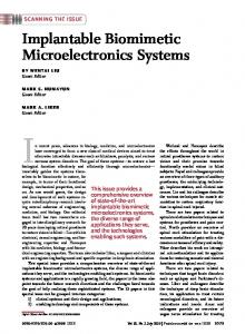
Fig. 1. Proposed closed-loop drug delivery system: (a) Block diagram, (b) Flow chart, and (c) Implant configuration.
manifestations (e.g. altered consciousness, tonic or clonic movements). An asynchronous device has inherent properties that work locally and activate all modules, if needed. Therefore, an asynchronous design holds significant promise in icEEG recording extraction applications, where complex analyses are needed for a short period (e.g. seizure onset). Therefore, power management pleads in favor of an asynchronous design. In this paper, an asynchronous device is designed and implemented to improve data-dependent computation and power management. Fig. 1 illustrates flowchart, block diagram, and implant configuration of the proposed closed-loop drug delivery
Fig. 2. Block diagram of closed-loop asynchronous drug delivery system. Copyright (c) 2011 IEEE. Personal use is permitted. For any other purposes, permission must be obtained from the IEEE by emailing
[email protected].
This article has been accepted for publication in a future issue of this journal, but has not been fully edited. Content may change prior to final publication.
system. The refilling port is made of a microtube (Ø 1.93 mm) coated with PDMS and sealed the entry port with PDMS so that a syringe easily goes through the PDMS to fill the reservoir without having a bacteria entry point. The functional block diagram in Fig. 2 presents the architecture of the closedloop DDS that includes total 11 modules, such as 1 amplifier, 4 voltage level detectors, 4 asynchronous frequency analyzers, 1 receiver- controller, and 1 DDS. First, the icEEG recording Vin is amplified and decomposed by seizure detectors. The extracted activities define an upcoming seizure. The responsive DDS must be adjusted by setting voltages (VT,i,a/b), time windows (Tf), and dose (drug injection period TPM and micropump control voltage VDP) labeled as corresponding to seizures and non-seizures. Optimum parameters are measured from time-amplitude and time-frequency analysis of 1 seizure from the EZ and normal signal activity. Clinical studies have shown that several electrographic patterns (e.g. low-voltage and high-voltage fast activities or rhythmic spiking) can be seen in EZ at seizure onset, depending on such factors as etiology, location, position of intracerebral electrodes [6], [12]. Generally, same patient-specific pattern and seizure frequency can also be seen in EZ which lead to a clinical seizure [12], [25], [28], [34].” 1) Front-end seizure detector Synchronous epileptic seizure detection algorithms have been suggested in our previous works [25], [28], [29]. In this paper, an asynchronous seizure detection algorithm is proposed for better power management. Table II compares the synchronous and asynchronous algorithms. Details of asynchronous frontend seizure detector are described below. 2) Asynchronous epileptic seizure detection algorithm Amplified input signal (Vamp) passes through voltage window detectors (VWD), and outputs Vw,i define the strength of neural signal activities in different amplitude windows (Eq. 1).
'1' , V wi = '0' ,
for VT ,i , a > V amp > VT ,i ,b for otherwise
(1) where i = 1, 2, 3…N. VT,i,a-b are the threshold voltage of VWDs. Frequency analyzers extract Vw,i and measure the signal TABLE II COMPARISON OF SYNCHRONOUS AND ASYNCHRONOUS EPILEPTIC SEIZURE DETECTION ALGORITHMS Synchronous [29]
Asynchronous (this work)
Modulates input signals before analysis Window detection in positive amplitude
Directly analyzes input signals without modulation Window detection in positive and negative amplitude
Signal analysis in high frequency band
Signal analysis in original frequency band
Demodulates to original frequency band
No demodulation
Frequency divider generates time frames
Externala device generates time frames
Clock controls frequency analysis
External device controls frequency analysis
a
External: On top of the skin
frequency (FS,i), as shown in Eq. (2). t =T f
∑V
W ,i
FS ,i =
t =0
(2)
Tf
If FS,i is greater than a specific seizure onset frequency (FSZ), the output of each channel VD,i is considered fast activity (Eq. 3).
'1' , V D ,i ( n ) = '0' ,
for FS ,i ≥ FSZ for
(3)
otherwise
Seizure detection logic analyzes VD,i and quantifies specific features characterized by a progressive increase in amplitude and signal frequency. Thus, a seizure onset is declared on the basis of the following conditions (Eq. 4).
'1' , V SD = '0' ,
for V D ,1 . V D , 2 . V D , 3 ... V D , N = 1 for
(4)
otherwise
VT,i,a-b and Tf are tuned to the patient’s specific FSZ to minimize false alarms. The parameters can optimize the decision boundary and enhance sensitivity and specificity. Fig. 3 shows maximum detection performance using 4 VWDs (2 VWDs monitor basal activities and the other 2 VWDs detect epileptic activities). 3) Asynchronous front-end detector Fig. 2 shows block diagram of the asynchronous front-end detector (AFED). The detector contains a neural signal amplifier, an asynchronous seizure detector, a controller and a receiver. Details are described below. • Amplifier The icEEG recording is generally characterized by lowamplitude signal (microvolts) and low-frequency bandwidth. Moreover, the instrumentation amplifier has relatively poor noise performance for amplifying the low spectral microvoltlevel signal due to the flicker and thermal noise. Thus, icEEG signal must be amplified very carefully before further analysis (e.g. detection, digitization). The proposed neural signal amplifier has 3 stages for amplification and noise reduction (Fig. 4(a)) [34]. A feedback noise reduction circuit reduces DC
Fig. 3. Asynchronous seizure detection performance analysis using optimum parameters of different VWDs based on 5 seizures and long normal icEEG of the test patient.
Copyright (c) 2011 IEEE. Personal use is permitted. For any other purposes, permission must be obtained from the IEEE by emailing
[email protected].
This article has been accepted for publication in a future issue of this journal, but has not been fully edited. Content may change prior to final publication.
Fig. 4. Neural signal amplifier: (a) Schematic diagram of amplifier, (b) Noise reduction method and corresponding frequency analysis of signals in different nodes, (c) Frequency response, and (d) Measured input-referred noise voltage spectral densities of the proposed and commercial amplifier.
offset VOS (Fig. 4(b)). Furthermore, flicker noise VnF and thermal noise VnT are reduced by high- and low-pass filters (Fig. 4(b)). The amplifier features high-amplification gain: 60 dB and 48 Hz (3 Hz to 51 Hz) bandwidth (Fig. 4(c)) with 6 µVrms input-referred voltage noise. Fig. 4(d) shows measured input-referred noise voltage spectral densities of the proposed and commercial amplifer. • Asynchronous seziure detector The detector comprises 4 VWDs and 4 asynchronous frequency analyzers. The VWDs have 8 tunable parameters to adjust their threshold voltages (VT,i,a-b). Asynchronous frequency analyzers detect high-frequency events by analyzing neural activities in Tf, which can be tuned externally and configured to the specific seizure onset frequency (FSZ) of a patient to reduce false alarms during icEEG monitoring. • Controller The proposed seizure detector allows to tune several parameters that are personalized in a specific manner to prevent false seizure detection. The controller includes 8 digital-toanalog converters, and 8 buffers. This controller can provide digitally-controlled, high-precision voltage VT,i,a/b and VDP. • Receiver The receiver is responsible for power and data (e.g. Tf) recovery. The power recovery circuit was introduced in our previous work [35] and Fig. 5(a) shows a schematic diagram of the data recovery. The external transmitter generates and transmits predefined Tf with inductive communication. A parallel LC network of the internal device receives Tf and controls the seizure detector. Graphic representation of the time-frame generation in Figs. 5(b)-(c) shows transmitted signal pattern VER and Tf recovered from VRST. 4) Drug delivery system Figs. 6(a)–(d) illustrate the operation of the DDS and demonstrate its timing process in response to seizure detections VSD. The duration of drug delivery (VPM) is TPM; if
Fig. 5. Time frame generation: (a) Schematic diagram of receiver, (b) Transmitted signal pattern VER from external coil, and (c) Recovered time frame VRST.
Fig. 6. Proposed drug delivery system: (a) Seizure detections VSD (b) Drug injection period TPM, (c) Focal treatment duration TON, (d) Two subsequent drug injections, and (e) – (h): Operation of micropump.
the injection does not terminate the seizure another dose is triggered by the detector after TON. VPM generates VP push-pull signal in the micropump (13 x 13 x 5 mm3) (Microbase technology Corporation) to drive the drug. 5) Hybrid subdural electrodes The electrodes fabrication process includes mould fabrication, electrodes assembly, and biomedical grade polydimethylsiloxane (PDMS) encapsulation. The mould was fabricated on an Aluminum substrate (Fig. 7(a)) using a
Fig. 7. Hybrid subdural electrodes fabrication process: (a) Dimension of mould; (b) Electrode, wire and fluidic channel assembly on mould; (c) Pour PDMS; (d) Cover up and heating with 110oC; (e) Side view of the released subdural electrodes; and (f) Top view of the electrodes.
Copyright (c) 2011 IEEE. Personal use is permitted. For any other purposes, permission must be obtained from the IEEE by emailing
[email protected].
This article has been accepted for publication in a future issue of this journal, but has not been fully edited. Content may change prior to final publication.
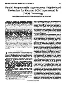
Fig. 10. Invasive study: 3-D images of implanted electrodes reconstructed with Stellate’s Gridview software and identification of epileptogenic zones.
Fig. 8. Proposed closed-loop drug delivery system: (a) – b) Top and bottom views of asynchronous front-end detector, (c) External transmitter, (d)–(e) Top and bottom views of drug delivery system, and (f) hybrid subdural electrodes.
computer numerically controlled machine. Electrodes (Platinum), wires (Platinum) and fluidic channel (Micro-tube: Ø 0.5 mm) were assembled on the mould (Fig. 7(b)). PDMS is poured (Fig. 7(c)) and the covered mould was heated to 110oC for 20 minutes (Fig. 7(d)). Once subdural electrode arrays become encapsulated with a thin PDMS substrate, they are released from the mould (Fig. 7(e) – (f)). B. Circuits and devices implementation The closed-loop DDS was assembled with discrete commercial components on circular shape printed circuit boards (PCB). Figs. 8 (a) – (c) show photographs of the asynchronous front-end detector. Photographs of the drug delivery system and hybrid subdural electrodes are presented in Figs. 8 (d) – (f). C. Patients description This study was conducted at Notre-Dame Hospital, Centre Hospitalier de l'Université de Montréal (CHUM). Seven patients with refractory focal epilepsy (age 15 to 49 years) were qualified for these studies based on electrographic seizure onset. The selected patients had previously undergone a comprehensive noninvasive assessment of brain. These complementary non-invasive studies failed to adequately localize the epileptogenic zone and invasive intracranial electrode studies were required to delineate with more precision the EZ. The seizure signals of cases 1, 4, 5, 6 and 7
were recorded using depth electrodes and the signals of cases 2 and 3 were recorded using subdural strip electrodes. These patients had seizure onsets characterized by progressive increase of low-voltage fast-activity in icEEG recordings. These patients also had frequent electrical seizures on the EZ. There is no consensus on what constitutes an appropriate definition of electrical seizure apart from the fact that they do not generate any clinical manifestations [36],[37]. Some electrical seizures are simply very brief (a few seconds), focal (without spread) high-frequency discharges which do not evolve significantly in time or frequency, and are hence clinically silent. Others are longer (several seconds) with some evolution. D. Device validation method The proposed implantable device was tested offline using icEEG recordings obtained from the 7 patients. The experimental procedure was approved by the University of Montreal’s Hospital Center (CHUM) and Polytechnique Montreal research ethics committees. Fig. 9 shows the proposed device validation procedure. These icEEG studies consist in implanting electrodes into or on the surface of the brain through a craniotomy or burr holes under general anaesthesia. After intracranial electrode implantation, patients are transferred to the Epilepsy Monitoring Unit for continuous video-EEG telemetry to record seizures. 128 channel icEEG recordings were performed using amplifiers (PRO-36 amplifiers, Stellate Harmonie System) at 200 Hz and 2000 Hz sampling frequency. Post-implantation MRI served to reconstruct 3-D representation of electrode positioning (via Stellate’s Gridview software) (Fig. 10). During the invasive study, several brief electrical seizures and electroclinical seizures were recorded
Fig. 9. The closed-loop epilepsy prosthesis: icEEG recorded from patients with medically refractory epilepsy were used to test the proposed seizure detector and the triggering of predefined drug dose was validated.
Copyright (c) 2011 IEEE. Personal use is permitted. For any other purposes, permission must be obtained from the IEEE by emailing
[email protected].
This article has been accepted for publication in a future issue of this journal, but has not been fully edited. Content may change prior to final publication.
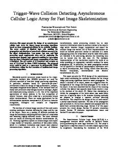
and marked by an epileptologist (DKN). All the seizures were found to originate from the red circle-marked electrodes in Fig. 10 with subsequent rapid spread to square-marked electrodes and then propagation to triangle-marked electrodes. Fig. 9 illustrates that the icEEG recordings from the two contacts positioned over the epileptogenic zone (Fig. 10) were selected, transferred to the signal conditioner (arbitrary signal waveform generator) for reproducing icEEG recording and fed into the closed-loop epilepsy prosthesis. The reproduced microvolt-level icEEG recordings were amplified by the amplifier of the AFED and processed for the seizure detection. The AFED detected seizure onset and triggered the DDS to deliver a predefined drug dose through the hybrid subdural electrodes. During the drug injection, the delivered drug was measured to validate the seizure suppression doses (Table I). In order to justify the effectiveness of the proposed responsive device, all the seizures were observed during the experiments and analyzed all channels to determine seizure progression and propagation. Fig. 11 shows typical electrographic seizures as recorded from the electrode contacts in the epileptogenic zone as well as nearest adjacent functional regions. Based on video-EEG analysis of this seizure, the epileptiform discharges propagated slowly to the nearest functional region (22 sec) and patient reported feeling an aura 33 sec after the electrographic seizure onset. Altered consciousness occurred a few seconds later. This propagation time varied from patient to patient. In this experiment, the minimum propagation time is 22 sec in case 1 (worst case scenario) and the maximum time is 110 sec in case 7 (worst case scenario). Furthermore, the pilot studies have shown epileptiform activities suppression within 5 sec due to the 3550 µL Diazepam drug injection on epileptogenic zone during an electrographic seizure [6]. Therefore, the key challenges for an automated drug delivery system are to detect seizure onset and deliver drug on epileptogenic zone before the epileptiform discharges propagating to others adjacent functional areas. E. Prosthesis pre-implantation procedure A prosthesis pre-implantation procedure was introduced to observe patients’ specific neurophysiology properties and configure the AFED and DDS. Ictal discharges from patients generally contained high-frequency activities, but not all high frequency discharges were seizures (i.e. some were physiological rhythms). Moreover, patients presented frequent, brief, low-voltage, high-frequency discharges without clinical manifestations (reminiscent of very Brief electrical seizures BES) which our clinician choose to ignore. As these discharges
Fig. 12. Prosthesis pre-implantation settings: (a) Training data Vin and (b) – (c): adjustment detection parameters FSZ and VT,i,a/b from time-frequency (FS) and time-amplitude (VMA) analysis of Vin.
did not frequently evolve into longer electro-clinical seizure, they were overlooked to prevent the triggering of non-clinically useful responsive drug deliveries that could rapidly deplete battery life and drug reserve. Hence, it was decided to configure the AFED to ignore these brief discharges. For proper AFED configuration, a normal signal, 2 BESs and an electro-clinical seizure from a patient were analyzed in the time-amplitude VMA (root mean square) and time-frequency FS domain. The obtained detection parameters from these training signals were used to adjust the AFED and consequently it detected the other seizures of the patient. Fig. 12 depicts the analysis of 30 minutes of icEEG recording to demonstrate optimum selection of the seizure detection parameters (i.e. FSZ in Fig. 12(b) and VT,i,a/b in Fig. 12(c)). Signal analysis of normal icEEG recordings Vin revealed no abnormalities in FS and VMA which define basal activity (|VT,2/3,a|). Brief high-frequency discharges or BES had high frequency contents in FS with limited amplitude increase (|VT.2/3,b|) in VMA. Analysis of an electro-clinical seizure onset demonstrated a sudden increment in FS with a progressive amplitude rise (|VT,1/4,a/b|) in VMA leading to clinical manifestations. Clinicians could set these parameters by visual inspection, but the pre-implantation procedure optimizes the trade off between detection delay and performance (i.e. sensitivity and specificity). III.
Fig. 11. The icEEG recordings from the epileptogenic zone as well as adjacent functional regions and the corresponding behavioural relation with the electrographical signals.
EXPERIMENTAL AND CLINICAL RESULTS
Measurements were undertaken separately on each block of the proposed asynchronous device and were compared with the synchronous system [29] assembled with discrete commercial components. Table III shows power dissipation of these devices in both active and power saving modes. These prototyped devices have been made using latest low-power discrete components. However, recently developed FPGA operated in freeze standby mode, discrete components or custom integrated circuits could reduce the power consumption
Copyright (c) 2011 IEEE. Personal use is permitted. For any other purposes, permission must be obtained from the IEEE by emailing
[email protected].
This article has been accepted for publication in a future issue of this journal, but has not been fully edited. Content may change prior to final publication.
TABLE III MEASURED POWER CONSUMPTION OF SYNCHRONOUS AND ASYNCHRONOUS PROTOTYPED DEVICES AT 3.3 V POWER SUPPLY Block
Comp
Synch Deviceb PCe (mW) Qtd AMf PSMg
Amplifier
IAh
1
2
2
1
2
2
Detector
VWDi
3
13.1
13.1
4
17.4
8.0
DFFj
3
6
6
0
0
0
FDk
3
19.3
19.3
4
25.7
12.1
a
l
Asynch Devicec PC e (mW) Qtd AMf PSMg
Clock
Osc
1
1.5
1.5
0
0
0
Modulator & Demodulator
SWsm CPn VWDi
4 3 3
0.1 5 13.1
0.1 5 13.1
0 0 0
0 0 0
0 0 0
TFGo
FDk
3
19.3
19.3
0
0
0
0 1
0 0
0 5
1 1
6 10
6 5
70.1
65.1
61.2
33.1
Receiver DDSp Total power consumptionq a
Comp: Component, bSynch Dev: Synchronous device, cAsynch Dev: Asynchronous device, dQt: Quantity, ePC: Power consumption, fAM: Active mode, gPSM: Power saving Mode, hIA: Instrumentational amplifier, iVWD: Voltage window detector, jDFF: Mater-salve D flip-flop, kFD: Frequency divider, lOsc: Oscillator, mSWs: Switches, nCP: charge pump, oTFG: Time frame generator, and pDDS: drug delivery system, and qpower dissipated by prototyped device based discrete components.
to microwatts. These prototyped devices have demonstrated smart operation to reduce power dissipation. The asynchronous prototyped device turned OFF 6 modules during the icEEG monitoring period and turned ON all 11 modules at seizure onset. With the synchronous device, the clock would have kept all modules running during the monitoring period. As a result, measured power consumption in the asynchronous detector was reduced by 49% in icEEG monitoring period (power saving mode) and 12% in active mode compared to the synchronous detector. The power dissipation densities were under maximum safety level (δPmax = 6mW/cm2 which may cause enough heat to damage surrounding tissue) [38]. Figs. 13 illustrate devices’ operation modes and their corresponding average power dissipation (PT) and power consumption densities (δp). Impedances of the proposed hybrid electrodes were electrically characterized in vitro using electrochemical impedance spectroscopy (Impedance analyzer HIOKI 3522 LCR) in standard physiological saline solution (0.9% NaCl).
Fig. 13. Demonstration of power dissipation of proposed system: (a) Electrographic seizure in icEEG recording and (b) Average power dissipation PT and densities δP of the devices, where PT_A and PT_S are power dissipation in asynchronous and synchronous devices, and δP_A and δP_s are power consumption densities, respectively.
Fig. 14. Electrical properties of the hybrid electrode: (a) SEM image of the proposed electrode contact and (b) Measured impedance sweep of the commercial and proposed electrodes.
Fig 14(a) shows scanning electron microscope (SEM) images of the Pt electrodes and Fig 14(b) illustrates comparative studies on impedance magnitude of commercial electrode (AdTech Medical Instruments, WI, USA) and the proposed electrode. The proposed electrodes have significantly lower impedance and better recording performances [39]. A. Validation of asynchronous front-end detector IcEEG recordings from two electrode contacts (Fig. 10) located in the EZ from 7 patients were fed into the proposed algorithm and device. The AFED was tested on an average of 6 electro-clinical seizures, 15 brief electrical seizures and 10 long normal signals (30 min randomly chosen) per patient. Fig. 15 shows validation of the proposed detection algorithm using Matlab tools. The recorded icEEG Vin from case 1 was uploaded in Matlab simulation platform, and analyzed in timeamplitude and time-frequency domain, and detected seizure onset. Fig. 16 illustrates measured seizure detection of the proposed device on icEEG recordings (Vin) from case 1 and the seizure detection delay TDET of VSD. The simulation and experimental results of synchronous and asynchronous detectors are presented in Table IV. The AFED detected the electro-clinical seizures of all 7 patients in an average of 16 sec after seizure onset. Sensitivity of the detectors has been defined according to [40] and both patient-specific seizure
Fig. 15. Validation of proposed algorithm: (a) icEEG recordings Vin, (b) seizure onset activity is characterized by progressive amplitude increase, (c) rapid fast-activity, and (d) seizure onset detections.
Copyright (c) 2011 IEEE. Personal use is permitted. For any other purposes, permission must be obtained from the IEEE by emailing
[email protected].
This article has been accepted for publication in a future issue of this journal, but has not been fully edited. Content may change prior to final publication.
Fig. 16. Measured seizure onset detection by AFED, where Vamp is icEEG of seizure from the epileptogenic zone, and VSD is seizure onset detection of case 1.
detectors had 100% sensitivity. The mean seizure detection delay was slightly longer than the one of the synchronous detector which took on average of 13 sec to detect seizures due to a lower noise effect in the device. B. Validation of drug delivery system The predefined drug dose was administrated in response to the early detection of seizure development. Fig. 17 shows 3 selftriggered drug injections in response to the seizure detection by the proposed system and illustrates the corresponding individualized dose. The doses are widely programmable by TPM and micropump control voltage VDP. Fig. 18 shows range of drug dose for different variables. The desired seizure suppression dose (Table I) was obtained by regulation TPM (Fig. 18(a)), however, more precise was achieved with different combinations of VDP and TPM (Fig. 18(b)). IV.
DISCUSSION
The proposed closed-loop epilepsy prosthesis offers promises of lower power dissipation, better data-dependent computation and higher detection performances compared to a synchronous design. Current commercially-implantable
epilepsy devices [14] and seizure detectors [24] – [32] are based on a traditional synchronous design. Thus, these devices continuously dissipate battery power for clock gating which ultimately shortens the interval for battery replacement or recharging. Open loop devices, such as the VNS or anterior thalamic stimulators, stimulate in a periodic manner with a built-in clock which defines the periodic action [1], [12], [13]. These devices do not require a change from traditional synchronous technology owing to the isolation of clock gating from the rest of the device in sleep mode. However, the RNS system [14] requires continuous long-term icEEG monitoring. Therefore, this system should replace traditional technology with asynchronous technology and improve data-dependent computation. Furthermore, the sensitivity of the proposed AFED are maximized 100% to prevent unwarranted drug delivery; however, sensitivity of [27], [31], and [32] are 94.35%, 95.3%, and 93%, respectively. Although the average detection of the proposed AFED is 8 sec (max) higher than the other detectors described in [27], [31], [32]; but the proposed device have showed well capability to detect seizure onset and deliver the antiepileptic drug well before the onset of clinical manifestations. For example, Fig. 11 shows minimum seizure
TABLE IV AVERAGE SEIZURE DETECTION DELAY (TDET) OF SYNCHRONOUS AND ASYNCHRONOUS DETECTORS Average TDET (s) Case Age Epileptogenic Syncha Asynchb no. /Gender zone c d Sim Exp Simc Expd
a
1
24/M
Right hippocampus
7
6
7
9
2
36/M
Left lateral temporal neocortex
12
15
10
16
3
44/M
7
15
7
18
4
41/M
10
21
12
22
5
49/F
24
12
18
15
6
32/M
12
12
11
13
7
15/M
25
20
19
22
Left pre-motor cortex Right amygdala/ hippocampus Left hippocampus Left hippocampus Right lingual gyrus
Synch: Synchronous, bAsynch: Asynchronous, cSim: Simulation results with Matlab, and dExp: Experimental results .
Fig. 17. Monitored drug delivery process triggered by seizure detector, where Vin is neural signal, Vamp is amplified neural signal, VSD is seizure detection, VPM is drug injection period TPM, and VON is focal treatment duration TON.
Copyright (c) 2011 IEEE. Personal use is permitted. For any other purposes, permission must be obtained from the IEEE by emailing
[email protected].
This article has been accepted for publication in a future issue of this journal, but has not been fully edited. Content may change prior to final publication.
Fig. 18. Controled widely programmable drug dose by: (a) injection period TPM and (b) micropump needed voltage VDP with different TPM.
progression time (~22 sec) for case 1 and subsequent aura reported at 33 sec; however, the proposed system detected this seizure at 8 sec (2nd row, 3rd column of Fig. 16) and delivered sufficient amount of drug in a short duration (Fig. 18), and consequently seizure may suppress within 5 sec [6]. The average detection delay of the detector is 16 sec, thus all the detections have sufficient time to inject drugs and block seizure progression. The direct drug delivery experiments for the seizure suppression in Table I were demonstrated using external prototypes (e.g. desktop computer and syringe pump); however, the proposed prototyped device is implemented on two PCB boards and expected to be reliable in an implantable device without risking false detections. V. CONCLUSION We have described the design and implementation of an asynchronous closed-loop drug delivery system that paves the way for improved power management of currently-available technologies. The proposed asynchronous seizure detector was validated using icEEG recordings of 7 patients, and detection performance was as accurate as that obtained with synchronous devices, but reduced power consumption by 49% in power saving mode. Data-dependent analysis of this asynchronous design kept 6 modules (out of 11) OFF in icEEG monitoring mode unless abnormal activity interrupted it. The AFED integrates a multiple feature-based algorithm for the detection of epileptic seizures. Due to the heterogeneity of seizure onset patterns, an individualized method is the best solution among general feature-based approaches. Furthermore, this method reduced device complexity. A computerized prosthesis pre-implantation procedure optimized individualized settings for adequate detection performance. Focusing on low-power dissipation, the proposed asynchronous detector is designed for progressively increasing low-voltage, fast-activity, seizure-onset patterns. Experimental results have demonstrated the feasibility of asynchronous design in a low-power, reliable, implantable device. Further validation in an animal model of epilepsy is warranted.
ACKNOWLEDGMENTS The authors are grateful for support from the Natural Sciences and Engineering Research Council of Canada (NSERC), the Canada Research Chair in Smart Medical Devices, Les Fonds Québécois de la recherche sur la nature et les technologies (FQRNT), and the EEG technicians at CHUM – Hôpital Notre-Dame, Montréal, Québec, Canada. REFERENCES [1] [2]
[3] [4]
[5] [6] [7] [8] [9]
[10] [11]
[12]
R. Terry, “Vagus nerve stimulation: a proven therapy for treatment of epilepsy strives to improve efficacy and expand applications,” Conf. Proc. IEEE Eng. Med. Biol. Soc., pp. 4631-4, 2009. S. V. Sarma, U. T. Eden, M. L. Cheng et al., “Using point process models to compare neural spiking activity in the subthalamic nucleus of Parkinson's patients and a healthy primate,” IEEE Trans. Biomed. Eng., vol. 57, no. 6, pp. 1297-305, 2010. D. Panescu, “Vagus nerve stimulation for the treatment of depression,” IEEE Eng. Med. Biol. Mag., vol. 24, no. 6, pp. 68-72, 2005. O. Celik, M. K. O'Malley, C. Boake et al., “Normalized movement quality measures for therapeutic robots strongly correlate with clinical motor impairment measures,” IEEE Trans. Neural. Syst. Rehabil. Eng., vol. 18, no. 4, pp. 433-44, 2010. E. J. Jerome, “The Treatment of Epilepsy: Overview of Surgical Treatment for Epilepsy,” pp. 4631-4: Wiley-Blackwell, 2009. S. S. Spencer, D. K. Nguyen, and R. B. Duckrow, “The Treatment of Epilepsy: Invasive EEG in Presurgical Evaluation of Epilepsy,” pp. 76798: Wiley-Blackwell, 2009. W. H. Theodore, and R. S. Fisher, “Brain stimulation for epilepsy,” Neurology, vol. 3, no. 8, pp. 111-8, 2004. P. Boon, R. Raedt, V. D. Herdt et al., “Electrical stimulation for the treatment of epilepsy,” Neurotherapeutics, vol. 6, no. 2, pp. 218-27, 2009. L. B. Good, S. Sabesan, L. D. Iasemidis, K Tsakalis, and D. M. Treiman, “Brain dynamical disentrainment by anti-epileptic drugs in rat and human status epilepticus,” Conf. Proc. IEEE Eng. Med. Biol. Soc., vol.1, pp. 176-9, 2004. A. G. Stein, H. G. Eder, D. E. Blum et al., “An automated drug delivery system for focal epilepsy,” Epilepsy Res., vol. 39, no. 2, pp. 103-14, 2000. G. R. Guerra, R. V. Davalos, P. A. Garcia et al., “Heat transfer model to characterize the focal cooling necessary to suppress spontaneous epileptiform activity,” Proc. of SPIE: Progress in Biomed. Optics and Imaging, pp. 240-6, 2005. M. T. Salam, M. Sawan, and D. K. Nguyen, “Low-power implantable device for onset detection and subsequent treatment of epileptic seizures: a review,” J. Healthc. Eng., vol. 1, no. 2, 2010.
Copyright (c) 2011 IEEE. Personal use is permitted. For any other purposes, permission must be obtained from the IEEE by emailing
[email protected].
This article has been accepted for publication in a future issue of this journal, but has not been fully edited. Content may change prior to final publication.
[13] F. Rychlicki, N. Zamponi, R. Trignani et al., “Vagus nerve stimulation: clinical experience in drug-resistant pediatric epileptic patients,” Seizure, vol. 15, no. 7, pp. 483-90, 2006. [14] F. T. Sun, M. J. Morrell, and R. E. Wharen, Jr., “Responsive cortical stimulation for the treatment of epilepsy,” Neurotherapeutics, vol. 5, no. 1, pp. 68-74, 2008. [15] T. L. Skarpaas, and M. J. Morrell, “Intracranial stimulation therapy for epilepsy,” Neurotherapeutics, vol. 6, no. 2, pp. 238-43, 2009. [16] David J. Anschel, Erik L. Ortega, Alli C. Kraus, Robert S. Fisher, “Focally injected adenosine prevents seizures in the rat,” Experimental Neurology, vol. 190(2), pp. 544-547, 2004 [17] D.C. Smith, S. E. Krahl, R. A. Browning, and E. J. Barea, “Rapid cessation of focally induced generalized seizures in rats through microinfusion of lidocaine hydrochloride into the focus,” Epilepsia, vol. 34, pp. 43-53, 1993. [18] Joseph Oommen, Alli C. Kraus, Robert S. Fisher, “Intraventricular administration of gabapentin in the rat increases flurothyl seizure threshold”, Neuroscience Letters, Vol.417(3), pp.308-311, 2007. [19] R. Meier, H. Dittrich, A. Schulze-Bonhage et al., “Detecting epileptic seizures in long-term human EEG: a new approach to automatic online and real-time detection and classification of polymorphic seizure patterns,” J. Clin. Neurophysiol., vol. 25, no. 3, pp. 119-131, 2008. [20] S. Ghosh-Dastidar, H. Adeli, and N. Dadmehr, “Principal component analysis-enhanced cosine radial basis function neural network for robust epilepsy and seizure detection,” IEEE Trans. Biomed. Eng., vol. 55, no. 2, pp. 512-8, 2008. [21] A. S. Zandi, M. Javidan, G. A. Dumont et al., “Automated real-time epileptic seizure detection in scalp EEG recordings using an algorithm based on wavelet packet transform,” IEEE Trans. Biomed. Eng., vol. 57, no. 7, pp. 1639-51, 2010. [22] M. E. Saab, and J. Gotman, “A system to detect the onset of epileptic seizures in scalp EEG,” Clin Neurophysiol., vol. 116, no. 2, pp. 427-4, 2005. [23] K. Lehnertz, and C. E. Elger, “Spatio-temporal dynamics of the primary epileptogenic area in temporal lobe epilepsy characterized by neuronal complexity loss,” Electroencephalogr. Clin. Neurophysiol., vol. 95, no. 2, pp. 108-17, 1995. [24] K. Abdelhalim, V. Smolyakov, and R. Genov, Phase-Synchronization Early Epileptic Seizure Detector VLSI Architecture, IEEE TBIOCAS, vol. 5, No. 5, 2011 [25] M. T. Salam, M. Sawan, and D. K. Nguyen, “Epileptic seizure onset detection prior to clinical manifestation,” Conf. Pro.c IEEE Eng. Med. Biol. Soc., pp. 6210-3, 2010. [26] M. T. Salam, M. Sawan, A. Hamoui, and D. K. Nguyen, “Low-power CMOS-based epileptic seizure onset detector,” IEEE NEWCAS-TAISA, pp. 1-4, 2009. [27] N. Verma, A. Shoeb, J. Bohorquez et al., “A micro-power EEG acquisition SoC with integrated feature extraction processor for a chronic seizure detection system,” IEEE J, Solid State Circuits, vol. 45, no. 4, pp. 804-16, 2010. [28] M. T. Salam, M. Sawan, D. K. Nguyen et al., “Epileptic low-voltage fast-activity seizure-onset detector,” IEEE BioCAS, pp. 169-72, 2009. [29] M. T. Salam, M. Sawan, and D. K. Nguyen, “A low-power implantable device for epileptic seizure detection and neurostimulation,” IEEE BioCAS, 2010. [30] M. T. Salam, M. Sawan, and D. K. Nguyen, “A novel low-power implantable epileptic seizure-onset detector,” IEEE TBIOCAS, vol. 5, no. 6, pp. 568 - 578, 2010. [31] S. Raghunathan, S. K. Gupta, M. P. Ward et al., “The design and hardware implementation of a low-power real-time seizure detection algorithm,” J. Neural Eng,. vol. 6, no. 5, 2009. [32] K. Patel, C. Chern-Pin, S. Fau et al., “Low power real-time seizure detection for ambulatory EEG,” 3rd International Conference on Pervasive Computing Technologies for Healthcare (Pervasive Health), pp. 7, 2009. [33] L. S. Nielsen, and J. Sparso, “Designing asynchronous circuits for low power: an IFIR filter bank for a digital hearing aid,” Proceedings of the IEEE, vol. 87, no. 2, pp. 268-81, 1999. [34] M. T. Salam, D. K. Nguyen, and M. Sawan, "A Multichannel Intracerebral EEG Monitoring System For Epilepsy Presurgical Evaluation," IEEE CCECE, 2011. [35] Mounaim and M. Sawan, “Miniature implantable system dedicated to bichannel selective neurostimulation”, IEEE ISCAS, pp. 2072-2075, 2007. [36] T. L. Babb, J. P. Lieb, W. J. Brown, J. Pretorius, P. H. Crandall, “Distribution of pyramidal cell density and hyperexcitability in the
[37] [38] [39]
[40]
epileptic human hippocampal formation,” Epilepsia, vol. 25, pp. 721728, 1984. J. Gotman, Seizure recognition and analysis. In Long-term Monitoring in Epilepsy, Electroencephalography and Clinical Neurophysiology, Suppl 37, Amsterdam: Elsevier, pp. 133-145, 1985. R. R. Harrison, "The Design of Integrated Circuits to Observe Brain Activity," Proceedings of the IEEE, vol. 96(7), pp.1203-1216, 2008. M. T. Salam, S. Desgent, S. Duss, L. Carmant, D. K. Nguyen, and M. Sawan, New Subdural Electrode Contacts for Intracerebral Electroencephalographic Recordings: Comparative Studies on Neural Signal Recording In Vivo, IEEE BioCAS, San Diego, USA, 2011. C. C. C. Pang, A. R. M. Upton, G. Shine, and M. V. Kamath, "A Comparison of Algorithms for Detection of Spikes in the Electroencephalogram", IEEE Trans. Biomed. Eng., vol. 50(4), 2003.
Muhammad Tariqus Salam is a Ph.D. candidate in Electrical Engineering at École Polytechnique de Montréal, Canada. He received the M.A.Sc degree in electrical and computer engineering from Concordia University, Canada in 2007 and the B.A.Sc. degree in Electrical and Electronics Engineering from IUT, Bangladesh in 2003. Currently, he is working in Polystim neurotechnologies laboratory and Epilepsy Monitoring Unit, Notre-Dame Hospital (Montreal), where his research focuses on implantable micro-device. Marjan Mirzaei received the B.Sc degree in electrical and communications engineering from Azad University, Najafabad unit, Iran in 2006. She is currently pursuing the M.A.Sc degree in Electrical Engineering at École polytechnique, Montréal, Canada. She joined Information and Communication Technology Institute from 2006 to 2009, where she was involved in design, implementation and test of various building blocks of advanced communication systems. My Sandra Ly is an undergraduate student in biomedical engineering at École Polytechnique de Montréal. She is an intern at Polystim Neurotechnologies Laboratory since 2010. She did testing on a seizure detector and worked on a device controlling a micropump for focal drug delivery. She has also worked on new subdural electrodes for epilepsy surgery. Dr. Dang Nguyen is an Associate Professor of medicine at the Université de Montréal with expertise in epilepsy. He obtained his MD degree and completed his neurology residency at the Université de Montréal. He is currently practising at Notre-Dame Hôspital where he acts as the director of the Epilepsy Monitoring Unit. His research interests focus on the study of medically intractable epilepsies. He and collaborators are developing and evaluating novel methods to better localize the epileptogenic zone and allowing its surgical resection in refractory cases. Mohamad Sawan (S’88–M’89–SM’96–F’04) received Ph.D. degree from Université de Sherbrooke, Sherbrooke, QC, Canada, in 1990, in electrical engineering. He joined Ecole Polytechnique, Montréal in 1991, where he is currently a Professor of microelectronics and biomedical engineering. His scientific interests are the design and test of analog and mixed-signal circuits and systems, signal processing, modeling, integration, and assembly. He holds the Canada Research Chair in Smart Medical Devices, and he is leading the Microsystems Strategic Alliance of Quebec (ReSMiQ). Dr. Sawan is Deputy Editor-in Chief of the IEEE Trans. on circuits and systems II (TCAS-II), and Associate Editor of the IEEE Trans. on biomedical circuits and systems (TBioCAS).
Copyright (c) 2011 IEEE. Personal use is permitted. For any other purposes, permission must be obtained from the IEEE by emailing
[email protected].