Mycologist, Volume 19, Part 4 November 2005. ©British Mycological Society Printed in the United Kingdom. DOI: 10.1017/S0269915XO5004015
An improved zymographic method for detection of amylolytic enzymes of fungi on polyacrylamide gels MUKESH K. UPADHYAY, RAHUL SHARMA*, AKHILESH K. PANDEY & RAM C. RAJAK Mycological Research Laboratory, Department of Biological Science, Rani Durgavati University, Jabalpur, Madhya Pradesh-482001, India. E-mail
[email protected] * Corresponding author
Zymography is an electrophoretic technique by which enzyme activity can be visualized directly on a polyacrylamide gel as discrete bands. A modified, more rapid technique for amylase zymography is described and compared with previously published methods. Whereas previous methods are based on 0.1 M acetate buffer as substrate buffer, our method utilizes 50mM Tris buffer containing Ca2+, Na+, NaN3 and Triton X-100 which helps rapid hydrolysis of the starch and stabilization of the enzyme. The staining procedure, previously requiring overnight incubation of the gel in iodine solution at 4 °C, has been reduced to 5 min at room temperature. Both methods gave rise to comparable levels of enzyme activity on polyacrylamide gels. Our modified method requires 8 h to complete the whole zymographical procedure instead of 18-20 h as in previous methods.
Keywords: Amylase; fungi; isozymes; zymography
morphologically similar isolates of four species of Aspergillus and one species of Lepiota.
Introduction Materials and Methods In biodiversity studies, morphological and cultural characteristics sometimes fail to recognize subgroups in a genus or within a species (Jones & Blackwell, 1996), and para-morphological criteria such as comparisons of isozyme variation or DNA sequence variability have attracted much attention by workers worldwide. A number of extracellular and intracellular enzymes have been used to characterize and study variation within morphologically similar but ecologically variant species of fungi (Bosland & Willams, 1987; Cruickshank & Pitt, 1987; Cruickshank, 1990; Elias & Schneider, 1992). Zymography of amylase isozymes is the method most commonly applied for taxonomic differentiation of fungi as well as other microorganisms (Leuchtmann & Clay, 1989; Paterson et al., 1989; Grigg & Lichtwardt; 1996; Skovgaard & Rosendahl, 1998). Since the prevailing method proposed by Cruickshank & Pitt (1987) for amylase zymography requires a long incubation period (18-20 h), a more rapid method was sought. In the present communication, such a modified method is described and tested for the differentiation of 138
Isolates of Aspergillus flavus, A. fumigatus, A. nidulans, and A. niger, as well as Lepiota mimica, were provided by the Fungal Germplasm Collection Centre (FGCC), Mycological Research Laboratory, R.D. University, Jabalpur, India. The Aspergillus isolates were maintained on Czapek-Dox agar whereas Lepiota mimica was maintained on malt extract agar. Blocks of agar were inoculated into 100 ml conical flasks containing 10 ml aliquots of liquid growth medium (50 g starch, 1.5 g K2HPO4, 0.025 g MgSO4.7H20, 0.025 g CaCl2, 0.015 g FeSO4.7H20 and 0.005 g ZnSO4.7H2O per litre distilled water) and incubated at 28 °C for 7 d (Rojanavanich et al., 1990). Before harvesting the enzymes, the flasks were kept at 4 °C for 15 min, followed by addition of 2 ml of 100 mM Tris buffer (pH 7.4) and vortexing (Gupta & Gautam, 1993). The culture was then centrifuged at 10,000 rpm and 4 °C for 1 min. The supernatant was taken as enzyme extract and stored at -20 °C until used for electrophoresis.
Mycologist, Volume 19, Part 4 November 2005
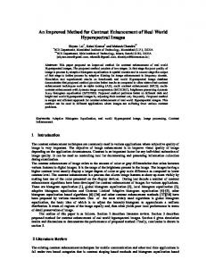
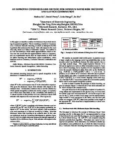
Fig 1 Comparison of amylase zymogram obtained in earlier method (A) and modified method (B); four Aspergillus niger isolates were taken as a reference.
Amylase isozymes were separated by SDS polyacrylamide gel electrophoresis in a discontinuous buffer system (Sambrook et al., 1989). The resolving gel contained 10% acrylamide (acrylamide/bisacrylamide 29:1) and 1.5 mg/ml soluble starch, whereas the stacking gel was made with 5% acrylamide lacking starch. Tris buffer was used at a concentration of 375 mM (pH 8.8) in the resolving gel and 126 mM (pH 6.8) in the stacking gel. The reservoir buffer (pH 8.3) contained 25 mM Tris and 192 mM glycine (Hames, 1990). Enzyme samples (15 µl aliquots) were loaded in the wells along with gel-loading dye. Gels were run at 80-100 V and 15-20 mA at 4 °C for 4 h. The gel was then washed for 10 min each with two washing buffers with a little agitation on a rotary shaker. The first washing buffer contained 2% Triton X-100 in 50 mM Tris buffer (pH 7.4) whereas the second buffer contained only 50 mM Tris-HCl (pH 7.4). The gel was then incubated for 3 h in modified substrate buffer (0.3 g CaCl2, 0.01 g NaCl, 0.3 g NaN3, 0.5 ml Triton X-100 in 50 ml of 50 mM Tris buffer pH 5.5) for substrate digestion. The gel was rinsed with distilled water before being stained for 5 min by incubation in 1 ml Lugol’s iodine stock solution (0.05 g I2, 0.1 g KI/ml) in 50 ml distilled water. The gel was finally rinsed with distilled water and photographed for documentation and analysis. Interpretation of bands was performed as described by Banke et al. (1997). The loci were interpreted as bands of distinct mobility classes or as bands with different appearance (intensity). Alleles were interpreted as bands with identical appearance and slightly different mobility
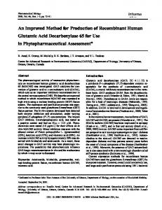
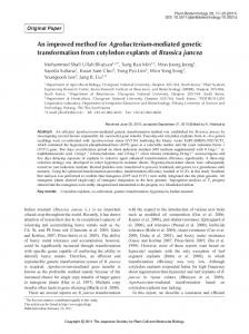
Results and Discussion Figure 1 shows that the bands visualized by our modified method (B) were very clear and similar to those obtained with the previous method (A). The substrate buffer used in the modified method contained Tris-HCl buffer with Ca2+, Na+, NaN3 and a small quantity of Triton X-100 instead of 0.1 M acetate buffer. Ca2+ ions activate and also stabilize amylases whereas Na+ at a concentration of 10 mM and NaN3 are known to enhance enzyme activity (Pierre & Tung, 1984; Rauscher, 1984). Moreover, Triton X-100 removes SDS from the gel left as a result of incomplete hydrolysis after washing with the first buffer. In contrast to other staining solutions (0.01 g I2 and 1.5g KI in 50 ml H2O used by Cruickshank & Pitt, 1987; 0.05M I2 used by Skovgaard & Rosendahl, 1998), the incubation time in our solution was reduced to only 5 min at room temperature in comparison to overnight staining period at 4 ºC in the previous methods. Moreover, the formation of brown spots on the gels due to undissolved iodine crystals was avoided. However, the greatest advantage of the modified method is that similar results can be obtained within the period of 8 h as compared to 18-20 h in the method of Cruickshank & Pitt (1987). When we tested our method by comparing morphologically similar strains isolated from different habitats, great variations in the presence or absence of specific bands were seen (Fig 2), and only one band with Rf 0.41 was common in most Aspergillus isolates, with two bands at Rf 0.27 and 0.87 shared by all isolates of Lepiota mimica (not shown). This indicates a 139
Mycologist, Volume 19, Part 4 November 2005
Rf value
10
119
12
101
102
107
105
109
110
111
112
118
114
0.06 0.21 0.29 0.41
0.41 0.51 0.59 0.67
Fig 2 Amylase zymogram of different strains of Aspergillus niger (A) and Aspergillus flavus (B)
diversity of loci or alleles encoding these enzyme forms in most isolates (Micales, 1986; Banke et al., 1997). The distribution of putative loci between isolates can be used to determine the genetic distance between them. Similar observations were recorded by Cruickshank (1990) and Upadhyay et al. (2003) in the zymogram pattern of Rhizoctonia and Agaricus bisporus, respectively. Zymographical techniques in the taxonomic differentiation of closely related fungal isolates were also employed by Elias & Schneider (1992) for the differentiation of strains of Fusarium oxysporum f. sp. lycopersici. The use of the amylase enzyme pattern as an aid in fungal taxonomy can be an important tool for the differentiation of morphologically similar but ecologically variable isolates and is easily demonstrated in the laboratory. Acknowledgement We thank Dr. R.W.S. Weber for his comments and editing of our manuscript, Prof. S. M. Singh (Department of Biological Science, Rani Durgavati University, Jabalpur) for providing laboratory facilities and the Department of Biotechnology (DBT), New Delhi, for providing financial assistance for the work.
References Banke, S., Frisvad, J. C. & Rosendal, S. (1997). Taxonomy of Penicillium chrysogenum and related xerophilic species based on isozyme analysis. Mycological Research 101: 617-624. Bosland, P. W. & Williams, P. H. (1987). An evaluation of Fusarium oxysporum from crucifers based on pathogenicity, isozyme polymorphism, vegetative compatibility and geographic origin. Canadian Journal of Botany 65: 2067-2073. Cruickshank, R. H. (1990). Pectin zymogram as criteria in taxonomy of Rhizoctonia. Mycological Research 94: 938-946. Cruickshank, R. H. & Pitt, S. I. (1987). Identification of species in Penicilium subgenus Penicillium by enzyme electrophoresis. Mycologia 79: 614-620.
140
Elias, S. K. & Schneider, R. W. (1992). Genetic diversity within and among race and vegetative compatibility group of Fusarium oxysporum f. sp. lycopersici as determined by isozyme analysis Phytopathology 82: 1421-1427. Grigg, R. & Lichtwardt, R. W. (1996). Isozyme patterns in cultured Harpellales. Mycologia 88: 219-229. Gupta, A. K. & Gautam, S. P. (1993). Production of extracellular amylases by thermophilic and thermotolerent fungi. Cryptogamic Botany 3: 303-306. Hames, B. D. (1990). One dimensional polyacrylamide gel electrophoresis In Gel Electrophoresis of Proteins: A Practical Approach II (ed. Hames, B.D. & Rickwood, D.), pp. 1-147. IRL Press: Oxford. Jones, K. G. & Blackwell, M. (1996). Ribosomal DNA sequence analysis places the yeast like genus Symbiotaphrina within filamentous ascomycetes. Mycologia 88: 212-218. Leuchtmann, A. & Clay, K. (1989). Isozyme variation in the fungus Aktinonella hypoxylon within and among population of its host grasses. Canadian Journal of Botany 67: 2600-2607. Micales, J. A. (1986). Use of isozyme analysis in fungal taxonomy and genetics. Mycotaxon 27: 405-449. Paterson, R. R. M., Bridge, P. D., Crosswhite, M. J. & Hawksworth, D. L. (1989). A reappraisal of the Penicillia using biochemical, physiological and morphological features, III. An evaluation of pectinase and amylase isozymes for species characterization. Journal of General Microbiology 135: 2979-2991. Pierre, K. J. & Tung, K. K. (1984). a-Amylase: UV-method with maltotetraose. In: Methods of Enzymatic Analysis, Vol. 4, third edition (Bergmeyer, H.U., ed.), pp. 146-151. WileyVCH: Weinheim. Rauscher, E. (1984). x-Amylase:colorimetric method. In: Methods of Enzymatic Analysis Vol. 4, third edition (ed. Bergmeyer, H.U.), pp. 157-161. Wiley-VCH: Weinheim. Rojanavanich, V., Yoshiike, T., Tasuboi, R., Takamori, K. & Ogawa, H. (1990). Purification and characterization of extracellular proteinase from Hendersonula toruloidea. Infection and Immunity 58: 2850-2861. Sambrook, J., Fritsch, E. F. & Maniatis, T. (1989). Molecular Cloning: A Laboratory Manual. Cold Spring Harbor Laboratory Press: New York. Skovgaard, K. & Rosendahl, S. (1998). Comparison of intraand extracellular isozyme banding pattern of Fusarium oxysporum. Mycological Research 102: 1077-1084. Upadhyay, Mukesh K., Pandey, A.K. & Rajak, R.C. (2003). Differentiation of Agaricus bisporus based on extracellular isozyme analysis. Journal of Basic and Applied Mycology 2: 6-9.