lymph node, ovary or, thymus, and miR-148 was detected in pancreas and ... Microarray platform ... analyzed for potential secondary structure and cross hybridization. .... Hartig, J.S., Grune, I., Najafi-Shoushtari, S.H., and Famulok, M. 2004.
METHOD
An optimized isolation and labeling platform for accurate microRNA expression profiling JACLYN SHINGARA,1 KERRI KEIGER,1 JEFFREY SHELTON, WALAIRAT LAOSINCHAI-WOLF, PATRICIA POWERS, RICHARD CONRAD, DAVID BROWN, and EMMANUEL LABOURIER Ambion, Inc., Austin, Texas 78744-1832, USA
ABSTRACT MicroRNAs (miRNAs) are small, noncoding RNAs that regulate gene expression in both plants and animals. miRNA genes have been implicated in a variety of important biological processes, including development, differentiation, apoptosis, fat metabolism, viral infection, and cancer. Similar to protein-coding messenger RNAs, miRNA expression varies between tissues and developmental states. To acquire a better understanding of global miRNA expression in tissues and cells, we have developed isolation, labeling, and array procedures to measure the relative abundance of all of the known human mature miRNAs. The method relies on rapid isolation of RNA species smaller than �40 nucleotides (nt), direct and homogenous enzymatic labeling of the mature miRNAs with amine modified ribonucleotides, and hybridization to antisense DNA oligonucleotide probes. A thorough performance study showed that this miRNA microarray system can detect subfemtomole amounts of individual miRNAs from 1) relative to the pool in at least one tissue are shown (78 out of 162 genesis (K. Keiger, J. Shelton, D. Brown, miRNAs, see Supplemental Table 1 at http://www.ambion.com/techlib/data/RNA05b.pdf for E. Labourier, in prep.). This approach, all miRNAs). Red illustrates higher expression in a given tissue, green corresponds to higher combined with genetic studies in C. expression in the pool, and gray represents microarray data that did not pass quality filters (see Materials and Methods). A magnification of the dendrogram is presented on the right. Brain elegans and functional studies in human cultured cells, recently helped identify and tissues with miRNA profiles clustering together are boxed. let-7 miRNA as a regulator of RAS oncogene expression and as a potential tumor example, we found that miR-125 is in fact more expressed in suppressor disrupted in lung tumors (Johnson et al. 2005). cervix, ovary, and uterus than in brain, miR-142 and -150 are Further, our microarray technology enables accurate miRNA not only expressed in spleen but are also very abundant in expression profiling in fixed tissues (P. Powers, R. Conrad, lymph node, ovary or, thymus, and miR-148 was detected in K. Keiger, J. Shelton, D. Brown, E. Labourier, in prep.), providpancreas and ovary in addition to liver and stomach. miRing access to a vast amount of information from archived clini181A was found highly expressed in brain, placenta, spleen, cal samples. If miRNAs are indeed involved in oncogenesis, and thymus, while miR-205 was identified in breast, prostate, inflammatory response, or viral infection, comparative miRNA and thymus. About 20 miRNAs, such as miR-126-AS, -143, expression studies in disease samples will certainly reveal novel -145, -189, or -321, were found highly expressed in one or therapeutic targets and diagnostic markers. www.rnajournal.org
Fig. 6 live 4/c
1467
Shingara et al.
according to the manufacturer’s instructions. For miRNA expression profiling in normal human tissues, miRNA certified FirstChoice Total RNA (Ambion) were used. To isolate miRNA fractions, total RNA samples were fractionated and cleaned up with the flashPAGE Fractionator and reagents (Ambion) per the manufacturer’s recommendation. Briefly, 1–20 mg of each RNA sample were loaded onto the top of a column filled with a denaturing acrylamide gel matrix and fractionated by applying an electrical current. A dye was loaded with the total RNA sample to track RNAs that are �40 nt in size. Electrophoresis was stopped when the dye reached the bottom of the column, and miRNAs were recovered from the bottom buffer chamber using a glass fiber filter-based cleaning procedure (flashPAGE Reaction Cleanup Kit, Ambion). Approximately 1 ng of miRNA was recovered per 10 mg of total RNA.
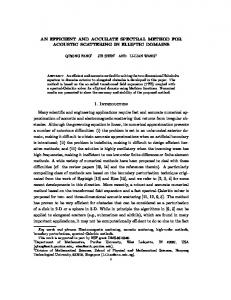
miRNA labeling and cleanup FIGURE 7. Validation of miRNA expression data. Total RNA was isolated from the 10 indicated tissues, independent from the ones used in the microarray profiling study (Fig. 6). Expression levels of let-7C and miR200B were analyzed by Northern blot (2 mg total RNA) or solution hybridization (1 mg total RNA). As a loading control, 5S and 5.8S ribosomal RNAs were also analyzed by Northern blot. The graphs under each autoradiograph show miRNA expression levels relative to the pool signal according to the microarray data (see Supplemental Table 1 at http://www.ambion.com/techlib/data/RNA05b.pdf). The value 1 corresponds to the average expression level of the corresponding miRNA in the pool sample, i.e., Log2(normalized ratio tissue/pool signal) = 0.
MATERIALS AND METHODS Microarray platform DNA oligonucleotide probes from the mirVana miRNA Probe Set (Ambion) were used to print custom microarrays. Each probe was designed using the sequence of its respective mature miRNA according to the miRNA Registry (Griffiths-Jones 2004) and was analyzed for potential secondary structure and cross hybridization. Negative control probes were designed using sequences from Escherichia coli, and each was functionally validated to have minimal cross-hybridization to a collection of chemically synthesized miRNAs as well as a series of tissue-derived miRNAs. Each probe carries a 15-nt linker at the 30 end of the miRNA complement sequence in addition to an amine group used to couple the probes to coated glass slides. Probes were resuspended in 33 SSC at 50 mM and spotted on Nexterion Slide E (Schott) at 55% humidity with the ArrayIt SpotBot (Telechem). Each feature was spotted using SMP4 Stealth pins with a spot diameter of �100 mm. The slides were rehydrated by incubation at room temperature in a humidity chamber for 30 min followed by a 30-min incubation at 60� C to dry. The slides were blocked in a solution containing 100 mM ethanolamine, 1 M Tris (pH 9.0), and 0.1% SDS for 15 min at 50� C, then thoroughly rinsed with water and spun dry.
RNA preparation Total RNA isolation and small RNA enrichment procedure were performed with the mirVana miRNA Isolation Kit (Ambion)
1468
RNA, Vol. 11, No. 9
Chemically synthesized oligoribonucleotides (Ambion), purified miRNAs, or fractions enriched in small RNAs were labeled with the mirVana miRNA Labeling Kit (Ambion) and amine-reactive dyes as recommended by the manufacturer. Poly(A) polymerase and a mixture of unmodified and amine-modified nucleotides were first used to append a poly-nucleotide tail to the 30 end of each miRNA. The amine-modified miRNAs were then cleaned up and coupled to NHS-ester modified Cy5 or Cy3 dyes (Amersham Bioscience). Unincorporated dyes were removed with a second glass fiber filter-based cleaning procedure.
Microarray hybridization and data analysis A 33 miRNA Hybridization Buffer (Ambion) was added to the fluorescently labeled miRNAs and the solution was heated at 95� C for 3 min. Slides were hybridized 12–16 h at 42� C in sealed cassettes using a water bath. Following hybridization, the slides were washed and dried prior to a high-resolution scan on a GenePix 4000B Array Scanner (Axon). Each element was located and analyzed using the GenePix Pro 5.0 software package (Axon). Data were filtered for quality and significance using the Longhorn Array Database (Killion et al. 2003). Filters were based on several data quality standards, including minimum intensity and pixel consistency. All data used for analysis had a signal-to-noise ratio >5, an average sum intensity 50% higher than that of the negative control spots, and a regression ratio >0.5. Data were normalized globally per array such that the average LogRatio was 0 after normalization. Hierarchical clustering was performed by Average Linkage using uncentered Pearson correlation (Eisen et al. 1998).
Microarray data validation miRNA expression levels in total RNA samples were measured by Northern blot (2–5 mg) or solution hybridization (1 mg) with the mirVana miRNA Detection Kit (Ambion) according to the manufacturer’s instructions. DNA or RNA probes were labeled and purified with the mirVana Probe & Marker Kit (Ambion) and [g-32P]ATP at 6000 Ci/mmol (PerkinElmer). For Northern analyses, RNA samples were resolved on denaturing 15% polyacrylamide gels and electroblotted on BrightStar-Plus Nylon membranes (Ambion).
microRNA expression profiling
Membranes were blocked in UltraHyb-oligo Hybridization Buffer (Ambion) for at least 1 h at 65� C, hybridized overnight at 42� C with the appropriate probe, and washed three times with NorthernMax Low Stringency Wash Buffer (Ambion) for 5 min at room temperature followed by 15 min at 42� C. Results were quantified on a PhosphorImager Storm 860 (Amersham Bioscience).
SUPPLEMENTARY MATERIAL Supplementary material can be found at http://www.ambion. com/techlib/data/RNA05b.pdf.
AKNOWLEDGMENTS We thank Eric Devroe for helpful comments on this article. This work was supported in part by National Genome Research Institute SBIR grant R43HG003430 to D.B. Received April 6, 2005; accepted June 3, 2005.
REFERENCES Allawi, H.T., Dahlberg, J.E., Olson, S., Lund, E., Olson, M., Ma, W.P., Takova, T., Neri, B.P., and Lyamichev, V.I. 2004. Quantitation of microRNAs using a modified Invader assay. RNA 10: 1153–1161. Ambros, V. 2004. The functions of animal microRNAs. Nature 431: 350–355. Babak, T., Zhang, W., Morris, Q., Blencowe, B.J., and Hughes, T.R. 2004. Probing microRNAs with microarrays: Tissue specificity and functional inference. RNA 10: 1813–1819. Barad, O., Meiri, E., Avniel, A., Aharonov, R., Barzilai, A., Bentwich, I., Einav, U., Gilad, S., Hurban, P., Karov, Y., et al. 2004. MicroRNA expression detected by oligonucleotide microarrays: System establishment and expression profiling in human tissues. Genome Res. 14: 2486–2494. Bartel, D.P. and Chen, C.Z. 2004. Micromanagers of gene expression: The potentially widespread influence of metazoan microRNAs. Nat. Rev. Genet. 5: 396–400. Baskerville, S. and Bartel, D.P. 2005. Microarray profiling of microRNAs reveals frequent coexpression with neighboring miRNAs and host genes. RNA 11: 241–247. Bohnsack, M.T., Czaplinski, K., and Gorlich, D. 2004. Exportin 5 is a RanGTP-dependent dsRNA-binding protein that mediates nuclear export of pre-miRNAs. RNA 10: 185–191. Cai, X., Hagedorn, C.H., and Cullen, B.R. 2004. Human microRNAs are processed from capped, polyadenylated transcripts that can also function as mRNAs. RNA 10: 1957–1966. Denli, A.M., Tops, B.B., Plasterk, R.H., Ketting, R.F., and Hannon, G.J. 2004. Processing of primary microRNAs by the Microprocessor complex. Nature 432: 231–235. Doench, J.G., Petersen, C.P., and Sharp, P.A. 2003. siRNAs can function as miRNAs. Genes & Dev. 17: 438–442. Eisen, M.B., Spellman, P.T., Brown, P.O., and Botstein, D. 1998. Cluster analysis and display of genome-wide expression patterns. Proc. Natl. Acad. Sci. 95: 14863–14868. Grad, Y., Aach, J., Hayes, G.D., Reinhart, B.J., Church, G.M., Ruvkun, G., and Kim, J. 2003. Computational and experimental identification of C. elegans microRNAs. Mol. Cell 11: 1253–1263. Gregory, R.I., Yan, K.P., Amuthan, G., Chendrimada, T., Doratotaj, B., Cooch, N., and Shiekhattar, R. 2004. The Microprocessor complex mediates the genesis of microRNAs. Nature 432: 235–240. Griffiths-Jones, S. 2004. The microRNA registry. Nucleic Acids Res. 32: D109–111.
Grishok, A., Pasquinelli, A.E., Conte, D., Li, N., Parrish, S., Ha, I., Baillie, D.L., Fire, A., Ruvkun, G., and Mello, C.C. 2001. Genes and mechanisms related to RNA interference regulate expression of the small temporal RNAs that control C. elegans developmental timing. Cell 106: 23–34. Hartig, J.S., Grune, I., Najafi-Shoushtari, S.H., and Famulok, M. 2004. Sequence-specific detection of MicroRNAs by signal-amplifying ribozymes. J. Am. Chem. Soc. 126: 722–723. Houbaviy, H.B., Murray, M.F., and Sharp, P.A. 2003. Embryonic stem cell-specific microRNAs. Dev. Cell 5: 351–358. Hutvagner, G., McLachlan, J., Pasquinelli, A.E., Balint, E., Tuschl, T., and Zamore, P.D. 2001. A cellular function for the RNA-interference enzyme Dicer in the maturation of the let-7 small temporal RNA. Science 293: 834–838. Johnson, S.M., Grosshans, H., Shingara, J., Byrom, M., Jarvis, R., Cheng, A., Labourier, E., Reinert, K.L., Brown, D., and Slack, F.J. 2005. RAS is regulated by the let-7 microRNA family. Cell 120: 635–647. Ketting, R.F., Fischer, S.E., Bernstein, E., Sijen, T., Hannon, G.J., and Plasterk, R.H. 2001. Dicer functions in RNA interference and in synthesis of small RNA involved in developmental timing in C. elegans. Genes & Dev. 15: 2654–2659. Killion, P.J., Sherlock, G., and Iyer, V.R. 2003. The Longhorn Array Database (LAD): An open-source, MIAME compliant implementation of the Stanford Microarray Database (SMD). BMC Bioinform. 4: 32. Krichevsky, A.M., King, K.S., Donahue, C.P., Khrapko, K., and Kosik, K.S. 2003. A microRNA array reveals extensive regulation of microRNAs during brain development. RNA 9: 1274–1281. Lagos-Quintana, M., Rauhut, R., Yalcin, A., Meyer, J., Lendeckel, W., and Tuschl, T. 2002. Identification of tissue-specific microRNAs from mouse. Curr. Biol. 12: 735–739. Lee, Y., Jeon, K., Lee, J.T., Kim, S., and Kim, V.N. 2002. MicroRNA maturation: Stepwise processing and subcellular localization. EMBO J. 21: 4663–4670. Lee, Y., Ahn, C., Han, J., Choi, H., Kim, J., Yim, J., Lee, J., Provost, P., Radmark, O., Kim, S., et al. 2003. The nuclear RNase III Drosha initiates microRNA processing. Nature 425: 415–419. Lee, Y., Kim, M., Han, J., Yeom, K.H., Lee, S., Baek, S.H., and Kim, V.N. 2004. MicroRNA genes are transcribed by RNA polymerase II. EMBO J. 23: 4051–4060. Lim, L.P., Lau, N.C., Weinstein, E.G., Abdelhakim, A., Yekta, S., Rhoades, M.W., Burge, C.B., and Bartel, D.P. 2003. The microRNAs of Caenorhabditis elegans. Genes & Dev. 17: 991–1008. Liu, C.G., Calin, G.A., Meloon, B., Gamliel, N., Sevignani, C., Ferracin, M., Dumitru, C.D., Shimizu, M., Zupo, S., Dono, M., et al. 2004. An oligonucleotide microchip for genome-wide microRNA profiling in human and mouse tissues. Proc. Natl. Acad. Sci. 101: 9740–9744. Llave, C., Xie, Z., Kasschau, K.D., and Carrington, J.C. 2002. Cleavage of Scarecrow-like mRNA targets directed by a class of Arabidopsis miRNA. Science 297: 2053–2056. Lund, E., Guttinger, S., Calado, A., Dahlberg, J.E., and Kutay, U. 2004. Nuclear export of microRNA precursors. Science 303: 95–98. Miska, E.A., Alvarez-Saavedra, E., Townsend, M., Yoshii, A., Sestan, N., Rakic, P., Constantine-Paton, M., and Horvitz, H.R. 2004. Microarray analysis of microRNA expression in the developing mammalian brain. Genome Biol. 5: R68. Murchison, E.P., and Hannon, G.J. 2004. miRNAs on the move: miRNA biogenesis and the RNAi machinery. Curr. Opin. Cell Biol. 16: 223–229. Nelson, P.T., Baldwin, D.A., Scearce, L.M., Oberholtzer, J.C., Tobias, J.W., and Mourelatos, Z. 2004. Microarray-based, high-throughput gene expression profiling of microRNAs. Nat. Methods 1: 155–161. Olsen, P.H. and Ambros, V. 1999. The lin-4 regulatory RNA controls developmental timing in Caenorhabditis elegans by blocking LIN14 protein synthesis after the initiation of translation. Dev. Biol. 216: 671–680. Overhoff, M., Wunsche, W., and Sczakiel, G. 2004. Quantitative detection of siRNA and single-stranded oligonucleotides: Relationship between uptake and biological activity of siRNA. Nucleic Acids Res. 32: e170.
www.rnajournal.org
1469
Shingara et al.
Pfeffer, S., Zavolan, M., Grasser, F.A., Chien, M., Russo, J.J., Ju, J., John, B., Enright, A.J., Marks, D., Sander, C., et al. 2004. Identification of virus-encoded microRNAs. Science 304: 734– 736. Rodriguez, A., Griffiths-Jones, S., Ashurst, J.L., and Bradley, A. 2004. Identification of mammalian microRNA host genes and transcription units. Genome Res. 14: 1902–1910. Schmittgen, T.D., Jiang, J., Liu, Q., and Yang, L. 2004. A highthroughput method to monitor the expression of microRNA precursors. Nucleic Acids Res. 32: e43. Schwarz, D.S., Hutvagner, G., Du, T., Xu, Z., Aronin, N., and Zamore, P.D. 2003. Asymmetry in the assembly of the RNAi enzyme complex. Cell 115: 199–208. Sempere, L.F., Freemantle, S., Pitha-Rowe, I., Moss, E., Dmitrovsky, E., and Ambros, V. 2004. Expression profiling of mammalian microRNAs uncovers a subset of brain-expressed microRNAs with possible roles in murine and human neuronal differentiation. Genome Biol. 5: R13. Sioud, M. and Rosok, O. 2004. Profiling microRNA expression using sensitive cDNA probes and filter arrays. Biotechniques 37: 574–576, 578–580. Sun, Y., Koo, S., White, N., Peralta, E., Esau, C., Dean, N.M., and Perera, R.J. 2004. Development of a micro-array to detect human and mouse microRNAs and characterization of expression in human organs. Nucleic Acids Res. 32: e188.
1470
RNA, Vol. 11, No. 9
Thomson, J.M., Parker, J., Perou, C.M., and Hammond, S.M. 2004. A custom microarray platform for analysis of microRNA gene expression. Nat. Methods 1: 47–53. Valoczi, A., Hornyik, C., Varga, N., Burgyan, J., Kauppinen, S., and Havelda, Z. 2004. Sensitive and specific detection of microRNAs by Northern blot analysis using LNA-modified oligonucleotide probes. Nucleic Acids Res. 32: e175. Yekta, S., Shih, I.H., and Bartel, D.P. 2004. MicroRNA-directed cleavage of HOXB8 mRNA. Science 304: 594–596. Yi, R., Qin, Y., Macara, I.G., and Cullen, B.R. 2003. Exportin-5 mediates the nuclear export of pre-microRNAs and short hairpin RNAs. Genes & Dev. 17: 3011–3016. Zeng, Y. and Cullen, B.R. 2003. Sequence requirements for micro RNA processing and function in human cells. RNA 9: 112–123. ———. 2004. Structural requirements for pre-microRNA binding and nuclear export by Exportin 5. Nucleic Acids Res. 32: 4776–4785. Zeng, Y., Wagner, E.J., and Cullen, B.R. 2002. Both natural and designed micro RNAs can inhibit the expression of cognate mRNAs when expressed in human cells. Mol. Cell 9: 1327–1333. Zeng, Y., Yi, R., and Cullen, B.R. 2005. Recognition and cleavage of primary microRNA precursors by the nuclear processing enzyme Drosha. EMBO J. 24: 138–148. Zhang, H., Kolb, F.A., Jaskiewicz, L., Westhof, E., and Filipowicz, W. 2004. Single processing center models for human Dicer and bacterial RNase III. Cell 118: 57–68.