J. of Biomed. & Clin. Sci. Dec 2018 Vol 3 (2), 10-17
F.A. Dzulkifli1, M.Y. Mashor1, H. Jaafar2
Original Article
An overview of recent counting methods for Ki67 IHC staining
1School
of Mechatronics Engineering, Universiti Malaysia Perlis (UniMAP), 02600, Arau, Perlis, Malaysia 2Department
of Pathology, School of Medical Sciences, Health Campus Universiti Sains Malaysia, 16150, Kubang Kerian, Kelantan, Malaysia Received 22 Sept 2018 Revised 15 Nov 2018. Accepted 30 Nov 2018. Published Online 5 Jan 2019
Abstract— Ki67, a nuclear protein presents in cells that is usually associated with tumor cell proliferation. Therefore, physical quantification of Ki67 is a direct measure cell proliferation activity. Immunohistochemical staining is known to be an effective method of assessing the proportion of tumor cells. Currently, there are various techniques for counting Ki67 in different types of cancer. This paper provides an overview of recent scoring methods of Ki67, which includes ‘eyeballing’ estimation, vision counting with a microscope or viewer software, manual counting of the camera-captured or digital image and automated counting.
Keywords: estimation.
Immunohistochemical
stain,
Ki67,
Proliferating
rate
*Corresponding author Fahmi Akmal Dzulkifli Email:
[email protected]
1 INTRODUCTION Cell proliferation refers to an increment in terms of cell numbers from the process of cell growth and cell division. Van Diest et al. [1] stated that cell proliferation is a biological process that is essential to all living organisms due to its role in the growth and maintenance of tissue homeostasis. Cell proliferation is a good indicator of cellular function and becomes one of the most important diagnostic and prognostic marker in cancer. It may reveal useful information about a patient prognosis [2]. A nuclear protein, known as Ki67, is generally associated with tumor cell proliferation Email: corresponingauthoraddress and growth. Due to this fact, Ki67 becomes an @email.com excellent marker because a higher proliferation rate is one of the characteristics of cancer cells [3]. In the early 1980s, Scholzen and Gerdes [4] incidentally identified Ki67 in an attempt to create a cancer-specific monoclonal antibodies. Previous data from in vitro experiments using Hodgkin’s lymphoma cells indicated that the Ki67 is an appropriate marker for cell proliferation [2][5]. The name of Ki67 was obtained from the city of origin (Kiel, Germany) and the number 67 was the number from which the hybridoma was first identified [2][4]. The Ki67 gene, was located at 10q25 position on the chromosome and has 15 exons, encodes two protein isoforms with a mass of 320 and 359kDa [6][7]. The Ki67 was localized mainly http://apps.amdi.usm.my/journal/
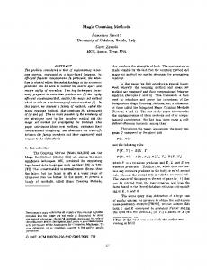
in nucleolus during the interphase stage of the cell cycle, where the cell spends most of the time in this phase before it leads to cell division process [3]. Other studies [8]&[9] had reported that Ki67 was also found on heterochromatic deoxyribonucleic acid (DNA) regions, in the early G1 phase of the cell cycle. Inside the nucleolus, Ki67 was generally located in the dense fibrillar component (DFC), which is one of the three major components in nucleolus that is free from the ribonucleic acid (RNA) polymerase I transcription apparatus [3][9]. Nevertheless, there was a small population of Ki67 which co-localized with components of RNA polymerase I transcription apparatus and newly synthesized ribosomal RNA in vivo, which contradicts with the Ki67 role in the early steps of rRNA synthesis [3][10]. Ki67 protein is presents only in growing and dividing phases of the cell cycle (GI, S, G2, and M) but absent during the resting phase (G0) [11]. Hence, Ki67 is a good proliferation marker for cancer cells since they aggressively grow and divide. Specifically, the expression of Ki67 gene began at the G1 phase and this expression increase during the S phase and reaches the highest expression during metaphase, or the M phase [12]. During the stage of anaphase and telophase, the Ki67 expression will start to decrease. Figure 1 shows the level of expression of Ki67 during the different phases of the cell cycle.
[10]
J. of Biomed. & Clin. Sci. Dec 2018 Vol 3 (2), 10-17
Original Article
Figure 1: Levels of Ki67 expression during the different phases of the cell cycle
During the mitosis stage, the Ki67 protein covered all the chromosomes, localizing it in the nuclear foci during G1 phase, which correlates with regions of satellite DNA [8]. In mitosis stage, prominent cellular distribution takes place [13]. During prophase, Ki67 formed a bright staining meshwork all over the nucleoplasm, which is associated with the chromatin [13]. At the metaphase stage, a reticulum of interconnected fluorescent fibrils was discovered, which was then transformed into a more granular pattern during anaphase and telophase [13]. In the late telophase stage, the Ki67 disengaged from the perichromosal layer and showed a speckled distribution pattern [13]. Ki67 immunohistochemical (IHC) staining or the immunoperoxidase stains method is an effective method of assessing the prognosis in a number of tumor type. Immunohistochemistry is an important application in monoclonal as well as polyclonal antibodies analyses to detect specific antigens in tissue sections [14]. IHC stains are widely used to determine the stage and grade of tumor and identifying cell type and origin of a metastasis to find the site of the primary tumor [14]. This method is also used by the pathologists as an aid in the differential diagnosis and classification of cancer and for certain diseases, including infection [15].
http://apps.amdi.usm.my/journal/
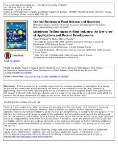
The result of the staining enables the pathologist to count the cancer cells and decides the grade of tumors. Figure 2 shows a sample of IHC stained Ki67 image from a meningioma. As shown in the figure, the positive cell nucleus was stained by diaminobenzidine (DAB) and the cell appeared in granular brown color, while the negative cell was stained by hematoxylin and appeared in blue color [16].
Figure 2: A Ki67 image of meningioma using IHC stains
Dowsett et al. [17] had stated there are a few aspects that could affect the results of IHC staining. The first condition that needs consideration is the method used by the observers to read the Ki67 slide. In this part, there are two variables that need to be examined, which are the
[11]
J. of Biomed. & Clin. Sci. Dec 2018 Vol 3 (2), 10-17
cellular component and staining intensity. Currently, there is no consensus on the staining intensity mentioned to define the positive Ki67 cells. The scoring method is carried out by counting all the nuclei that have been stained, which supposedly represents the positive Ki67 cells. Next is the determination of the area of the slide for the scoring process. Based on previous studies, several techniques can be applied to access the Ki67 LI. Jang et al. [18] analyzed the comparison between the average method with 'hot-spot' method in assessing Ki67 LI in luminal breast cancer. This study showed both methods have good predictive performances for tumor recurrence in luminal/HER2-negative breast cancer. However, in this study, the authors preferred the average method because of its greater reproducibility. In a study by Leung et al. [19] three scoring methods were evaluated to perform a standardized scoring protocol for Ki67 in breast cancer. These methods consist of a global method (4 fields of 100 cells), weighted global method (similar as global but weighted by estimated percentages of total area), and assessing the Ki67 in 'hot-spots' area (single field of 500 cells). Due to the heterogeneity of the study findings, the authors could not conclude the best method for standardization of Ki67 IHC assay for use in breast cancer. 2.0 Ki67 COUNTING TECHNIQUES Previous studies have shown that the Ki67 becomes a reliable marker in guiding the diagnosing of different types of cancer [20]–[23]. High proliferative activity is a characteristic of the aggressive tumor that is also characterized by high tissue heterogeneity [23][24]. Proliferating rate estimation (PRE) is one of the prognostic indicators that help pathologists to determine the type of treatment for a suspected cancer patient. PRE is the percentage of Ki67 positive cells among the cell population. Normally, pathologists will find the ‘hotspot area’, which is the area where there is increased tumor proliferation. The ‘hotspot areas’ is refers to the selection area of malignant cells with a high proliferative activity be potentially associated with a more aggressive biological behavior that represented in the whole of the tumor [26]. Many types of research had been carried out in order to improve the quality and the accuracy for
http://apps.amdi.usm.my/journal/
Original Article
Ki67 counting and scoring methods. These methods are further discussed below. 2.1 ‘Eye-Balling’ Estimation The ‘eye-balling’ estimation method was done by looking at the slide under a microscope, which is usually at low power magnification of 10x. The percentage of positive tumor cells will be estimated and this estimation does not involve the counting of individual cells. This method was widely used by the pathologists and it was reported as a timesaving measure [26][27]. A.Vörös [29] a doctoral candidate from the department of pathology, University of Szeged had applied the ‘eye-balling’ method to estimate Ki67 labeling index in breast cancer. In this study, three pathologists were involved to count the Ki67 cells. The immunostained slide images were captured under magnification of 200X. Parallel grid lines were drawn on the digital images to define the stained and unstained cells for pathologists consideration during counting. The pathologists made the Ki67 estimation based on the approximation from a hot spot area containing 100 cells. Based on kappa statistics, the kappa values relating to one of the pathologists were lower than other observers. This was due to different methods that were used by the pathologists when evaluating the Ki67 labeling index (LI). Zhong et al. [30] had applied ‘eye-balling’ method to compare with the automated digital image analysis (DIA) for Ki67 labeling index on breast cancer. ‘Eye-balling’ method was assessed by five pathologists. The value was obtained by finding the mean value of five pathologists for each case. The score for each case was decided based on a percentage of positive tumor cells among all cells in the tumor area with 10% intervals (from 10% until 100%). Each pathologist used two different technique to determine the scoring fields, which are the hot-spot scoring method and using an average score method. The automated DIA system was used by scanning all the stained slides into digital slides by using VENTANA iScan HT System version 1.0. Then, the slides were analyzed using VENTANA Virtuoso Digital Pathology Image Analysis Software. Based on the result, both methods showed a good agreement in determining the Ki67 LI. Fulawka and Halon [26] had implemented the ‘eye-balling’ estimation technique to assess the daily approach to define the PRE of Ki67 cells in breast cancer. Four pathologists were recruited
[12]
J. of Biomed. & Clin. Sci. Dec 2018 Vol 3 (2), 10-17
for this study to execute the estimation technique. Three pathologists used the ‘eye-balling’ method while another pathologist counted the cells individually. The results showed that there was a significant difference in scoring the tumor cells among the pathologists. This was due to several issues such as different selection areas of counting, different magnification used among the pathologists and different perception in determining the tumor cells due to different Ki67 staining intensity between the nuclei in the tumor. 2.2 Visual Counting Using a Microscope or Viewer Software This method was well known as ‘real-time’ counting and the counting process was done under a microscope at an intermediate magnification power of 200X. The counting process focused on identifying the ‘hot-spot’ area. This method involved the use of the grid and other counting tools that are normally used by the pathologists. Desmeules et al. [31] had investigated the comparison between a visual scoring method with DIA method on digital images to calculate Ki67 in prostate cancer prognosis after prostatectomy. Two observers did the visual scoring. The digital images were based on IHC stained slides and that was obtained at 200X magnification using a slide scanner. These images were then visualized using viewer software (ndpi.viewer) for visual scoring and digital image analysis. DIA method was operated using Calopix software, where this software is able to recognize each nuclei by isolating brown (DAB stained) and blue (hematoxylin stained). As a result, this study showed that the DIA method was comparable to visual scoring for Ki67 quantification for prostate cancer. 2.3 Manual Counting of Camera-Captured or Digital Images This method manually counts the cells by looking either at a printout of cell images or a screen capture of a section previously visualized from the microscope. Usually, it was done under magnification of 100X. The observers will mark manually the positive tumor cells on the printed image. Kroneman et al. [32] had implemented three different Ki67 quantification methods to select the best Ki67 quantification method for pancreatic neuroendocrine tumors. The methods consist of
http://apps.amdi.usm.my/journal/
Original Article
manual counting, ‘eye-balling’ estimation, and DIA technique. Images of 8 to 10 hot spot areas were used in the manual counting and DIA method. Manual counting method was done by the cytotechnologist that counted at least 500 tumor cells of the brown and blue staining. In the DIA method, cytotechnologist used the Automated Cellular Imaging System (ACIS) for image analysis. Three pathologists were involved in this study to apply the ‘eye-balling’ estimation for Ki67 quantification. The pathologists will provide a percentage of positive tumor cells that were present in the cell population. The results were then averaged. Concordance correlation coefficient had been used to measure the agreement between the pathologists and the three different methods of Ki67 labeling index. ‘Eyeballing’ estimation showed a good agreement among three pathologists where the agreement was 0.86 (95%, CI 0.81-0.90). The agreement between manual counting with DIA technique was 0.97 (95% CI 0.96-0.98). The concordance correlation coefficient between eyeball estimation with manual counting delivered the results of 0.88 (95% CI 0.84-0.92). Mu et al. [33] had designed a standardized quantification method for Ki67 and cyclin A immunohistochemistry in breast cancer. Each image was captured using a digital camera (with 400X magnification) in at least 5 to 14 random tumor areas of 613 breast carcinomas. Two types of counting had been selected for this study, which were the average counting and hot-spot counting methods. In average counting, the percentage of positive tumor cells was calculated from at least 1000 tumor cells in the selected areas, whereas in hot-spot counting, the positive tumor cells were counted between 200 to 400 cells in the hot-spot area. The hot-spot area was determined by the area of 25mm2 around the point of the highest concentration of cells immunoreactive for Ki67 or cyclin A. According to the results, the hot-spot counting has been selected to be a prognostic marker for a clinical decision due to the factor of reliability and time efficiency. Cottenden et al. [28] had compared between cytotechnologist manual counting and pathologists manual count estimation. This study applied the manual counting method for counting Ki67 in neuroendocrine tumors of the pancreas and gastrointestinal tract. Four pathologists and three cytotechnologists have been selected to perform the estimation of the Ki67 index on 20
[13]
J. of Biomed. & Clin. Sci. Dec 2018 Vol 3 (2), 10-17
printed images. Each digital image was estimated consists of at least 1000 cells for assessment. The images were printed in color at a size of 8 inches in height by 10 inches in width with 96 dots per inch resolution [28]. The result showed that for manual counting method, the time taken for the pathologists to count for each slide was 17.0 minutes, faster than the time taken by cytotechnologist at 18.8 minutes. Pathologists eyeball estimation using glass slides took an average of 57.0 seconds per case, while for digital image eyeball estimation took an average of 42.0 seconds per case. This study also reveals a discrepancy in terms of the agreement in WHO tumor grade between the eyeball estimates and manual count. 2.4 Automated Counting System This method was divided into two parts, either using an automated counting device or image analysis software. The hot-spot area was selected manually before automated scoring Ki67 can be executed. Markiewicz et al. [34] had introduced a computerized system for cell counting on Ki67 IHC staining of selected types of primary brain tumors such as meningioma and oligodendroglioma. The algorithm of the proposed system was based on mathematical morphology that was developed to execute a quantitative evaluation of slides. The algorithm together with Support Vector Machine (SVM) was used to classify the cell immunoreactivity. The analysis showed that mean relative discrepancy between the proposed method and the human expert score was 8%. Therefore, this system could be utilized to support a histopathologic diagnosis. Al-Lahham et al. [35] had designed a fully automated computer-aided system for proliferation rate estimation (PRE) from Ki67 histopathology images. The proposed method used Ki67 images from breast cancer for nuclei counting and evaluate the proliferation rate. This method consists of three steps, namely image preprocessing, image clustering and followed by nuclei segmentation and counting. Color modification and color transformation have been implemented in the image processing section. The image was converted from RGB to L*a*b color spaces. The image was then clustered into 3 regions, which were brown nuclei, blue nuclei, and remaining tissue. The counting algorithm consisted of three steps, which were applying http://apps.amdi.usm.my/journal/
Original Article
global thresholding, implementing the morphological operations and counting the connected objects. Based on the result, the average percentage difference of brown nuclei, blue nuclei and proliferation rate between manual and automated processes were 13%, 26%, and 25%, respectively. Mohammed et al. [36] had compared between the visual and automated assessment of Ki67 proliferative activity for breast cancer histological images. Slidepath Image Analysis System was used to obtain digitized slides. These slides were then evaluated using the nuclear scoring algorithm, which automatically determines the Ki67 Labelling Index (LI). Ki67 IHC method was used to stain the slides. Thresholding and segmentation algorithms were implemented in this system for nuclei stained identification. The nuclear Ki67 staining was classified as positive or negative based on observer-specified intensity thresholds. It showed that the Intraclass Correlation Coefficient (ICC) and Spearman’s Rank Correlation (ρ) between the visual and automated system were (ICC = 0.96) and (P˂ 0.001). Hence, it showed that the correlation between the visual Ki67 LI and automated assessment were comparable. Nielsen et al. [37] had investigated the diagnostic performance of automated dermal Ki67 indices and the ability of the system to predict sentinel lymph node (SLN) status. The Ki67 indices were executed into three different areas which were epidermis, dermis and a combination of epidermis and dermis and dermal hotspot. SD filter and polynomial Laplace filter were assigned to enhance the structural differences of epidermis and dermis. The images were segmented using Kmeans clustering and a polynomial blobs filter were implemented to enhance the circular shape of nucleus structures. The Bayesian classifier was implemented to identify the image classes that associated with the brown Ki67, blue hematoxylin, and red MART1. Thereafter, the system will differentiate between the positive Ki67 and negative Ki67 cells based on size, MART1 surrounding, and nuclear irregularity. According to the Wilcoxon-Mann-Whitney test, there was a significant difference in the Ki67 index between the SLN-positive and SLN negative patients. However, it was reported that the system still could be useful for predicting the SLN status of melanoma patients.
[14]
J. of Biomed. & Clin. Sci. Dec 2018 Vol 3 (2), 10-17
Fuyong Xing et al. [38] had proposed an automatic Ki67 counting using robust cell detection and learning based framework for neuroendocrine tumor (NET). The proposed method was divided into three stages. The first part was detecting the geometric centers (seeds) of the cells for tumor and non-tumor cells localization. The second part was the learningbased classification that consists of three methods. At the first stage, there were 112 features were extracted for each sample that will be used for classification. The second stage focused more on the challenging cases for tumor and non-tumor detection. For the third stage, the immunopositive and immunonegative tumor cells were separated. The system analysis is further tested using four methods, which were the Laplacian-of-Gaussian (LoG) filter, Iterative Radial Voting (IRV), Single-pass voting and Image-Based Tool for Counting Nuclei (ITCN). Based on the results, it showed that the proposed method was able to perform Ki67 counting and the accuracy of detecting tumor and non-tumor cells were reasonably high, compare to other methods. Shi et al. [16] had formulated an automated
Original Article
Ki67 quantification based on IHC staining image of human nasopharyngeal carcinoma. K-means clustering was utilized to group local correlation features. In image pre-processing, Gaussian Filter was used for smoothing the image and Median Filter was applied for enhancement. Scatter noises were also eliminated and the local color intensity of pigment aggregate was enhanced. There were 3 features that had been selected from the images, which were blue color intensity in RGB color space (B-channel), and hue (H) intensity from huesaturation-value color space (HSV), mean (µ) and standard deviation (σ) value from B and H color channel and the local texture features including kurtosis and skewness. These features were selected to determine between pixel samples in the feature space. K means clustering had been chosen for nucleus segmentation. In this method, the watershed algorithm had been applied to solve issues of touching cells. The method conferred an accuracy of 91.8% for the segmentation process. This method was promising since the error rate of nucleus segmentation showed that the False Acceptance Rate (FAR) was 5.5% and the False Rejection Rate (FRR) was 2.7%.
Table I: Comparison of Ki67 Counting Techniques
3.0 CONCLUSION The expression of Ki67 shows a strong association with tumor cell proliferation growth and is widely used among pathologists as a proliferation marker to measure proliferation of tumor cells. In view of the specific characteristics that Ki67 exhibits in different phases of the cell cycle (G1, S, G2 and http://apps.amdi.usm.my/journal/
M), hence Ki67 proved to be a good immunohistochemical marker of proliferating cells. Table I shows the comparison between the different techniques of Ki67 quantification. The 'eye-balling' technique is generally known and is fast and simple. Regrettably, this method has a poor accuracy in scoring the Ki67 cells, which
[15]
J. of Biomed. & Clin. Sci. Dec 2018 Vol 3 (2), 10-17
Original Article
makes the method unacceptable among pathologists. Manual counting on printed image is considered to be the most reliable since this method is feasible and allows the observers to avoid duplicate scoring. However, the type of scoring through individual counting of the Ki67 cells makes this method becomes time-consuming and tedious. The high cost of automated counting devices was an issue of the past. However, free image analysis software for counting cells like ImmunoRatio, Cell Profiler, and ImageJ are currently available online. The main issue that occurs during the automated counting process is the miscounting of non-target cells. Besides that, the image analysis software also has pitfalls in interpreting different staining intensities, which could lead to misdetection of unwanted objects like stained artefacts. However, these problems can be solved by providing an additional step to enhance the color and contrast of the images before performing the counting process. Hence, this step could help the pathologists to solve the issue of low contrast images. In the nutshell, we recommend the use of automated counting method as this technique demonstrates improved cell detection accuracy as compared to the conventional method. Besides that, this technique also helps in reducing the time taken of scoring the Ki67 cell compared as compared to the manual counting.
[8]
REFERENCES
[17]
[1]
[2]
[3]
[4]
[5]
[6] [7]
P. J. Van Diest, G. Brugal, and J. P. A. Baak, “Proliferation markers in tumours: Interpretation and clinical value,” Journal of Clinical Pathology, vol. 51, no. 10. pp. 716–724, 1998. M. Juríková, Ľ. Danihel, Š. Polák, and I. Varga, “Ki67, PCNA, and MCM proteins: Markers of proliferation in the diagnosis of breast cancer,” Acta Histochemica, vol. 118, no. 5. pp. 544–552, 2016. M. Takagi, Y. Nishiyama, A. Taguchi, and N. Imamoto, “Ki67 antigen contributes to the timely accumulation of protein phosphatase 1γ on anaphase chromosomes,” J. Biol. Chem., vol. 289, no. 33, pp. 22877–22887, 2014. T. Scholzen and J. Gerdes, “The Ki-67 protein: From the known and the unknown,” Journal of Cellular Physiology, vol. 182, no. 3. pp. 311–322, 2000. J. Gerdes, U. Schwab, H. Lemke, and H. Stein, “Production of a mouse monoclonal antibody reactive with a human nuclear antigen associated with cell proliferation.,” Int. J. cancer, vol. 31, no. 1, pp. 13–20, 1983. D. Coppola, Molecular pathology and diagnostics of cancer, 1st ed. Springer Netherlands, 2014. E. Endl and J. Gerdes, “The Ki-67 protein: Fascinating forms and an unknown function,” Exp. Cell Res., vol. 257, no. 2, pp. 231–237, 2000.
http://apps.amdi.usm.my/journal/
[9]
[10]
[11]
[12]
[13]
[14]
[15]
[16]
[18]
[19]
[20]
[21]
[22]
[23]
J. M. Bridger, I. R. Kill, and P. Lichter, “Association of pKi-67 with satellite DNA of the human genome in early G 1 cells,” Chromosom. Res., vol. 6, no. April 1997, pp. 13–24, 1998. I. R. Kill, “Localisation of the Ki-67 antigen within the nucleolus. Evidence for a fibrillarin-deficient region of the dense fibrillar component.,” J. Cell Sci., vol. 109 ( Pt 6, pp. 1253–1263, 1996. J. Bullwinkel, B. Baron-Lühr, A. Lüdemann, C. Wohlenberg, J. Gerdes, and T. Scholzen, “Ki-67 protein is associated with ribosomal RNA transcription in quiescent and proliferating cells,” J. Cell. Physiol., vol. 206, no. 3, pp. 624–635, 2006. L. T. Li, G. Jiang, Q. Chen, and J. N. Zheng, “Ki67 is a promising molecular target in the diagnosis of cancer (Review),” Mol. Med. Rep., vol. 11, no. 3, pp. 1566–1572, 2015. M. Starborg, K. Gell, E. Brundell, and C. Höög, “The murine Ki-67 cell proliferation antigen accumulates in the nucleolar and heterochromatic regions of interphase cells and at the periphery of the mitotic chromosomes in a process essential for cell cycle progression.,” J. Cell Sci., vol. 109 ( Pt 1, pp. 143–53, 1996. R. Verheijen et al., “Ki-67 detects a nuclear matrixassociated proliferation-related antigen. II. Localization in mitotic cells and association with chromosomes.,” J. Cell Sci., vol. 92 ( Pt 4), pp. 531– 540, 1989. K. Kaliyappan, M. Palanisamy, J. Duraiyan, and R. Govindarajan, “Applications of immunohistochemistry,” J. Pharm. Bioallied Sci., vol. 4, no. 6, p. 307, 2012. K. Petersen and H. C. Pedersen, “Detection Methods,” in Education Guide-Immunohistochemical Staining Methods, 6th ed., L. R. Clive R. Taylor, Ed. Dako Denmark A/S, 2009, p. 79. P. Shi, J. Zhong, J. Hong, R. Huang, K. Wang, and Y. Chen, “Automated Ki-67 Quantification of Immunohistochemical Staining Image of Human Nasopharyngeal Carcinoma Xenografts,” Sci. Rep., vol. 6, no. August, pp. 1–9, 2016. M. Dowsett et al., “Assessment of Ki67 in Breast Cancer: Recommendations from the international Ki67 in breast cancer working Group,” J. Natl. Cancer Inst., vol. 103, no. 22, pp. 1656–1664, 2011. M. H. Jang, H. J. Kim, Y. R. Chung, Y. Lee, and S. Y. Park, “A comparison of Ki-67 counting methods in luminal Breast Cancer: The Average Method vs. the Hot Spot Method,” PLoS One, vol. 12, no. 2, pp. 1–15, 2017. S. C. Y. Leung et al., “Analytical validation of a standardized scoring protocol for Ki67: phase 3 of an international multicenter collaboration,” npj Breast Cancer, vol. 2, no. 1, p. 16014, 2016. S. W. Sorbye et al., “Prognostic impact of lymphocytes in soft tissue Sarcomas,” PLoS One, vol. 6, no. 1, 2011. N. Ciancio, M. G. Galasso, R. Campisi, L. Bivona, M. Migliore, and G. U. Di Maria, “Prognostic value of p53 and Ki67 expression in fiberoptic bronchial biopsies of patients with non small cell lung cancer,” Multidiscip. Respir. Med., vol. 7, no. 4, 2012. A. Josefsson et al., “Low endoglin vascular density and Ki67 index in Gleason score 6 tumours may identify prostate cancer patients suitable for surveillance,” Scand. J. Urol. Nephrol., vol. 46, no. 4, pp. 247–257, 2012. B. Grala, T. Markiewicz, W. Kozłowski, S. Osowski, J.
[16]
J. of Biomed. & Clin. Sci. Dec 2018 Vol 3 (2), 10-17
[24]
[25]
[26]
[27]
[28]
[29]
[30]
[31]
[32]
[33]
[34]
[35]
[36]
[37]
Słodkowska, and W. Papierz, “New automated image analysis method for the assessment of Ki-67 labeling index in meningiomas.,” Folia Histochem. Cytobiol., vol. 47, no. 4, pp. 587–592, 2009. X. M. Lopez et al., “Clustering methods applied in the detection of Ki67 hot-spots in whole tumor slide images: An efficient way to characterize heterogeneous tissue-based biomarkers,” Cytom. Part A, vol. 81 A, no. 9, pp. 765–775, 2012. A. Persson and E. Englund, “Different assessments of immunohistochemically stained Ki-67 and hTERT in glioblastoma multiforme yield variable results: a study with reference to survival prognosis.,” Clin. Neuropathol., vol. 27, no. 4, pp. 224–33, 2008. L. Fulawka and A. Halon, “Ki-67 evaluation in breast cancer: The daily diagnostic practice,” Indian J. Pathol. Microbiol., vol. 60, no. 2, p. 177, 2017. M. D. Reid et al., “Calculation of the Ki67 index in pancreatic neuroendocrine tumors: A comparative analysis of four counting methodologies,” Mod. Pathol., vol. 28, no. 5, pp. 686–694, 2015. J. Cottenden et al., “Validation of a cytotechnologist manual counting service for the Ki67 index in neuroendocrine tumors of the pancreas and gastrointestinal tract,” Arch. Pathol. Lab. Med., vol. 142, no. 3, pp. 402–407, 2018. A. Vörös, “Various Aspects of Ki-67 Immunohistochemistry Determined Proliferation in Breast Cancer,” University of Szeged, 2014. F. Zhong, R. Bi, B. Yu, F. Yang, W. Yang, and R. Shui, “A comparison of visual assessment and automated digital image analysis of Ki67 labeling index in breast cancer,” vol. 11, no. (Zhong, Bi, Yu, Yang, Yang, Shui) Department of Pathology, Fudan University Shanghai Cancer Center, Shanghai, China, p. no pagination, 2016. P. Desmeules et al., “Comparison of digital image analysis and visual scoring of KI-67 in prostate cancer prognosis after prostatectomy,” Diagn. Pathol., vol. 10, no. 1, pp. 1–10, 2015. T. N. Kroneman, J. S. Voss, C. M. Lohse, T.-T. Wu, T. C. Smyrk, and L. Zhang, “Comparison of Three Ki-67 Index Quantification Methods and Clinical Significance in Pancreatic Neuroendocrine Tumors,” Endocr. Pathol., vol. 26, no. 3, pp. 255–262, 2015. K. Mu et al., “A standardized method for quantifying proliferation by Ki-67 and cyclin A immunohistochemistry in breast cancer,” Ann. Diagn. Pathol., vol. 19, no. 4, pp. 243–248, 2015. T. Markiewicz, B. Grala, W. Kozlowski, and S. Osowski, “Computer system for cell counting in selected brain tumors at Ki-67 immunohistochemical staining,” Anal. Quant. Cytol. Histol., vol. 32, no. 6, pp. 323–332, 2010. H. Z. Al-lahham, R. Alomari, H. Hiary, and V. Chaudhary, “Automating Proliferation Rate Estimation from Breast Cancer Ki-67 Histology Images,” SPIE 8315,Medical Imaging Comput. Diagnosis, vol. 1, no. 1, p. 7, 2012. Z. M. A. Mohammed et al., “Comparison of Visual and automated assessment of Ki-67 proliferative activity and their impact on outcome in primary operable invasive ductal breast cancer,” Br. J. Cancer, vol. 106, no. 2, pp. 383–388, 2012. P. S. Nielsen, E. Spaun, R. Riber-Hansen, and S. Torben, “Automated quantification of MART1-verified Ki-67 indices: Useful diagnostic aid in melanocytic lesions,” Hum. Pathol., vol. 45, no. 6, pp. 1153–1161, 2014.
http://apps.amdi.usm.my/journal/
Original Article [38]
Fuyong Xing, Hai Su, J. Neltner, and Lin Yang, “Automatic Ki-67 Counting Using Robust Cell Detection and Online Dictionary Learning,” IEEE Trans. Biomed. Eng., vol. 61, no. 3, pp. 859–870, 2014.
[17]


![Counting Regulations: An Overview of Rulemaking, Types of Federal ... [PDF]](https://m.moam.info/img/260x300/counting-regulations-an-overview-of-rulemaking-typ_647d980d098a9ef8478b4584.jpg)