Key words: Ultradian clock, Cellular clock, Paramecium tetraurelia,. Locomotor ..... Intracellular timekeeping: epigenetic oscillations reveal the functions of the ...
Journal of Cell Science 109, 867-873 (1996) Printed in Great Britain © The Company of Biologists Limited 1996 JCS8953
867
An ultradian clock controls locomotor behaviour and cell division in isolated cells of Paramecium tetraurelia Fred Kippert Institute of Physiological Chemistry, University of Tübingen, Hoppe-Seyler-Strasse 4, 72076 Tübingen, Germany Present address: Institute of Cell, Animal and Population Biology, University of Edinburgh, West Mains Road, Edinburgh EH9 3JT, UK.
SUMMARY An ultradian clock operates in fast growing cells of the large ciliate, Paramecium tetraurelia. The period of around 70 minutes is well temperature-compensated over the temperature range tested, i.e. between 18°C and 33°C. The Q10 between 18°C and 27°C is 1.08; above 27°C there is a slight overcompensation. The investigation of individual cells has revealed that two different cellular functions are under temporal control by this ultradian clock. First, locomotor behaviour, which is an alternation between a phase of fast swimming with only infrequent turning, and a phase of slow swimming with frequent spontaneous changes of direction. In addition, the ultradian clock is involved in the timing of cell division. Generation times are not randomly distributed, but occur in well separated clusters. At all of the six temperatures tested, the clusters are separated by around 70 minutes which corresponds well to the period of
the locomotor behaviour rhythm at the respective temperatures. Whereas the interdivision times were gradually lengthened both above and below the optimum growth temperature, the underlying periodicity remained unaffected. Also cells of different clonal age had identical periods, suggesting that neither the differences in DNA content, not other changes associated with ageing in Paramecium have an effect on the clock. A constant phase relationship was observed between the rhythm in locomotor behaviour and the time window for cell division; this strongly suggests that the same ultradian clock exerts temporal control over both processes.
INTRODUCTION
subcycles of about 4 hours (Klevecz and King, 1982; Klevecz et al., 1985). Gating of cell division was also reported for the ultradian clocks of the eukaryotic microbes Tetrahymena pyriformis (Kippert, 1996) and Schizosaccharomyces pombe (Kippert and Lloyd, unpublished). Ultradian clocks have almost exclusively been studied in cultures of eukaryotic microbes (for review see Lloyd and Kippert, 1987; Lloyd, 1993). It is a well known phenomenon that cultures of microorganisms under particular conditions tend to display spontaneous oscillations. Therefore, the general possibility exists that the observed oscillations may be due to interactions between individual organisms. The only way to definitely exclude possible interaction effects is to study isolated cells. Since it appears that ultradian clocks have temporal control over cell functions in rapidly growing cells that are under control of the circadian clock in slowly growing or stationary phase cells (Kippert, 1992), a large unicellular eukaryote which is capable of both rapid and slow growth seemed best suited for a single cell study of ultradian clocks. Here, large ciliates, members of the genus Paramecium, should be good candidates because a variety of studies on circadian rhythms have already been carried out with three different species. Moreover, Paramecium species have been the subject of studies of various cell biological aspects (for reviews see Görtz, 1988), among those the cell division cycle (for review see Berger, 1988).
Ultradian clocks are a distinguished category of cellular oscillations (for review see Lloyd and Kippert, 1987; Lloyd, 1993). They display temperature-compensation and, moreover, a general homeostasis of their period. They are involved in the temporal control of multiple cellular functions including the cell division cycle. These characteristics suggest that they are equivalent to circadian rhythms, operating on a shorter time scale. As a consequence, it has been suggested that they can be taken as a model system for the study of cellular clock mechanisms in general (Kippert, 1992). Of particular interest is the interaction between cellular clocks - both circadian and ultradian - and the eukaryotic cell cycle. There are many examples of temporal control exerted by the circadian clock (for review see Edmunds, 1988), which ‘gates’ cell division. This means that cell division is confined to a certain phase of the day; a cell that has not attained other requirements, e.g. a minimum cell size, towards the end of the gate must wait until the next day to divide. As a consequence, generation times are quantized, i.e. values can only be integer multiples of the circadian period. In a few cases gating of cell division has also been reported for ultradian clocks. A well studied example is mammalian cells in culture; Klevecz and colleagues studied quantized cell cycles with ultradian
Key words: Ultradian clock, Cellular clock, Paramecium tetraurelia, Locomotor behaviour, Cell division cycle
868
F. Kippert
Circadian rhythms in mating reactivity or mating type reversal have been studied in P. aurelia (Karakashian, 1968), P. multimicronucleatum (Barnett, 1966) and P. bursaria (Miwa et al., 1987). Cell division was found to be under control of a circadian clock in P. multimicronucleatum (Barnett, 1969) and in P. bursaria (Volm, 1964). Also for P. bursaria, Johnson et al. (1989) described a rhythm in photoaccumulation and Nakajima and Nakaoka (1989) studied circadian changes in photosensitivity (monitored as circadian rhythms in photoaccumulation, swimming velocity, resting membrane potential and membrane conductance). A circadian rhythm of locomotor behaviour in P. multimicronucleatum (Hasegawa and Tanakadate, 1984; Hasegawa et al., 1984) has also been investigated in isolated cells (Hasegawa et al., 1990). Another study on single cells, this time with P. bursaria, has demonstrated that damping out of a circadian rhythm in a culture can be due to increasing desynchrony amongst individual organisms whereas the clock itself continues running (Miwa et al., 1987); underlining the value of single cell studies. For the present study, I have chosen the species Paramecium tetraurelia because it seemed to be a good compromise of size and capability for fast growth. I here report that a temperature-compensated ultradian clock is involved in the control of both locomotor behaviour and cell division in this ciliate. MATERIALS AND METHODS Stock cultures Paramecium tetraurelia, stock 51S of syngen 4 (Sonneborn, 1975) was cultured in a Cerophyl infusion medium buffered at pH 6.8; with Aerobacter cloacae as food source (Sonneborn, 1970). Small cultures were transferred to the respective experimental temperatures the day before the experiment was performed. The clonal age of Paramecium is known to cause differences in physiology in a variety of aspects; therefore some data on generation times have been obtained with cells which can be assigned to either the young (up to 30 fissions), or the old cell stage (more than 120 fissions). Since clonal age apparently does not have an effect on period, it was not strictly controlled nor monitored for the remainder of the present study. Recording of locomotor behaviour For the recording of locomotor behaviour a square observation vessel with a chessboard pattern on its bottom was constructed. The size of the vessel was 3 cm × 3 cm, small squares were 0.5 cm × 0.5 cm (see Fig. 1). Locomotor behaviour was monitored under darkfield illumination as the number of lines crossed by the cell within a 5 minutes interval. If during the phase of frequent turning a cell tumbled along a line, this was taken as only a single crossing. Individual large cells were transferred to the observation vessel. Immediately after division, one of the sister cells was removed and the monitoring of the other started. Medium was to a height of a maximum of 2 mm in order to restrain motility strictly to two dimensions. Determination of generation times For the determination of generation times, the tips of ordinary 1.5 ml microfuge tubes were used. An individual large cell was transferred to such a tube into a volume of 150 to 200 µl. Immediately after division, one of the sister cells was transferred to a second tube. Cells were monitored at 1 minute intervals; the maximum uncertainty was thus ±2 minutes. For both types of experiments, the observation vessels were submersed into a waterbath in which the temperature was kept constant with ±0.2°C. All experiments were carried out in a moist chamber in order to avoid evaporation of the medium.
Determination of period length For determination of the period of the locomotor behaviour rhythm a maximum entropy spectrum analysis (MESA) program was used (Kippert and Lloyd, 1995). For determination of the period underlying the quantization of generation times, individual generation times were divided by the obvious number of ultradian subcycles (i.e. the mean of the respective cluster divided by 70 minutes). Values given are the mean of these determinations.
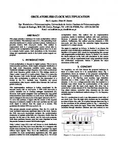
RESULTS A minor part of the results has been presented previously, in a preliminary form (Kippert, 1987). An ultradian clock controls locomotor behaviour In Paramecium species, two different circadian rhythms have been studied in isolated cells: a rhythm in mating reactivity in P. bursaria (Miwa et al., 1987) and a rhythm in locomotor behaviour in P. multimicronucleatum (Hasegawa et al., 1990). Only the latter type of rhythmicity seemed, on a shorter time scale, feasibly to occur also in fast growing Paramecium. Ultradian rhythms in motility have already been described for Euglena gracilis (Balzer and Hardeland, 1992). Hasegawa and colleagues characterized in both populations (Hasegawa and Tanakadate, 1984; Hasegawa et al., 1984) and individual cells (Hasegawa et al., 1990) the pattern of locomotor behaviour that underlies the observed circadian rhythm in P. multimicronucleatum. They found that organisms swam fast and unidirectionally during the day, while during the night they swam slowly and turned frequently. A comparable pattern was described for P. bursaria (Nakajima and Nakaoka, 1989). After initial observations that indicated that a similar alternation of locomotor patterns occurs on a shorter, i.e. ultradian time scale, an observation vessel was constructed for the easy monitoring of an ultradian rhythm of locomotor behaviour in isolated cells of P. tetraurelia. The principle is shown in Fig. 1. On the bottom of a 3 cm × 3 cm observation vessel a checked pattern is imprinted. In this vessel, locomotor activity can easily be monitored as the number of lines crossed by the isolated cell within a certain time interval. Fig. 1A shows the track of a cell during a 5 minute interval close to a maximum whereas the track in Fig. 1B is from a 5 minute interval close to the minimum of the locomotor rhythm. As was earlier found for the circadian rhythm of P. multimicronucleatum (Hasegawa and Tanakadate, 1984), one phase is characterized by swimming at a high velocity that is only infrequently interrupted by changes of direction; in the other phase tumbling predominates and swimming is much slower. Fig. 2 shows the locomotor activity for an individual cell between two successive divisions. An oscillation with a period of approximately 70 minutes is clearly apparent. The smaller peak towards the end of the cell cycle is due to the relative immobility that cells show when commencing division. Unfortunately, this example was one of only a few such clearcut patterns. There are two problems with chance elements that are negligible in circadian studies but become more predominant when the time interval is only 5 minutes. First, a cell can have at any time, including the maxima of motility, a rest lasting 2 to 4 minutes, which reduces the motility value for this time interval considerably. Second, during the phase with frequent turning, the respective position
Ultradian clock in Paramecium tetraurelia
869
Fig. 1. Track of an isolated Paramecium cell during a 5 minute interval in the course of the ultradian rhythm in locomotor behaviour. (A) Close to the maximum, (B) close to the minimum.
Fig. 3. Temperature compensation of the ultradian rhythm. Locomotor behaviour was monitored at 33°C (C, n = 17), 27°C (B, n = 16), and 24°C (A, n = 12). Data are the mean of the indicated number of recordings and are arranged from a reference point, i.e. a pronounced maximum, into both directions of the time axis. Fig. 2. Rhythm in locomotor behaviour of an isolated cell at 27°C. The number of lines crossed by the cell was monitored for 5 minute intervals from one division to the next.
of the cell can lead to different numbers of lines crossed for the same motility. However, there were two reasons to adhere to this very simple method of analysis. On the one hand, periodogram analysis did in all cases reveal a significant periodicity with the typical period. On the other hand, I found a striking constancy of the rhythm which enabled the plotting together of the data from many individual organisms. The most clearcut result was obtained when the data were plotted from a reference point, i.e. a pronounced maximum, into both directions on the time axis. This presentation of data has been employed in all further experiments. A general homeostasis of the period is a characteristic distinguishing a clock from other oscillations (Kippert, 1992). This homeostasis is best exemplified, and easily tested, as the independence of period from ambient temperature. The ultradian rhythm has been monitored at six different temperatures from 18°C to 33°C. Data for three of the temperatures are shown in Fig. 3. It is immediately apparent that period is almost the same at all three temperatures. Periodogram analysis gave values of 71.7, 68.9, and 70.5 minutes at 21, 27 and 33°C, respectively. The temperature dependence for all temperatures is given in Table 1. The Q10 between 18°C and 27°C is 1.08, which is a value in the range typically found for circadian rhythms. Above 27°C, there is a slight overcompensation. A general homeostasis of period is also indicated by the observation that period is not altered in different media. This was e.g. found for the ultradian clock of Schizosaccharomyces pombe (Kippert, unpublished). P. tetraurelia can be cultured in axenic medium (Thiele et al., 1980), which means a rather different environment to the cells, and results in increased
Table 1. Temperature dependence of ultradian period and generation times Temperature 18°C 21°C 24°C 27°C 30°C 33°C
Ultradian period* (motility rhythm) (minutes)
Ultradian period† (quantal cell cycles) (minutes)
Generation time‡ (minutes)
74.2 72.4 70.8 68.0 69.7 71.2
73.6 71.7 70.5 68.9 69.3 70.5
619 542 464 373 309 398
*The ultradian period of the rhythm in locomotor behaviour was determined by periodogram analysis of the mean of the data obtained for the respective temperatures. †The ultradian period of the rhythm underlying the quantal cell cycle times is the mean of the values obtained by dividing the individual generation times by the obvious number of ultradian subcycles. ‡Generation time is the mean of all interdivision times at the respective temperatures.
doubling times. However, experiments using this medium produced periods which were indistinguishable from those obtained with bacterized medium (data not shown). An ultradian clock is involved in the timing of cell division
Both circadian and ultradian clocks have temporal control over multiple cellular functions (Kippert, 1992). Most basic for the life history of any cell is the duplication of its material with subsequent division. Gating of cell division has frequently been reported for the circadian clock and has also been demonstrated for the ultradian clocks of the microbes T. pyriformis (Kippert, 1996) and S. pombe (Kippert and Lloyd, unpublished). A result of that control is the quantization of generation times which manifests as a clustering instead of a random distribution. With its comparably long period of 70
870
F. Kippert
Fig. 5. Generation times of cells of different clonal age. Combined values for the temperatures 24°C to 33°C for young cell with less than 30 fissions (A), or old cells with more than 120 fissions (B). For presentation, numbers were grouped into 8 minute intervals.
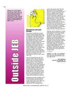
Fig. 4. Clustering of generation times for cells grown at six different temperatures. Interdivision times were determined for 708 isolated cells with an accuracy of ±2 minutes.
minutes, P. tetraurelia seemed well suited for a single cell study. It is also of advantage that the cell division cycle of P. tetraurelia has already been studied in some detail (Berger, 1988; Tang et al., 1994). Generation times were monitored for over 700 cells with an accuracy of ±2 minutes. Fig. 4 shows the distribution of generation times at six temperatures between 18°C and 33°C. This temperature range has been chosen because it allows relatively fast growth. Below 18°C, growth rate drops rapidly; a condition not suited for the study of ultradian clocks. The generation times are clearly clustered with the clusters being approximately 70 minutes apart. This strongly indicates that the interdivision time cannot have any value whatever but is restrained to an integer multiple of the ultradian cycle. The temperature dependence of the ultradian period underlying this quantal pattern is given in Table 1. It is virtually identical to that found for the ultradian rhythm in locomotor behaviour. In contrast, the length of the cell cycle shows a strong dependency on temperature, where 33°C is already a suboptimal temperature. In Paramecium species clonal age, i.e. the number of fissions since last conjugation or autogamy, is known to affect a variety of physiological parameters, and in particular the macronuclear DNA content. To look for a possible effect on the ultradian clock, data from Fig. 4 were separated for young cells (with not more than 30 fissions since last autogamy) and old cells (more than 120 fissions). These data are shown in Fig. 5A and B, respectively. Since period is only slightly different at these temperatures, the data obtained from 24°C to 33°C were pooled. As expected, at increased clonal age interdivision
Fig. 6. Differences in the generation times of sister cells. Values were obtained for 218 pairs. Accuracy was ±2 minutes for the individual determinations. For presentation, numbers were grouped into 4 minute intervals.
times become somewhat prolonged, i.e. from 368 minutes to 406 minutes for the mean value. In contrast, for period length no significant difference could be found; the mean values are 69.5 and 70.4 minutes, respectively. The temporal control exerted by the ultradian clock over the cell division cycle is also shown in the differences in the generation times of sister cells which cluster at around 0, 70 and 140 minutes (Fig. 6.) In Fig. 7 the data from Figs 4 and 6 are presented as α- and β-curves (Smith and Martin, 1973), i.e. as the percentage of undivided cells, and the percentage of undivided sister cells, respectively, plotted on a logarithmic scale against time. Also here, the clustering becomes well apparent as an oscillation around a straight line which would be expected in the case of a constant division probability. The fact that the pattern is so clearcut can be ascribed to the relatively long period which is at the upper border for ultradian clocks in the 30 to 70 minutes range found in eukaryotic microbes. This allows a much better resolution as occurs in organisms with shorter periods. In the literature there are numerous examples where α- or β-curves show considerable deviations from an exponential line. In the light of the increasing evidence for the wide distribution of ultradian clocks it
Ultradian clock in Paramecium tetraurelia
Fig. 7. α- and β-plots of the cell cycle time data. (A) β-plot of the data from Fig. 6. The percentage of undivided sister cells is plotted on a logarithmic scale against the time elapsed since division of the first sister. (B) α-plot of the data from Fig. 4. The percentage of undivided cells is plotted on a logarithmic scale against generation times.
seems reasonable to assume that at least in some cases the observed patterns may be attributed to such a clock. Locomotor behaviour and cell division are controlled by the same ultradian clock
The apparent identity of period length of the locomotor rhythm and of the period derived from the generation time data suggested that both phenomena are caused by the same ultradian clock. Both parameters were therefore monitored simultaneously. Fig. 8 shows the history of 12 cells from one division to the next. It is obvious that in all cases there is a constant phase relationship between the time window during which cell division can occur and the locomotor rhythm. This is a further strong indication that both processes are under control of a single ultradian clock. DISCUSSION In the present study an ultradian clock has for the first time been demonstrated for individual specimens of a eukaryotic microbe. These results provide direct proof that ultradian clocks are not due to interactions amongst cells within a culture. Instead, like their circadian counterparts, they occur on the single cell level. The period of 70 minutes is at the upper border of the range of the hourly microbial rhythms where periods range from 30 minutes for T. pyriformis (Kippert, 1996) to 70 minutes for Acanthamoeba castellanii (Lloyd et al., 1982). In the temperature range in which the phenomenon can be studied, i.e. from 18°C to 33°C the rhythm is well temperature-compensated. The Q10 does not fall behind that found for circadian rhythms. Such compensation has now been found for ultradian rhythms of several phylogenetically unrelated species, i.e. T. pyriformis (Kippert, 1996; Michel and Hardeland, 1985), Acanthamoeba castellanii (Lloyd et al., 1982), S. pombe (Kippert and Lloyd, 1995), and mammalian cells in culture (Klevecz et al., 1985). A further demonstration of the general homeostasis of period is the observation that period length is not affected by the type and composition of the medium, as has now been found for S. pombe (Kippert, unpublished) and P. tetraurelia (this study).
871
Fig. 8. Phase relationship between the ultradian rhythm in locomotor behaviour and cell division. Data for the motility rhythms are the mean of 12 recordings at 27°C and are arranged from a reference point, i.e. a pronounced maximum, into both directions of the time axis. Divisions of the 12 cells are indicated by the bars and are assigned to the respective 5 minute interval.
Very impressively, this homeostasis is also shown by the constancy of period in cells of different clonal age. In P. tetraurelia, ageing is a well studied phenomenon (for reviews see Smith-Sonneborn, 1981; Tagaki, 1988): along with the ageing process, i.e. the increasing number of fissions since the last autogamy, several parameters of cellular physiology change; in particular there is a decrease in macronuclear DNA content. As shown in Fig. 5, the period length of young and old cells is indistinguishable, whereas generation time increases. It thus appears that gene dosage does not affect period length. This can also be inferred from the fact that variability in period is at most 3 minutes (as discussed below) while there are, in addition to the age effect, also considerable inter- and intraclonal variations (e.g. Kimball, 1967) in macronuclear DNA content to be encountered. This suggests that there may be some gene dosage compensation as proposed for the Drosophila melanogaster period gene (Cooper et al., 1994). Changes in macronuclear DNA content are by far not the only alterations on cell physiology associated with ageing, rather there are several further parameters subject to considerable changes (listed by Smith-Sonneborn, 1981; Tagaki, 1988). From the results presented here, it can be concluded that these parameters, too, do not affect period length. These results indicate that constancy of the period is an important property of cellular clocks. For circadian rhythms the significance of compensation is obvious; for a clock adapted to the daily changes in the environment it is crucial that it does not run at different speeds, e.g. faster in summer and more slowly in winter. A corresponding selection pressure is not immediately apparent for ultradian clocks. Instead, it may be assumed that the constancy of period is an important general feature of a timekeeping device. It may be of advantage that the temporal structure of a cell is unaltered both under different environmental conditions, and with changes in internal cell physiology. The central role of a timekeeping device is to exert temporal control, and thereby temporal coordination, over a variety of cellular processes. This has in many cases been shown for circadian rhythms. P. tetraurelia is now another example for an ultradian clock that controls at least two different processes. Both parameters have previously been shown to oscillate with a circadian rhythm in Paramecium cells which are growing very slowly or are in the non-growing state. The ultradian rhythm in locomotor behaviour shows striking similarities to
872
F. Kippert
those of the circadian rhythm found in P. multimicronucleatum. Although these rhythms have been studied in two different species, and a circadian rhythm of motility has not yet been looked for in slowly growing or stationary phase cells of P. tetraurelia, this is another indication that ultradian clocks exert temporal control over exactly the same function as do circadian clocks, with the only difference being the time scale. It suggests that the underlying mechanisms may be very similar. There is now considerable knowledge about the molecular mechanisms, both electrophysiological and biochemical, which are responsible for the pattern of locomotion (Kung and Saimi, 1982; Saimi and Kung, 1987). It can be postulated that changes in these properties must occur concomitantly with the locomotor rhythm. This assumption is supported by the results obtained by Nakajima and Nakaoka (1989) on circadian changes in resting potential and membrane conductance in P. bursaria. Paramecium species may be well suited organisms to study how these processes are coupled to the ultradian clock. Gating of cell division by an ultradian clock has been shown for S. pombe (Kippert and Lloyd, unpublished), T. pyriformis (Kippert, 1996), and mammalian cells (Klevecz et al., 1985). The present study has now demonstrated gating also for P. tetraurelia; moreover, it has convincingly proven the consequence of gating, quantized cell cycles. Whereas interdivision times were progressively prolonged by lowering the temperature, they always remained integer multiples of the ultradian subcycle with its temperature compensated period. In the light of the present results, the question can be raised why this phenomenon has not been reported in earlier studies on interdivision time distributions. One major reason for the clustering becoming so apparent in the present study certainly is the relatively long period. Only if the period is considerably longer than the sum of the length of the time window plus the naturally occurring variability, can such a clearcut pattern be expected. In general, it can be assumed that in most earlier studies the relationship between cell cycle duration, period length of the ultradian rhythm, and sampling interval may not have been so fortuitous as in the present investigation. A second aspect is probably of equal importance. In the case of Paramecium, the variability in period length between individual cells is apparently very low. This becomes obvious looking at the distribution of generation times. Any variability in period length must result in a progressive broadening of the clusters as interdivision times, and thus the number of subcycles, increase. The width of the short generation time clusters is about 30 minutes, and increases to about 45 minutes. If one assumes that cell division is constricted to a time window of 15 minutes, the remaining 15 to 30 minutes could account for the variability in period. From this it can be calculated that variability in period length is at most 3 minutes. If any cells had periods outside this range, this would become apparent as intermediate values in-between the clusters. Fig. 4 shows that this is definitely not the case. Thus, the period of Paramecium must be accurately regulated with a variability of less than 5%. This is considerably less than found for S. pombe where the periods of individual clones show a variability of more than 15% (Kippert, unpublished). In general, clustering is much more likely to become apparent when generation times are dispersed over a wide range, as was achieved in the present study by using different
temperatures with correspondingly different growth rates. Notably, the curves shown in Fig. 7A and B incorporate data obtained at six different temperatures. In contrast, most of the previous studies have been carried out only at one particular growth rate. It can also be speculated that specialities of Paramecium may have contributed. The cell division cycle of Paramecium tetraurelia shows some peculiarities when compared to other eukaryotes (for review see Berger, 1988); e.g. the commitment to cell division is set in the previous cell cycle. Perhaps clock control in Paramecium is not (only) on mitosis but (also) on cell division; the ciliate may be a good experimental organism for future comparative studies of clock control mechanisms. In general, it should be noted that there are several examples where at least two clearly identifiable clusters have been observed previously, e.g. in the ciliate T. pyriformis (Williams, 1964). Although the phenomenon was not recognized at that time, such examples may well be cases of gating of cell division by an ultradian clock. When investigated in an appropriate way, quantized cell cycles, as an inevitable consequence of temporal control by an ultradian clock, may be found in fast growing cells of other species as well. Moreover, it may turn out to be a general pattern that ultradian clocks control cell division in fast growing cells whereas circadian clocks take over this role when generation time exceeds 24 hours. It will now be interesting to study the effect of lower temperatures on the interdivision times of P. tetraurelia, and probably observe the transition of ultradian clock control to circadian clock control when generation time exceeds 24 hours. I am grateful to Profs. G. Cleffmann and F. Jauker, Institute of Animal Physiology, University of Gießen, for providing laboratory space and support for the initial stages of this work. I thank Prof. D. Lloyd, University of Wales College of Cardiff for critical reading of the manuscript.
REFERENCES Balzer, I. and Hardeland, R. (1992). Multiple ultradian frequencies in dark motility of Euglena. J. Interdiscipl. Cycle Res. 21, 47-55. Barnett, A. (1966). A circadian rhythm of mating type reversals in Paramecium multimicronucleatum, syngen 2, and its genetic control. J. Cell. Physiol. 67, 239-270. Barnett, A. (1969). Cell division: a second circadian clock system in Paramecium multimicronucleatum. Science 164, 1417-1419. Berger, J. D. (1988) The cell cycle and regulation of cell mass and macromolecular DNA content. In Paramecium (ed. H.-D. Görtz), pp. 98119. Springer-Verlag, Berlin. Cooper, M. K., Hamblen-Coyle, M. J., Liu, X., Rutila, J. E. and Hall, J. C. (1994). Dosage compensation of the period gene in Drosophila melanogaster. Genetics 138, 721-732. Edmunds, L. N. Jr (1988). Cellular and Molecular Bases of Biological Clocks. Models and Mechanisms for Circadian Timekeeping. Springer, New York. Görtz, H.-D., ed. (1988). Paramecium. Springer-Verlag, Berlin. Hasegawa, K., Katakura, T. and Tanakadate, A. (1984). Circadian rhythm in the locomotor behavior in a population of Paramecium multimicronucleatum. J. Interdiscipl. Cycle Res. 15, 45-56. Hasegawa, K. and Tanakadate, A. (1984). Circadian rhythm of locomotor behavior in a population of Paramecium multimicronucleatum: Its characteristics as derived from circadian changes in the swimming speeds and the frequencies of avoidance response among individual cells. Photochem. Photobiol. 40, 105-112. Hasegawa, K., Tanakadate, A. and Ishikawa, H. (1990). Circadian locomotor activity in an isolated cell of Paramecium. In Chronobiology: Its
Ultradian clock in Paramecium tetraurelia Role in Clinical Medicine, General Biology, and Agriculture, Part B. (ed. D. K. Hayes, J. E. Pauly and R. J. Reiter), pp. 707-715. Wiley-Liss, New York. Johnson, C. H., Miwa, I., Kondo, T. and Hastings, J. W. (1989). Circadian rhythm of photoaccumulation in Paramecium bursaria. J. Biol. Rhythms 4, 405-415. Karakashian, M. W. (1968). The rhythm of mating in Paramecium aurelia, syngen 3. J. Cell Physiol. 71, 197-210. Kimball, R. F. (1967). Persistent intraclonal variation in cell dry mass and DNA content in Paramecium aurelia. Exp. Cell Res. 48, 378-394. Kippert, F. (1987). Temperature-compensation of ultradian rhythms in ciliates. In Chronobiology & Chronomedicine (ed. G. Hildebrandt, R. Moog and F. Raschke), pp. 60-64. Verlag Peter Lang, Frankfurt. Kippert, F. (1992). Ultradian and circadian clocks - two sides of one coin? J. Interdiscipl. Cycle Res. 23, 192-196. Kippert, F. (1996). A temperature-compensated clock of Tetrahymena: Oscillations in respiratory activity and cell division. Chronobiol. Int. 13 (in press). Kippert, F. and Lloyd, D. (1995). A temperature compensated ultradian clock ticks in Schizosaccharomyces pombe. Microbiology 141, 883-890. Klevecz, R. R. and King, G. A. (1982). Temperature compensation in the mammalian cell cycle. Exp. Cell Res. 140, 307-318. Klevecz, R. R., Kauffman, S. A. and Shymko, R. M. (1985). Cellular clocks and oscillators. Int. Rev. Cytol. 86, 97-128. Kung, G. and Saimi, Y. (1982). The physiological basis of taxes in Paramecium. Annu. Rev. Physiol. 44, 519-534. Lloyd, D., Edwards, S. W. and Fry, J. C. (1982). Temperature-compensated oscillations in respiration and cellular protein content in synchronous cultures of Acanthamoeba castellanii. Proc. Nat. Acad. Sci. USA 79, 37853788. Lloyd, D. and Kippert, F. (1987). A temperature-compensated ultradian clock explains temperature-dependent quantal cell cycle times. Symp. Soc. Exp. Biol. 4, 135-155. Lloyd, D. (1993). Intracellular timekeeping: epigenetic oscillations reveal the
873
functions of the ultradian clock. In Ultradian Rhythms in Life Processes (ed. D. Lloyd and E. L. Rossi), pp. 5-22. Springer, London. Michel, U. and Hardeland, R. (1985). On the chronobiology of Tetrahymena. III. Temperature compensation and temperature dependence in the ultradian oscillation of tyrosine aminotransferase. J. Interdiscipl. Cycle Res. 16, 17-23. Miwa, I., Nagatoshi, H. and Horie, T. (1987). Circadian rhythm within single cells of Paramecium bursaria. J. Biol. Rhythms 2, 57-64. Nakajima, K. and Nakaoka, Y. (1989). Circadian change of photosensitivity in Paramecium bursaria. J. Exp. Biol. 144, 43-51. Saimi, Y. and Kung, C. (1987). Behavioral genetics of Paramecium. Annu. Rev. Genet. 111, 433-445. Smith, J. A. and Martin, L. (1973). Do cells cycle? Proc. Nat. Acad. Sci. USA 70, 1263-1267. Smith-Sonneborn, J. (1981). Genetics and aging in protozoa. Int. Rev. Cytol. 73, 319-354. Sonneborn, T. M. (1970). Methods in Paramecium research. In Meth. Cell Biol., vol. IV, pp. 241-339. Academic Press, New York. Sonneborn, T. M. (1975). The Paramecium aurelia complex of fourteen sibling species. Trans. Am. Microsc. Soc. 94, 155-178. Tagaki, Y. (1988) Aging. In Paramecium (ed. H.-D. Görtz), pp. 131-140. Springer-Verlag, Berlin. Tang, L., Pelech, S. E. and Berger, J. D. (1994). A cdc2-like kinase associated with commitment to division in Paramecium tetraurelia. J. Euk. Microbiol. 41, 381-387. Thiele, J., Honer-Schmid, O., Wahl, J., Kleefeld, G. and Schultz, J. E. (1980). A new method for axenic mass cultivation of Paramecium tetraurelia. J. Protozool. 27, 118-121. Volm, M. (1964). Die Tagesperiodik der Zellteilung von Paramecium bursaria. Zeitschr. Vergl. Physiol. 48, 157-180. Williams, N. E. (1964). Relations between temperature sensitivity and morphogenesis in Tetrahymena pyriformis GL. J. Protozool. 11, 566-572. (Received 21 April 1995 - Accepted 2 January 1996)