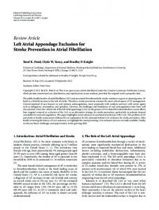
CASE REPORT DOI: 10.12710/cardiometry.2017.7779
Aneurysm left atrial appendage Cindy Guignan1, Francys Guerra1, Cristian Ramirez1 1
Latin American Children's Cardiology
Submitted: 7.1.2017, Accepted: 9.2.2017, Published online: 25.5.2017
Clinical case
Preschool female 3 years old, asymptomatic cardiovascular, who was heart murmur auscultation as incidental finding. When R1 single physical examination, breath holosystolic I / VI mitral area radiating to the armpit, physiological split S2, R3 and R4 not.
Lateral left thorax Retrocardiac space occupied in three thirds compressing the barium column due to growth of Ai and VI. Transthoracic echocardiography 1. aneurysmal dilation of the left atrial appendage. 2. Mitralinsufficiency moderate – severe + cleft of the posterior leaflet.
Hospital Dr. Gilberto Rodríguez Ochoa Venezuela, Caracas *
Chest X-ray
Corresponding author: phone: +58 (212) 407-50-40, e-mail:
[email protected]
Abstract Aneurysms of the atrial appendages are rare and extremely rare clinical entities. It can be confused with pericardial cyst, coronary artery aneurysm, left ventricular pseudo-aneurysm and partial or complete congenital absence of the pericardium. Usually clinically asymptomatic, diagnosed as incidental finding by the presence of heart murmur or cardiomegaly on chest radiograph, it can also manifest in the
Figure 1.
presence of supraventricular arrhythmias or systemic embolism. The treatment of this pathology makes aneurysmectomy. For 3-year preschool heart murmur and echocardiographic finding of insufficiency
ChestPA ICT: 0.63. Right edge, with double contour image, vascular image in the left edge of cardiac silhouette.
Mitral and aneurysmal dilation of the left atrial appendage it is presented.
Keywords Aneurysm, Atrial appendage, Echocar diography, Cardiac murmur
Imprint Cindy Guigñan, Francys Guerra, Cristian Ramirez. Aneurysm left atrial appendage; Cardiometry; No.10 May 2017; p.77–79; DOI:10.12710/cardiometr y.2017.10. 7779; Available online: www.cardiometry.net/issues/no10-may-2017/aneu rysm-left-atrial-appendage
Figure 2.
Figure 3. Parasternal long axis Issue 10. May 2017 | Cardiometry | 77
Operative findings Under Cardiopulmonary Bypass. Aneurysmal base level of the left atrium of approximately 8x6 cm dilation. Appendage of normal size and shape. Left atrial aneurysmectomy was performed to find healthy tissue resection 3 mm from the mouth of the left pulmonary veins and 4 mm mitral ring in the anterior segment, then placing an autologous pericardial patch. With satisfactory evolution after surgery.
Discussion
Aneurysms of the atrial appendages, are rare and extremely rare clinical entities. But there can be confused knowledge about the disease with other conditions such as pericardial cyst, coronary artery aneurysm, left ventricular pseudo-aneurysm and partial or complete congenital absence of the pericardium [1]. Historically in 1938, Semans and Taussig, reported the saccular dilation of the left atrium in a girl of 5 years with dextrocardia, without involving the appendage. In 1962, Parmley reported dilation of the left atrial appendage 2 children, a 11 year old patient who presented with atrial arrhythmia and two episodes of systemic embolism, and a second patient 7 years old, with an aneurysm of the atrial appendage left associated with a congenital anomaly of the left renal artery [2]. In 1963, Williams reported a case of dilatation of the wall of the left atrial appendage in a 27 years old. Subsequently they cited several articles which show the presence of aneurysmal dilation of the atrial appendage [3]. In 1999, Zhao and colleagues reported the case of a 27 year old with an aneurysm of the left atrial appendage, 78 | Cardiometry | Issue 10. May 2017
Figure 4. 3D reconstruction
which was removed in 1996 without cardiopulmonary bypass, approached by lateral thoracotomy [2]. Among the etiologies of this pathology are mentioned which may be congenital or acquired causes, among the causes acquired history of chest or secondary trauma is a mitral valve disease and among congenital causes congenital dysplasia pectineus or muscle due to pericardial defects [3, 4]) These congenital aneurysm of the left atrium may be extrapericardicos or intrapericardial. The extrapericardial type is associated with defects of the pericardium through which the atrial appendage or any portion of the left atrium hernia. Intrapericardial type is always associated with an intact pericardium [5]. Morphologically it can also be due to dysplasia pectineus bands or the presence of connection on the handset or weakness in the wall of the atrial appendage [4]. Clinically observed in patients with normal, asymptomatic phenotype, which are diagnosed as incidental finding by the presence of heart murmur during a physical examination, presence of
cardiomegaly on chest radiograph, arrhythmias, usually supraventricular tachycardia, due to the significant expansion of the left atrium, dyspnea, symptoms of congestive heart failure or left chest pain. Other manifestations include the generation of systemic embolisms produced inside the dilated atrium [3, 6–8]. The diagnostic approach to this condition can be done in as first tool chest radiograph which is evidenced an increase in the upper left border of the cardiac silhouette as an incidental finding, echocardiography as initial study for the diagnosis of this disease, where evidence the presence of the aneurysmal portion, also studies Cardiac Magnetic resonance tomography or to define the image and discard if it is other clinical entities (see Figures 1–4) [4, 7, 9]. Treatment of this condition is surgical aneurysmectomy made with reconstruction of the atrium under extracorporeal circulation [10]. In other patients with arrhythmogenic foci Maze procedure is used. However arrhythmias in most patients tend to disappear in the immediate postoperative period. [4, 11, 12].
Statement on ethical issues
Research involving people and/or ani mals is in full compliance with current national and international ethical standards.
Conflict of interest None declared.
Author contributions
The authors read the ICMJE criteria for authorship and approved the final manuscript.
References
1. Vásquez C, Cruz O, Ruiz E. Aneurisma gigante auricular izquierdo asociado a comunicación interauricular. Rev EspCardiol. 2014; 67(1): 61. 2. Solomon V, Vijaya M. Nayak, MS. Aneurysm of the Left Atrial Appendage. Tex Heart Inst J. 2001;28:111–8.
3. Bramlet D, Edwards J. Congenital aneurysm of left atrial appendage. Brit.Heart J., 1963.25,637. 4. Alok M, Kenton J, Lawrence J, Robert F. Left Atrial Appendage Aneurysm. Ann ThoracSurg 2005; 79: 1392–3. 5. Jinping Z, Youxin G, Hua Y, Youmin P, Yongde L. Treatment of Congenital Aneurysms of the Left Atrium and Left Atrial Appendage. Tex Heart Inst J. 1999; 26: 136–9. 6. Perafán S, Ascione G, Parra L, Jiménez L, Aneurisma congénito gigante del apéndice de la aurícula izquierda. Una entidad potencialmenteletal. Rev. Col. Cardiol. 2004; 11: 174–177. 7. Yoshihisa T, Hideaki K, Yuichi S, Koichi S. Left Atrial Appendage Aneurysm in a Child. Ann ThoracSurg 2004;77:721–3. 8. Otero J, Villanueva E, Becker P, Parra R, Zelada P. Aneurisma congénito
de la orejuela izquierda. Caso clínico. Cir. Cardiov. 2008;15(2):201–3. 9. Munarriz A, Escribano E, Urchaga A, Olaz F, Beunza M, De La Fuente A, Cantabrana S, et al. Congenital aneurysm of the left atrial appendage. European Journal of Echocardiography. 2008;9:152–4. 10. Aryal M, Hakim F, Ghimire S, Ghimire S, Giri S, Pandit A, et al. Left Atrial Appendage Aneurysm: A Systematic Review of 82 Cases. Echocardiography 2014;31:1312–8. 11. Nakai Y, Asano M, Nomura N, Mishima A. Surgical management of an aneurysm of the left atrial appendage to prevent potential sequelae. Interactive CardioVascular and Thoracic Surgery 17 (2013) 586–7. 12. Ashish Ashtekar, Abhijit Patil, Sahil Garg. Left Atrial Appendage Aneurysm: A Case Report. J of Evolution of Med and Dent Sci. Nov 03, 2014;3(58).
Issue 10. May 2017 | Cardiometry | 79