JBR–BTR, 2014, 97: 30-32.
Incidentally detected congenital giant left atrial appendage aneurysm IN a child: MRI findings A. Oz1, B. Oguz1, M. Karcaaltincaba1, M. Yilmaz2, M Haliloglu1 Left atrial appendage aneurysms are usually congenital and are very infrequent anomalies of the heart. They are very rarely diagnosed during childhood, with most cases symptomatic between the 2nd and 4th decades of life. Diagnosis is vitally important due to potential life-threatening complications. Surgical excision is the treatment of choice. Surgery reduces the risks of cardiac arrest, respiratory distress, arrhythmia, heart failure, thromboembolism, or rupture. We report the case of a 3-year-old boy with incidental diagnosis of a giant aneurysm of the left atrial appendage that was confirmed with magnetic resonance imaging and treated with surgery. Key-word: Cardiac anomalies, atrial appendage, aneurysm, MRI.
A left atrial aneurysm is a very rare cardiac anomaly. There are two types: intrapericardial and extrapericardial. In the intrapericardial type, the pericardium is intact, whereas the extrapericardial type is associated with pericardial defects. Intrapericardial type aneurysms are congenital, and they can bulge from the left atrial wall or from the left atrial appendage (1, 2). Although initially small, they are thought to grow over decades and cause symptoms when they reach a certain size (2). Patients are usually symptomatic after the 2nd decade, and the most frequent symptom is arryhythmia (1). Surgical excision is indicated even in asymptomatic patients because of potentially life-threatening complications, including arrhythmias, systemic thromboemboli, and congestive heart failure. There are only a few reports of this anomaly in children diagnosed by echocardiography. The aneurysm causes an abnormal cardiac border on routine chest radiography mimicking a tumor or a cyst. We report the case of a child with a giant left atrial appendage aneurysm (LAAA) suspected on chest radiography and detected and confirmed by cardiac magnetic resonance imaging (MRI) without the need for conventional angiography and treated with surgery. Case report A 3-year-old boy was admitted to the pediatrics department with coughing of 5 months’ duration. A cardiac murmur was not detected on physical examination. There was en-
Fig. 1. — Chest radiography shows enlargement of the left heart border at the level of the left atrial appendage segment (arrows).
largement of the left heart border at the level of the left atrial appendage segment on chest radiography (Fig. 1). No previous chest radiograph was available for comparison. An ECG showed a normal sinus rhythm. Echocardiography revealed a left atrial aneurysm. Cardiac MRI was performed with a 1.5T scanner (Symphony; Siemens, Erlangen, Germany). ECG gating was used to confirm the exact size of the aneurysm and to exclude the possibility
From: Department of 1. Radiology, 2. Thorax and Cardiovascular Surgery, Faculty of Medicine, Hacettepe University, Hacettepe Universitesi Tip Fakültesi Radyoloji Anabilim Dali Sihhiye Ankara, Turkey. Address for correspondence: Dr A. Oz, M.D., Department of Radiology, Faculty of Medicine, Hacettepe University, Hacettepe Universitesi Tip Fakültesi Radyoloji Anabilim Dali Sihhiye Ankara, Turkey. E-mail:
[email protected]
oz.indd 30
of any associated cardiac lesions. Black blood HASTE sequences on 3 orthogonal planes, short axis 4-chamber cine images, and sagittal oblique MR angiography sequences were obtained before intravenous contrast media administration. Sagittal oblique MR angiography and transverse plane fat-suppressed T1weighted images were then obtained. The MRI revealed a 4.2 × 3 × 3.5 cm sized left atrial appendage dilatation compatible with an LAAA. The aneurysm was compressing the left ventricle wall (Fig. 2). The aneurysm was connected to the left atrium with a large opening. The aneurysm was larger than the left ventricle. There was no other cardiac anomaly. The patient was managed surgically with aneurysmectomy and
24/02/14 10:24
INCIDENTALLY DETECTED CONGENITAL GIANT LEFT ATRIAL APPENDAGE ANEURYSM — OZ et al
31
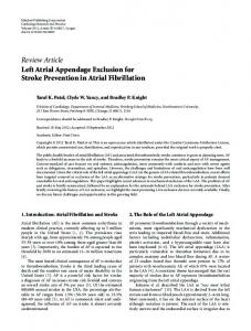
A
C Fig. 2. — A: Axial cine image shows giant aneurysm (*) c ommunicating with left atrium (a), indenting left ventricule (v) lateral wall. Sagittal oblique MR angiography (B) and volume rendering technique (VRT) (C) images show giant left atrial appendage aneurysm (*).
B rimary repair. The surgical findings p revealed a 4 × 3 cm sized aneurysm at the left atrial appendage. Histopathologically, all 3 cardiac wall layers were intact. The postoperative course was uncomplicated, and the patient was discharged on the 7th postoperative day. At 6-month follow up, he was asymptomatic with no complications and with no evidence of recurrence. Discussion Intrapericardial LAAAs are congenital but usually asymptomatic. Asymptomatic patients are diagnosed during adulthood incidentally. In some cases, acute symptoms and complications may occur, such as supraventricular arrhythmia, pulmonary and systemic embolism, mitral regurgitation, cardiac arrest, respiratory distress, heart failure, and cardiac tamponade (1, 2). Compression of the left coronary artery or its branches causes supraventricular arrhythmias and atypical chest pain
oz.indd 31
episodes (3). Left atrial dilatation with annular deformity of the mitral valve caused by the aneurysm leads to mitral regurgitation. The risk of possible complications rises with the increase in the size of the aneurysm (1, 2). When an enlarged abnormal upper left heart border is detected on a chest radiograph, a left atrial aneurysm should be considered in the differential diagnosis. Other condi tions that can cause a similar abnormality are a mediastinal mass, a pericardial cyst, a bronchogenic cyst, an epicardial lipoma, a cardiac or paracardiac tumor, a pericardial defect, or valvular heart disease. Fetal echocardiography, transthoracic echocardiography, transesophageal echocardio graphy, chest CT, nuclear molecular imaging, and CT and conventional angiography are other invasive and noninvasive methods useful for the diagnosis of left atrial aneurysms (1). Transthoracic echocardiography is the most useful noninvasive method because
it typically shows the cyst-like structure connected to the left atrium. Transesophageal echocardiography is a useful diagnostic tool for the detection of thrombi inside the left atrium or the aneurysm, but it is a more invasive technique. Angiography is also an invasive procedure, which should be reserved for patients with concomitant cardiac anomalies or for cases in which echocardiography or MRI are inconclusive. Cardiac multidetector CT is valuable for evaluating the anatomy of the coronary artery when compression of the left coronary artery or its branches is suspected (4, 5). MRI has the highest temporal resolution among the various diagnostic methods. This quality makes MRI the optimum approach for evaluating the surrounding structures, revealing the cardiac anatomy, and obtaining reproducible measurements of systolic and diastolic function. The absence of ionizing radiation also makes MRI superior in the evaluation of cardiac anomalies of (6). children and young patients Complications are more likely to occur with intrapericardial aneurysms than with extrapericardial aneurysms. Differentiation between an
24/02/14 10:24
32
extrapericardial- and an intrapericardial-type aneurysm by echocardio graphy is usually not possible. Smaller aneurysms can mimic left pulmonary artery anomalies. In such cases, axial and coronal sections are recommended (7). Pericardial cysts show high signal intensity on T2-weighted images when the left atrial wall is not involved. Central bronchogenic cysts that have no contact with the pericardium have high signal intensity on T2-weighted images. The presence of air or fluid in these cysts may also be detected. Epicardial lipomas have typical high signal intensity on both T1- and T2-weighted MR images. Cardiac tumors can be differentiated by the presence of a flow signal (7). The goal of treating an LAAA is to prevent life-threatening complications, the likelihood of which increases with the size of the aneurysm. Particularly in children, aneurysmectomy through a median sternotomy with a cardiopulmonary bypass is a conventional and safe method (2). An anurysmal thrombus is detected in one-third of patients with a LAAA (1).
oz.indd 32
JBR–BTR, 2014, 97 (1)
On the MRI, signal voids can be seen within the sac, suggestive of thrombus. In this case, a thrombus was not observed on either the echocardio graphy, MRI, or during surgery. Moderate to severe mitral regurgitation is associated with the absence of thrombus (8). In conclusion, this rare cardiac pathology should be kept in mind in the differential diagnosis of lesions adjacent to the left heart. Both noninvasive and invasive diagnostic methods are useful for detecting a left atrial aneurysm (1). MRI can reveal morphological and functional para meters of the aneurysms while excluding other cardiac abnormalities without ionizing radiation (6). To prevent complications and high mortality and morbidity, early diagnosis and aneurysmectomy are mandatory. References 1. Park J.S., Lee D.H., Han S.S., Kim M.J., Shin D.G., Kim Y.J., Shim B.S.: Incidentally found, growing congenital aneurysm of the left atrium. J Korean Med Sci, 2003, 18: 262-266.
2. Cho M.J., Park J.A., Lee H.D., Choo K.S., Sung S.C.: Congenital left atrial appendage aneurysm diagnosed by fetal echocardiography. J Clin Ultrasound, 2010, 38: 94-96. 3. Gupta S., Agarwal S., Pratap H., Datt V., Banerjee A. Congenital aneurysm of left atrial appendage: a case report. J Card Surg, 2010: 25: 3740. 4. Plonska-Gosciniak E., Larysz B., Jurczyk K., Kasprzak J.D.: Five- chambered heart: a 20-year story of left atrial appendage aneurysm. Eur Heart J, 2009, 30: 1014. 5. Smeglin A., Merchan J., Maysky M., Johnstone M., Pastore J.O.: Images in cardiovascular medicine: giant left atrial appendage aneurysm. Circulation, 2008, 118: 2393-2394. 6. Kroft L.J., de Roos A.: MRI diagnosis of giant right atrial aneurysm. AJR Am J Roentgenol, 2007, 189: W94-95. 7. Hoffmann U., Hamed N., Herold C., Globits S.: Radiological signs of a left atrial aneurysm. Eur Radiol, 2000, 10: 1332-1334. 8. Bilge M., Yasar A.S., Bozkurt M., Karakas F., Bilen E., Yuksel I.O.: Left atrial appendage aneurysm secondary to eccentric severe ischemic mitral regurgitation. Echocardiograph, 2009, 26: 1225-1227.
24/02/14 10:24