tative of the 3 different phases showed increased expression of clusterin/apoJ (clust/apoJ) in the involuting samples. Clust/apoJ is a multifunctlonal glycoprotein ...
Clusterin/ApoJ Expression During the Development of Hemangioma QURRATULAIN HASAN, PHD, BEATE M. ROGER, MMLSc, SWEE T. TAN, MBBS, FRACS, JASON GUSH, BSc, AND PAUL F. DAVIS, PHD Hemangioma is the most common tumor of infancy. This vascular tumor is characterized by an initial rapid proliferation followed by an inevitable regression. The life cycle of hemangioma is divided into proliferative, involuting, and involuted phases. The cellular and molecular mechanisms responsible for controlling the biological behavior of hemangioma are largely unknown. Differential display analysisusing mRNAisolated from biopsy specimens representative of the 3 different phases showed increased expression of clusterin/apoJ (clust/apoJ) in the involuting samples. Clust/apoJ is a multifunctlonal glycoprotein that has been associated with apoptosis. Reverse transcription-polymerase chain reaction (RT-PCR)and immunohistochemistry showed that both the transcription and protein expression of clust/apoJ were increased in hemangioma as the tumor progressed from the proliferative to the involuting and involuted phases. This suggests that clust/apoJ is involved in regulating apoptosis during the spontaneous regression of hemangioma. It has been suggested that mast cells (MC) play a role in the regression of
hemangioma. The increase in the number and proportion of d u s t / apoJ-positive MC with progression of hemangioma, along with the
H e m a n g i o m a , the most c o m m o n t u m o r of infancy, is a p r i m a r y t u m o r o f the microvasculature. 1 T h e natural history of h e m a n g i o m a is characterized by an initial excessive angiogenesis followed by inhibition and regression o f the newly f o r m e d b l o o d vessels. 2 T h e life cycle of the h e m a n g i o m a has b e e n arbitrarily divided into 3 phases. 3 T h e proliferative phase, which features a rapid postnatal growth for 8 to 12 months, is followed by the involuting phase as the t u m o r u n d e r g o e s regression over the next 1 to 5 years. T h e involuted phase, in which the lesion continues to regress, may last until the patient is 12 years o f age. These clinical phases can be distinguished by certain cellular and extracellular markers. ~ Although h e m a n g i o m a can be categorized into 3 phases, it is a c o m m o n clinical observation that the tumors progress at different rates a n d that within a single lesion there are areas representative o f different stages of development. TM T h e cellular and molecular m e c h a n i s m s controlling the p r o g r a m m e d biological behavior of hemangio m a are largely unknown. To identify genes that may be involved in the natural progression o f h e m a n g i o m a , m R N A was isolated f r o m biopsy specimens representa-
five of the 3 different phases of the t u m o r and analyzed by differential m R N A display. O n e polymerase chain reaction (PCR) product, which showed an increased expression, in an involuting h e m a n g i o m a , was seq u e n c e d and f o u n d to have h o m o l o g y to the h u m a n c l u s t e r i n / a p o L i p o p r o t e i n J (clust/apoJ) gene. Clust/apoJ, also known as testosterone repressed prostatic message 2 and sulfated glycoprotein 2, is a widely expressed, highly conserved, secreted glycoprotein for which m a n y functions have b e e n proposed. 9 It is induced during processes of tissue differentiation a n d regression that involve p r o g r a m m e d cell d e a t h or apoptosis. ]° It has b e e n suggested that apoptosis may be an i m p o r t a n t regulatory event in the s p o n t a n e o u s regression of h e m a n g i o m a . TM Razon et all 2 showed that apoptosis increases 5-fold during the involuting phase of h e m a n g i o m a . Because clust/apoJ m R N A a n d protein have b e e n consistently detected in apoptofic cell death paradigms, clust/apoJ expression has b e e n considered as a p r o m i n e n t m a r k e r of apoptofic cell loss. is In the c u r r e n t study, we have identified a n d investigated the expression of c l u s t / a p o J in different phases o f h e m a n g i oma.
From Swee Tan Plastic Surgery Trust, the Department of Medicine, Wellington School of Medicine; and Malaghan Institute of Medical Research, Wellington, New Zealand. Accepted for publication February 23, 2000. Supported by the Reconstructive Plastic Surgery Research Foundation. Address correspondence and reprint requests to Swee T. Tan, MBBS, FRACS, Swee Tan Plastic Surgery Trust, Bowen Hospital, Churchill Dr, Crofton Downs, Wellington, New Zealand. Copyright © 2000 by W.B. Saunders Company 0046-8177/00/3106-0010510.00/0
doi: 10.1053/hupa.2000.7638
691
localization of clust/apoJ to MC granules, supports this hypothesis. We suggest that MC may be synthesizing/releasing this apoptoti¢ modulator, leading to the regression of the tumor. Better understandhag of the pathogenesis of hemangioma by identification of the relevant factors involved in its regression such as clnst/apoJ will result in the development of novel therapies for this condition and tumors that do not undergo spontaneous regression. HUM PATHOL31:691697. Copyright © 2000 by W.B. Saunders Company Key words: hemangloma, involution, clusterin, apolipoprotein J, apoptosis. Abbreviations: clust/apoJ, clusterin/apolipoprotein J; EC, endothelial cells; DD, differential display; Ig, immunoginbnlin~MC, mast cells; mAb, monoclonal antibodies; pAb, polyclonal antibodies; PCNA, proliferating cell nuclear antigen; RT-PCR, reverse transcriptasepolymerase chain reaction.
METHODS
Samples Biopsy materials were obtained from patients with he.mangioma according to a protocol approved by the Wellington Ethics Committee. The hemangiomas were categorized as being in proliferative, involuting, or involuted phase based on clinical findings and histology rather than the age of the patient. Typically, each lesion showed a spectrum of developmental changes clinically and histologically. However, they were individually assigned to 1 of the 3 phases according to
HUMAN PATHOLOGY
Volume31, No. 6 (June 2000)
their predominant features. Rapid growth during the neonatal period, out of proportion to the child's growth, is the hallmark of hemangioma during the proliferative phase. The tumor assumes a bright crimson color, is tense on palpation, and is often tender. During the involuting phase, the growth of hemangioma reduces and for a time appears to be proportional to that of the child. The lesion begins to shrink and soften. The crimson color begins to fade, becoming dull purple, and the surface shows a mottled, grayish mande with tiny white flecks. These cutaneous signs of involution, which start centxally, spread to the periphery. In the involuted phase, the child's growth is proportionately greater than that of the hemangioma. Typically the involved skin that remains shows mild atrophy, has a wrinkled quality with few telangiectatic vessels, and is slightly paler than the surrounding skin. Often there is fibrofatty tissue residuum. The demographic data of the patients and the characteristics of their hemangiomas are shown in Table 1.
Differential Display Reverse Transcriptase Polymerase Chain Reaction Fresh biopsy material was snap frozen in liquid nitrogen and stored at -70°C. mRNA was isolated using an mRNA direct kit (Qiagen, Hilden, Germany) according to the manufacturer's instructions, and differential display (DD) reverse transcriptase polymerase chain reaction (RT-PCR) was performed as previously reported. TM Briefly, mRNA was reverse transcribed with anchored poly-dT primers, and the resulting cDNA was amplified with a 3-stage PCR using a primer mix comprising oligo-dT and 5 arbitrary primers) 4 The differentially expressed bands were excised, eluted, amplified, and sequenced as described. 14
RNA Isolation and RT-PCR RNA was isolated from fresh biopsy samples using Trizol reagent (GIBCO-Life Technol, Gaithersburg, MD) as described above. The hemangioma from patient 8 (Table 1) provided an opportunity to isolate RNA from distinctive "proliferative," "involuting," and "involuted" regions. RNA was equalized using hypoxanthine phosphoribosyl transferase
TABLE 1. Demographic Data of the Patients and Details of Their Hemangiomas and the Analytical Techniques Applied to Each Patient No.
Sex
Age
Site
Phase
Technique
1 2 3 4 5 6 7 8 9 10 11 12 13 14 15 16 17 18
M F M F F F M F F M M F F M F F F F
3 mo 5 mo 4 mo 2 wk 3.5 mo 5 mo 12 mo 13 mo 2 yr 9 mo 5 yr 1 mo 5 mo 8 mo 9 yr 3 yr 6 mo l0 yr 2 yr 9 mo 8 yr 6 mo 5 yr 1 rno
Knee Buttock Scalp Perineum Axilla Back Scalp Elbow Forehead Eyebrow Forehead Chest Submentum Lip Chin Lip Forehead Lip
Proliferative Proliferative Proliferative Proliferative Proliferative Involuting Involuting Involuting Involuting Involuting Involuting Involuting Involuting Involuted Involuted Involuted Involuted Involuted
IH IH IH RT IH, RT IH IH, RT IH, RT IH IH RT IH, RT RT IH, RT IH IH, RT IH IH, RT
gene as controlJ 5,16 The transcript expression of clust/apoJ was analyzed using the Perkin Elmer rTth kit as described previously.14,17Briefly, a 10-minute RT step was followed by 45 cycles of 3-stage PCR with annealing at 60°C. The primers were designed using the Oligo program, based on the h u m a n clust/apoJ sequence Is and synthesized by GIBCO-Life Technologies. The sense primer 5' CTCGCCCTrCTACI~CTGGA 3' and anti-sense primer 5' GGTCATCGTCGCC'rTCTCGT3' amplified a 404-bp product.
Histology Two- to 3-pm sections were attached to poly-L-lysinecoated glass slides, deparaffinized, and stained with hematoxylin and eosin, as described by Tan et al. 6.a7
Antibodies Peptide-specific goat polyclonal antibody (pAb) directed against h u m a n apoLipoproteinJ (Rockland Inc., Gilbertsville, PA) was used at a dilution of 1:350. To assess the n u m b e r of proliferating cells, a mouse monoclonal antibody (mAb) to the proliferating cell nuclear antigen (PCNA) (Dakopatts, Carpinteria, CA) was used at a dilution of 1:80. The primary antibodies were diluted in 50 m m o l / L Tris HCI, pH 7.5/1% bovine serum albumin.
Immunohistochemistry Immunostaining was performed on 2- to 3-pm paraffin sections from formalin-fixed hemangioma specimens. For the detection of clust/apoJ, the tissue sections were predigested with 0.05% protease XXIV (Sigma, St Louis, MO) in 50 m m o l / L "Iris HCI, pH 7.5, for 10 minutes at 37°C. Labeling with anti-PCNA was performed after microwave pretreatment of sections for 10 minutes in 10 m m o l / L citrate buffer, pH 6.0. Endogenous peroxidase activity was quenched using 0.3% hydrogen peroxide in methanol for 30 minutes at room temperature. The primary antibodies were incubated for either 2 hours at room temperature (clust/apoJ) or overnight at 4°C (PCNA). Controls included omission of primary antibody and substitution with an irrelevant mAb of the same isotype, or with normal goat serum for the pAb. Primary antibody binding was detected with a sensitive 3-step immunoperoxidase technique as described previously.15A9,2° The reactivity with clust/apoJ pAb was shown using rabbit anti-goat immunoglobulins (Ig) (Dako) (1:80) as bridging reagent, followed by goat peroxidase antiperoxidase (PAP) (Dako) (1:50), both incubated for 30 minutes at room temperature. PCNA-positive cells were detected with biotinylated antimouse Igs (BioGenex, San Ramon, CA), diluted 1:50 in 0.5% casein/Tris-buffered saline buffer, and streptavidin-peroxidase complex (BioGenex) (1:50 dilution). The sections were exposed to 0.05% diaminobenzidine in 50 m m o l / L "Iris HC1, pH 7.5, counterstained with Meyer's Haemalum, and mounted in Glycergel m o u n t i n g medium (Dako).
Double Staining To identify clust/apoJ-positive mast cells (MC), and to assess the n u m b e r and proportion of proliferating MC, sections labeled by immunohistochemistry with clust/apoJ and PCNA antibodies were counterstained with Csaba stain or with anti-tryptase as described previouslyfi,15,17
Cell Counting In each reaction, MC were counted in 10 fields at 40× magnification by 2 observers (Q.H. and B.M.R.). The percent-
Abbreviations: IH, immunohistochemistry; RT, RT-PCR.
692
CLUSTERIN/ApoJ EXPRESSION(Hasan et al)
age of MC positive for PCNA and positive for clusterin/apoJ were calculated. RESULTS D D RT-PCR a n d RT-PCR
DD RT-PCR analysis of the hemangioma samples showed 5 bands that varied in intensity. One of the PCR products, which showed increased expression in the involuting hemangioma sample, was excised and sequenced. GenBank BLASTN and BLASTX searches showed that the sequence of this cDNA was homologous with the human clust/apoJ sequence. Because this gene is considered as a marker of apoptosis and its expression is elevated in an involuting tissue, primers were designed to study its transcription in hemangioma biopsy specimens at different developmental stages. RT-PCR showed a varied transcript expression of clust/apoJ between samples (Fig 1). The transcripts were relatively abundant in all involuting and all but 1 involuted sample compared with proliferative specimens. In this exception (patient 16 in Table 1), the hemangioma had undergone complete regression both clinically and histologically. Histology
Hemangioma in the proliferative phase was highly cellular and showed plump proliferating endothelial cells (EC), which were organized into lobules with tiny lumens (Fig 2A). The cellular elements were progressively replaced by interlobular and intralobular fibrosis with channel dilatation during the involuting phase (Fig 2B). A few scattered thick-walled vessels lined with flat endothelium and an abundance of interstitial fibrofatty tissue were seen in the involuted phase (Fig 2C). Distinct areas displaying proliferation or varying degrees of involution were evident in a single sample, indicating a continuous spectrum of structural changes occurring during the natural progression of hemangioma (Fig 3A).
entered the involuting phase. Clust/apoJ was detected in the endothelium of the dilated vessels (Fig 2E). In the involuted phase, few small vessels showed intense staining for clust/apoJ (Fig 2F). Although the EC showed high levels of expression in the involuting and involuted phases, the surrounding fibrofatty tissue was devoid of clust/apoJ (Fig 2E, F). Within a single specimen, there were regions that showed a varying degree of clust/apoJ staining, with the intensity ranging from totally negative to strong staining (Fig 3B). This staining pattern corresponded to the histological and clinical observations, with the proliferative region showing less staining, whereas there was an increase in the involuting region (Fig 3A, B). In samples in which both RT-PCR and immunohistochemistry were performed (Table 1), a positive correlation of clusterin/apoJ transcription and protein expression were observed. In addition to its localization to EC, clust/apoJ was detected in cells scattered in the interstitium. These were identified as MC by both tryptase staining (data not shown) and Csaba staining (Fig 4A), although not all MC showed positive staining for clusterin/apoJ (Fig 4B). Clust/apoJ was localized to the granules in the positive MC. Within the vicinity of clusterin/apoJpositive MC, positive granules also were detected in the adjacent EC and capillary lumens (Fig 4A). During the proliferative phase, only a small proportion of MC were positive for clust/apoJ protein (Fig 2D). However, the proportion of MC expressing clust/apoJ increased with the regression of hemangioma (Fig 2E, F; Fig 5). PCNA was detected in most EC during the proliferative phase (Fig 2G), in fewer EC during the involuting phase (Fig 2H), and in a negligible number of EC in the involuted phase (Fig 2I) of hemangioma. Although the total number of MC was highest in the involuting phase (data not shown), the proportion of proliferating MC was highest in the proliferative phase and lowest in the involuted phase, thus showing an inverse staining pattern to that for clust/apoJ (Fig 5).
Immunohistochemistry
During the proliferative phase, staining for clust/ apoJ was negative (Fig 2D). Positive granular staining for the protein became apparent once the lesions M
1
2
3
4
5
6
7
8
9
10
11
12 13
II
bp
FIGURE 1. Ethidlum bromide stained agarose gel showing RT-PCR products (404 bp) of clust/apoJ in hemangioma biopsy specimens. Proliferative specimens from patients 4 and 5 (lanes 4 and 5, respectively) and the proliferative region from patient 8 (lane 7) did not show expression of the clust/apoJ transcript. Although the involuting hemangloma samples from patients 7, 8, 11, 12, and 13 (Table 1) (lanes 10, 8, 11, 12, and 13, respectively) and Involuted samples from patients 8, 14 and 18 (lanes 9, 3 and 1, respectively) show a high level of clust/apoJ transcription, patient 16 (lane 2) with an involuted hemangioma did not give an RT-PCRproduct (see text for details). Lane 6 Is a lane with no RTstep (negative control). 693
DISCUSSION
Using differential mRNA display analysis, we showed increased expression of clust/apoJ in hemangioma during the involuting phase. RTopCR confirmed a high level of transcription of clust/apoJ in all the hemangioma samples in the involuting phase, and all but 1 of the samples in the involuted phase compared with those in the proliferative phase. The absence of clust/apoJ transcription and protein in the involuted hemangioma in 1 patient may be attributable to the lesion having completely regressed. Immunohistochemistry was used to evaluate the expression and localization of the clust/apoJ protein in hemangioma specimens during the different phases of development. The absence of clust/apoJ protein expression in proliferating hemangiomas and its increasing detection with the progression of the disease parallels the ongoing involution. In the involuted samples, clust/ apoJ staining was intense in the endothelium of the vessels scattered amongst the fibrofatty tissue. The
HUMAN PATHOLOGY
Volume 31, No. 6 (June 2000)
®
©
@
O
finding of reduced PCNA positivity with progression of hemangioma confirms previous observations that proliferation is markedly decreased as the tumor undergoes regression, s,6,n This is in contrast to the observation of Razon et a112 that cell proliferation remained prominent in the involuting phase. The elevated number of MC during the involuting phase 3,6 suggests that these cells play a role in the regression of hemangioma, x4 This is in contrast to the large body of evidence showing the pro-angiogenic role of MC. 21,22 The number of PCNA-positive MC was highest in the proliferative phase and showed an inverse relationship with both the number and the proportion of clust/apoJ-positive MC, which increased with ongoing regression of hemangioma. Although the life cycle of the hemangioma has 694
FIGURE 2. Representative sections of hemangiomas during the proliferative (A, D, G), Involuting (B, E, H), and Involuted (C, F, I) phases. H&E stain shows proIlferative hemangioma with plump endothelial cells (EC) organized Into Iobules with tiny lumens (A). In the InvolutIng hemangloma, the dilated channels are surrounded by loose fibrofatty tissue (B), whereas the involuted hemangioma shows interlobular and Intralobular fibrosis with few thick-walled vessels lined with fiat endothellum (C). Staining for clust/apoJ was negative in the proliferative phase (D; MC: labeled with Csaba), was present within the endothellum of the dilated vessels in a granular fashion In the involuting phase (E), and was strong in the few scattered vessels in the involuted phase (F). Note that although the EC showed high levels of expression In the involuting and Involuted phases, the fibrofatty tissue was devoid of clust/apoJ (E, F). PCNA staining was positive for most of the EC and MC in the proliferative phase (G; arrows: MC stained with Csaba); in contrast, fewer EC and MC were positive in the involuting phase (H; arrows: MC), whereas the PCNA expression was negligible in the involuted phase (I). (A-C: H&E stain; D-I: Immunoperoxidase/Csaba double staining; original magnification A-I, x 290).
been arbitrarily divided into the proliferative, involuting, and involuted phases and can be distinguished by certain cellular and extracellular markers, s,6 it is a common clinical and histological observation that individual hemangiomas progress at different rates, s Furthermore, within a single lesion, there are areas representative of different stages of progression, s This has been observed by us both in the variation of transcript expression using RT-PCR and by clust/apoJ staining using immunohistochemistry. In this article, the hemangiomas were staged according to their predominant clinical and histological features rather than the age of the patients. Evidence for apoptosis in hemangioma has been reported. 7,n Razon et al a2 have recently shown that apoptosis is highest during the involuting phase. How-
CLUSTERIN/ApoJ EXPRESSION(Hasan et al)
sels in the epidermal region. In contrast, most vessels in the hemangioma showed positive granular staining (Rfiger BM, Hasan Q, Tan ST, and Davis PF, unpublished data). Our observatitms that clust/apoJ upregulation occurs in the EC of hemangioma indicate that at least in this tissue it does not act as a cytoprotective agent, because the EC are destined to be eliminated as the tumor regresses. Furthermore, we have shown that the fibrofattY tissue that subsequently replaces the proliferating EC is devoid of clust/apoJ, whereas the adjacent EC show high levels of expression. We believe that the presence of clust/apoJ represents a form of stress response similar to the synthesis of a heat shock protein, 26,2sand its upregulation may be an indicator of the pro-apoptotic mechanisms in the process of remod, eling in hemangioma. Another significant finding is the synthesis/accumulation of clust/apoJ within the MC granules. This is the first report, to our knowledge, of clust/apoJ within MC, and given the multifunctional nature of the clust/apoJ protein, it is difficult to predict the exact role it plays in relation to these cells. However, the presence of clust/
O
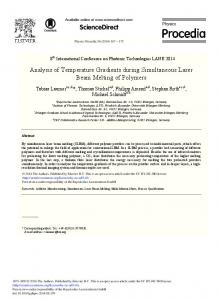
FIGURE 3. A hemangioma on the elbow of an 13-month-old girl (patient 8 in Table 1). P, Proliferative, Ig, involuting, and Id, involuted regions representing different stages of development within a single lesion (A). Clust/apoJ stain of the biopsy specimen from A, showing clearly defined regions with proliferative (upper) and involuting (lower) regions (B).
ever, the factor(s) responsible for this are unknown. Our observation that clust/apoJ is increased during the involuting phase of hemangioma suggests that it may be a factor responsible for the spontaneous regression of the tumor. The localization of the clust/apoJ protein in MC granules indicates that it may be synthesized/ released by MC, which play an important role in the spontaneous regression ofhemangioma. 3'14 Clust/apoJ has been implicated in a variety of biological processes, including lipid transport, inhibition of complement attack, sperm maturation, and membrane remodeling during apoptosis. 18This gene is known to be induced de novo during the regression of the prostate and other hormone-dependent tissues and t u m o r s . 1°,23,24 Although traditionally clust/apoJ upregulation has been shown in a number of cell paradigms involving cell stress and apoptosis, a cytoprotective role for clust/apoJ has also been proposed more recently.25-29The evolutionary conservation of clust/apoJ together with its expression in a multitude of biological processes suggests that the protein is involved in more than 1 essential biological function.9 Although it has been proposed that apoptotic processes are responsible for increased clust/apoJ expression, s° the exact molecular mechanism involved in the induction of clust/apoJ remains unclear. A skin sample taken from a site adjacent to the hemangioma showed only a few clust/apoJ-positive ves-
695
FIGURE 4. Clust/apoJ expression by M(~. Double-labeling of sections with apoJ Ab and Csaba stain showing presence of clust/apoJ protein within MC in an involuting hemangiom6 (A). The consecutive section control incubated with normal goat serum shows negative staining (insert; arrows show corresponding cells). Clust/apoJ is localized to MC granules. However, not all MC showed positive staining (B; arrowheads: clust/apoJ staining of MC, arrows: negative MC). Note the positive granules in the lumen of the capillary vessels. (Original magnification A, x290; B, x725).
HUMAN PATHOLOGY
Volume 31, No. 6 (June 2000)
90
80
,- P C N A + v e MC • apoJ+ve M C
7O
FIGURE 5. H e m a n g i o m a sections were stained for either PCNA or clust/apoJ and counterstalned with Csaba and counted as described in Materials and Methods. Individual patient d a t a are presented In the bar graph; samples 1 to 3, 4 to 8, and 9 to 12 refer to patients 1 to 3, 6 to 10, and 14 to 17, respectively, in Table 1.
o
u
20
10
I
0
Sample
i 1
2 i
3
4 I
I
5
6
7
I
8
I
I
9
10
11 i
12
I
9. J e n n e DE, Tschopp J: Clusterin: The intriguing guises of a widely expressed glycoprotein. Trends Biochem Sci 17:154-159, 1992 10. Wong P, PineauhJ, LakinsJ, et al: Genomic organization and expression of the rat TRPM-2 (clusterin) gene, a gene implicated in apoptosis.J Biol Chem 268:5021-5031, 1993 11. Mancini AJ, Smoller BR: Proliferation and apoptosis within juvenile capillary hemangiomas. Am J Dermatopathol 18:505-514, 1996 12. Razoq MJ, Kraling BM, Mulliken JB, et ah Increased apoptosis coincides with onset of involution of infantile hemangioma. Microcirculation 5:189-195, 1998 13. Ahoja HS, Tenniswood M, Lockshin R, et ah Expression of cluste,in in cell differentiation and cell death. Biochem Cell Biol 72:523-530, 1994 14. Hasan Q, Tan ST, Gush J, Peters SG, Davis PF: Steroid therapy of a proliferating hemangioma: Histochemical and molecular changes. Pediatrics 105:117-121, 2000 15. ROger B, Dunbar PR, Hasan Q, et ah Human mast cells produce type VIII collagen in vivo. IntJ Exp Pathol 75:397-404, 1994 16. Hasan Q, Dunbar PR, Murray-McIntosh RP, et ah Transforming growth factor 13 isoforms in human glomerulonephropathies. Nephrology 4:353-359, 1998 17. Tan ST, Hasan Q, Velickovic M, et ah A novel in vitro human model of hemangioma. Mod Pathol 13:92-99, 2000 18. Wong P, Taillefer D, LakinsJ, et al: Molecular characterisation of human TRMP-2/clusterin, a gene associated with sperm maturation, apoptosis and neurodegeneration. EurJ Biochem 221:917925, 1994 19. ROger BM, Hasan Q, G,eenhill NS, et ah Mast cells and type VIII collagen in human diabetic nephropathy. Diabetologia 39:12151222, 1996 20. ROger BM, Hasan Q, Erb KJ, et ah Progression of renal disease in interleukin-4 transgenic mice: Involvement of transforming growth facto1=13. IntJ Exp Pathol 80:113-123, 1999 21. Metcalfe DD, Baram D, Mekori YA: Mast cells (review). Physiol Rev 77:1033-1079, 1997 22. Fang KC, Wohers PJ, Steinhoff M, et ah Mast cell expression of gelatinases A and B is regulated by kit ligand and TGF-13.J Immunol 162:5528-5535, 1999 23. Tenniswood ME Guenette RS, Lakins J, et ah Active cell death in hormone-dependent tissue. Cancer Metastasis Rev 11:197220, 1992 24. French LE, Chonn A, Ducrest D, et ah Murine clusterin: Molecular cloning and mRNA localization of a gene associated with
apoJ-positive MC granules both in the adjacent EC and in capillary lumens suggests that MC may be secreting this apoptotic modulator to promote the regression of hemangioma. Upregulation and synthesis of clust/apoJ appears to be involved with the spontaneous regression of hemangioma. Most published reports address the role of angiogenic factors in the progression of hemangioma. 3.5.6 This is the first report in which a specific apoptotic factor, clust/apoJ, has been associated with the spontaneous regression of hemangioma. Whether this intriguing protein triggers apoptosis, leading to regression of hemangioma or whether it is a factor induced during the apoptotic process requires further elucidation. REFERENCES 1. Vikkula M, Boon LM, Mulliken JB, et al: Molecular basis of ~tscular anomalies. Trends Cardiovasc Med 8:281-292, 1998 2. Mulliken JB: Diagnosis and natural history of hemangiomas, in Mulliken JB, Young AE (eds): Vascular birthmarks: Hemangiomas and vascular malformations. Philadelphia, PA, Saunders, 1988, pp 41-62 3. Takahashi K, Mulliken JB, Kozakewich HPW, et ah Cellular markers that distinguish the phases of hemangioma during infancy and childhood.J Clin Invest 93:2357-2364, 1994 4. Jang Y-C, Arumugam S, Ferguson M, et al: Changes in matrix composition of human hemangiomas.J Surg Res 80:9-15, 1998 5. Chang J, Most D, Bresnick S, et ah Proliferative hemangiomas: Analysis of cytokine gene expression and angiogenesis. Plast Reconstr Surg 103:1-9, 1999 6. Tan ST, Velickovic M, ROger BM, et ah Cellular and extracellular markers of hemangioma. Plast Reconstr Surg (in press) 7. lwata J, Sonobe H, Furihata M, et al: High frequency of apoptosis in infantile capillary haemangioma. J Pathol 179:403-408, 1996 8. Hasan Q, Tan ST, Gush J, et ah Altered mitochondrial cytochrome b gene expression during the regression of hemangioma. (submitted for publication)
696
CLUSTERIN/ApoJ EXPRESSION(Hasan et al) 28. Humphreys DT, Carver JA, Easterbrook-Smith SB, et al: Clusterin has chaperone-like activity similar to that of small heat shock proteins.J Biol Chem 274:6875-6881, 1999 29. Viard I, Wehrli P, Jornot, L, et al: Clusterin gene expression mediates resistance to apoptotic cell death induced by heat shock and oxidative stress.J Invest Dermatol 112:290-296, 1999 30. Klock G, Storch S, RickertJ, et al: Differential regulation of the clusterin gene by Ha-ras and C-myc oncogenes and during apoptosis. J Cell Physiol 177:593-605, 1998
epithelial differentiation processes during embryogenesis.J Cell Biol 122:1119-1130, 1993 25. Bruchovsky NR, Snoek R, Rennie PS, et ah Control of tumor progression by maintainence of apoptosis. Prostate 6:13-21, 1996 (suppl) 26. Michel D, Chatelain G, North S, et al: Stress-induced transcription of the clnsterin/apoJ gene. Biochem J 328:45-50, 1997 27. Truong LD, Sheikh-Hamad D, Chakr-aborty S, et ah Cell apoptosis and proliferation in obstructive uropathy. Semin Nephrol 18:641-651, 1998
697