s that the resulting tree overfits the data and is too sensitive on the sample noise. Th ... Massey LA, Micallef C, Paviour DC, O'Sullivan SS, Ling H, Williams DR.
MRI acquisition and segmentation of subcortical regions The MRI protocol comprised a coronal T1-weighted 3D magnetization prepared rapid gradient echo (TR 1600 ms; TE 3.44 ms; inversion time, 800 ms; slice thickness, 1.2 mm; matrix, 256 x 224 pixels; number of excitations, 1; flip angel, 15°; field of view 220 x 192mm), and a transversal double-echo fast spin echo sequence with T2 and proton density contrast (TR, 3270 ms; TE1-2, 12 and 85 ms; slice thickness, 3-5 mm; spacing between slices, 3.6-6 mm; matrix, 256 x 200; number of excitations, 1; flip angle, 150°; field of view, 220 x 172 mm; echo train length, 5). Segmentation of subcortical regions from T1-weighted 3D structural MRI data and estimation of structure volumes was performed by using the FreeSurfer toolkit (version 5.1.0 available at http://surfer.nmr.mgh.harvard.edu/). The procedure automatically segments and labels brain structures based on i) the prior probabilities of anatomical classes throughout an atlas space, ii) the prior probability obtained from frequency histograms within the atlas space providing the likelihood that a given anatomical class occurs at a given atlas location, and iii) the modelling of local spatial relationships between labelled structures as anisotropic non-stationary Markov random fields.1 The processing of T1-weighted 3D structural MRIs included correction of motion artefacts, removal of non-brain tissue, transformation into the Talairach reference space, segmentation of the subcortical white matter and deep gray matter volumetric structures, intensity normalization, tessellation of the gray matter, white matter boundary, automated topology correction and surface deformation following intensity gradients. 1-5The procedure generated the intracranial volume as well as the volumes of the putamen, caudate, globuspallidum, thalamus, hippocampus, amygdala, brainstem, cerebellar white and gray matter and the 3rd and 4th ventricle. By using the software package tkmedit, included in the FreeSurfer toolkit, the brainstem was manually divided into the midbrain and pons as there is
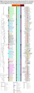
consistent evidence from imaging and neuropathologically observations that these brain areas are affected in MSA and PSP (Figure e-1). 6In addition, the previous preprocessing steps were visually inspected to ensure that no misalignment of brain structures had occurred. All MRI scans were included into the study. MRI acquisitions were processed on a Dell Studio XPS 435 T workstation with 8 cores, each with a 2.93 GHz Intel 7 processor.
Decision tree algorithm and classification performance: We have chosen the C4.5. classifier in order to obtain a decision tree providing a ran ked visualization of predictors that can be applied in the clinical work-up of patients wi th a parkinsonian syndrome.7
The C4.5 decision tree method is a classification algorithm which input consists of a s et of training data with an a-priori list of independent variables (features) and a class attribute (e.g. disease groups).8 The method examines the normalized information gai n (difference in entropy) that results from choosing an attribute for splitting the data. T o make the decision, the attribute with the highest normalized information gain is use d and the algorithm recurs on the smaller subsets. The algorithm separates the traini ng-set into smaller parts such that the data in each of the descending parts are “narro wer” than the data in the parent set. Each split is based on an optimal threshold value of a single feature. The splitting procedure stops if all instances in a subset belong to the same class. Then a leaf node is created in the decision tree indicating to choose t hat class. The result is a tree in which each leaf represents a class and each node sp ecifies a split on a particular independent attribute. The tree constructed in the trainin g phase can be visualized to understand the predictive capability of the features and splitting criteria which the algorithm has used to separate the data set. It often appear
s that the resulting tree overfits the data and is too sensitive on the sample noise. Th erefore the size of the decision tree is pruned by removing sections of the tree that pr ovide little power to classify instances. The result is a pruned decision tree showing cl assified cases. The final tree can be used to classify unseen cases where the class la bel is unknown.7 For the decision tree in our analysis all volumetric measures were normalized by the subject's intracranial volume. Data were further processed by z-transformation using mean centering and unit-variance scaling of the gender adjusted healthy control coho rts.
Two thirds of each patient group were assigned to the training set and one third to th e test set, respectively. Additionally, the training and the test set were controlled for a ge. The classification performance is expressed by the percentage of correctly classif ied patients, the percentage of total correct classifications (accuracy), the area under the receiver operating characteristic curve, which is 0.5 for completely random predict ions and 1.0 for accurate classification as well as sensitivity and specificity.
Reference: 1.
Fischl B, Salat DH, Busa E, et al. Whole brain segmentation: automated
labeling of neuroanatomical structures in the human brain. Neuron 2002;33:341–355.
2.
Fischl B, Dale AM. Measuring the thickness of the human cerebral cortex from
magnetic resonance images. ProcNatlAcadSci USA 2000;97:11050–11055.
3.
Sled JG, Zijdenbos AP, Evans AC. A nonparametric method for automatic
correction of intensity nonuniformity in MRI data. IEEE Trans Med Imaging 1998; 17:87–97.
4.
Fischl B, van der Kouwe A, Destrieux C, et al. Automatically parcellating the
human cerebral cortex. Cereb Cortex 2004;14:11–22.
5.
Ségonne F, Dale AM, Busa E, et al. A hybrid approach to the skull stripping
problem in MRI. Neuroimage 2004;22:1060–1075.
6.Massey LA, Micallef C, Paviour DC, O'Sullivan SS, Ling H, Williams DR Conventional magnetic resonance imaging in confirmed progressive supranuclear palsy and multiple system atrophy. Mov Disord 2012;27:1754–1762.
7. Mudali D, Teune LK, Renken RJ, Leenders KL, Roerdink JB. Classification of Parkinsonian syndromes from FDG-PET brain data using decision trees with SSM/PCA fe atures. Comput Math Methods Med 2015:1-10.
8. Quinlan JR. C4.5: Programs for Machine Learning. Morgan Kaufmann, Publ Inc., San Mateo; 1993