2011 International Conference on Bioscience, Biochemistry and Bioinformatics IPCBEE vol.5 (2011) © (2011) IACSIT Press, Singapore
Application of a Surface Displayed Receptor Platform for Screening Biological Ligands of Cannabinoid Receptor2 Mei-Li Wu
Ru-Ling Jiang
Department of Food Science National Pingtung University of Science and Technology Pingtung, Taiwan
[email protected]
Department of Food Science National Pingtung University of Science and Technology Pingtung, Taiwan
Show-Mei Chuang
Hso-Chi Chaung
Institute of Biomedical Sciences National Chung-Hsing University Taichung, Taiwan
[email protected]
Department of Veterinary Medicine National Pingtung University of Science and Technology Pingtung, Taiwan
[email protected] 'peripheral cannabinoid receptor', particularly abundant in immune tissues, with an expression level 10-100-fold higher than that of CB1 [2], and was initially presumed absent from the central nervous system. However, it has been found that CB2-selective agonists show potential for the treatment of acute pain without CNS side effects; AM1241, a CB2-selective agonist, inhibits acute nociception [3]. CB2 receptor mRNA and proteins have been located in brainstem neurons [4], perivascular microglial cells in the cerebellum [5], and on cerebrovascular endothelial cells [6]. Recent studies have demonstrated anti-nociceptive effects of selective CB2 receptor agonists in animal models of pain with the absence of CNS side effects [7-8]. Thus, selective CB2 agonists and peripherally restricted CB1 or CB1/CB2 dual agonists are being developed for the treatment of inflammatory and neuropathic pain, as they demonstrate efficacy in treatment of a range of pain models [9]. The cannabinoid receptors are G protein-coupled receptors (GPCRs) recognized by a variety of endogenous ligands [1] and are known to activate multiple signaling pathways. CB2 activation is responsible for the antinociceptive effects of AM1241 [10], which are prevented by the CB2 receptor-selective antagonist AM630 [3]. The physiological functions of the endocannabinoid system are controlled by the complex multiplicity of ligands and intracellular transduction mechanisms, which require a finely tuned regulation of the signaling events triggered by receptor activation [11]. Therefore, we established the surface-displayed receptor platform for screening biologically active ligands of CB2 with selectively physiological functions through the activation of defined signaling cascades for the development of efficacious and safe cannabinoid-based therapeutics.
Abstract—Cannabinoid (CB) receptor subtype CB2 is expressed in high quantities in the cells of the immune system, such as the human spleen and tonsils, and thus CB2 endogenous ligands can possibly act as immnomodulators. The objective of this study is to establish a competitive binding assay for quantification of the affinity of endogenous/exogenous ligands to human CB2 (hCB2) by using an intracellular cyclic AMP (cAMP) accumulation assay and a MAP kinase activity assay in the stable clone of HEK293 cells expressing human CB2 on the cell surface. Results showed that the molecular weight of the hCB2 recombinant protein expressed on HEK293 is about 46kDa, andexpression on the cell surface was further confirmed using confocal microscopy. This recombinant hCB2 was able to bind with CP55,940, a synthetic ligand, and lead to the inhibition of cAMP accumulation and the activation of MAP kinase. By using this cell-based platform, this competitive binding assay has been successfully established to screen for quantities of a natural active component of Morinda citrifolia, (Noni) fruit, with binding affinity for hCB2. Keywords- Cannabinoid receptor; cAMP accumulation assay; MAP kinases activity assay; HEK293
I.
*
INTRODUCTION
Techniques for expressing proteins on the surfaces of typical cells, or cell-surface display technologies, have played important roles in the elucidation of ligand-receptor interactions among cells, and also in the development of research leading to new drug discovery. Mammalian cell-based display systems have been well established and applied in the screening of bioactive ligands, a function that has proven significant to the pharmaceutical industry. Two types of cannabinoid receptor proteins (CB) have been identified: the central cannabinoid receptor, CB1, and the peripheral cannabinoid receptor, CB2 [1]. CB1 and CB2 were initially described as observed in the rat brain and spleen, respectively. CB2 was first considered to be the
48
II.
MATERIALS AND METHODS
cells were transfected with 5μg of the hCB2-expression plasmids using jetPEITM transfection reagent (Polyplus-transfection; Illkirch, France). On the day of transfection, 5x10⁴ cells/well were seeded in 48 multi-well tissue culture plates in 0.5mL of DMEM containing 10% FBS and antibiotics. For a triplicate assay, DNA (hCB2-expression plasmid) and various amounts of jetPEI were diluted separately in 150µL of 0.15M NaCl. After 10 min, the jetPEI-containing solution was added to the DNA and left for 30 min at room temperature. The jetPEI/DNA complexes (100mL per well) mixed with HEK293 cells were incubated at 37°C/5% CO2 for 2 h. Then, cells were collected, washed 3 times with phosphate buffer solution (PBS) and resuspended in DMEM containing 10% FBS and antibiotics. After 24 h, G418 sulfate (600μg/mL) (Calbiochem; San Diego, CA , USA) was added to the culture medium, selection was performed for 6 wks in a final concentration of 2000 μg/mL G418, and each of the isolated colonies were picked up for continuous culture. The mRNA expressions of HEK293 without any transfection or hCB2-transfected cells following G418 selection were examined. Total RNA was extracted using 1 mL Trizol reagent (Invitrogen) and immediately converted into cDNA using the RT kit (Promega Corp.; Madison, WI, USA). Then, amplification of each cDNA of hCB2 or GAPDH was performed in a mixture containing 0.5 μL of each pair of hCB2 or GAPDH primers (Table 1), 2.5μL of 10x PCR buffer, 2.5 μL of dNTP (2mM), 0.1 μL of Taq/Pfu polymerase (5 U/μL), and 1 μL of cDNA; the mixture was subject to the cycling parameters for PCR described previously. The final reaction product was then stained with 0.5μg/mL ethidium bromide in a 2% agarose gel. The sequences of the PCR products were confirmed to be 100% homologous to those of hCB2 and GAPDH, respectively, using the ABI PRISMTM 377 automated DNA sequencer.
A. Cloning the expression vectors of hCB2 The hCB2 and human glycerol-3-phosphate dehydrogenase (GAPDH), a house keeping gene to be used as the internal control, were cloned according to the sequences published with accession numbers of NM_001841.2 and AF261085 in GenBank, respectively. Specific primers for amplification of hCB2 and GAPDH were designed using the Expert Sequence Analysis software (Table 1) and applied in polymerase chain reactions (PCR) with templates of human kidney cDNA (Clontech; Palo Alto, CA, USA). Then, amplification of hCB2 and GAPDH was performed in a mixture containing 0.5μL of each pair of hCB2 or GAPDH primers, 2.5μL of 10x PCR buffer, 2.5μL of dNTP (2mM), 0.1μL of Taq/Pfu polymerase (5 U/μL), and 1μL of cDNA; the mixture was adjusted to a final volume of 25μL with pure water and subject to the following cycling parameters for PCR: pre-heating at 95℃ for 5 min, followed by 30 cycles of denaturing at 95℃ for 30 s, annealing at 55℃ for 30 s, and extension at 72℃ for 30 s, and concluding with a final extension step at 72℃ for 10 min. The final reaction product was then transferred into the well of a 2% agarose gel stained with 0.5μg/mL ethidium bromide in 0.5x TBE buffer. The amplified products of hCB2 and GAPDH (1083 bp and 800 bp, respectively) were purified and separately ligated to an expression vector obtained from the pcDNA3.1/V5-his TOPO TA Expression Kit® (Invitrogen; Carlsbad, CA, USA). Then, the constructed hCB2-expression plasmid was transformed into Escherichia coli XL1-Blue (Stratagene, USA), the colonies containing the hCB2-expression plasmid were selected, and the nucleotide sequence was confirmed using the ABI PRISMTM 377 automated DNA sequencer (Life Technologies Corporation, Carlsbad, CA, USA). TABLE I. Target gene
Accession No.
hCB2
NM_001841.2 F CGCGGTACCTCGATTATGGAG
Oligonucleotide sequence 5′-3′
R GADPH
AF261085
C. Expression of hCB2 on HEK293 cell surface For examination of hCB2 expressed on the surface of HEK293 cells, the transfected cells (1 x10⁵ cells/mL) were seeded in a 12-well dish overnight. Then, cells were labeled with 200μL of 1:300 diluted rabbit anti-CB2 affinity purified polyclonal antibody (Abcam, Cambridge, MA, USA) as the 1st antibody. After 1 h incubation at room temperature, 200μL of Alex Fluor 488® goat anti-rabbit (Invitrogen) as the 2nd antibody at a 1:500 dilution was added as a red-fluorescent probe, and then cells were incubated for another 1 h. After washing 3 times with PBS to remove unbound antibodies, the fluorescent-labeled cells were analyzed using a laser confocal microscope (CARV II Confocal Imager; BD Bioscience, San Jose, CA, USA).
PRIMER SEQUENCES DESIGNED FOR THESE EXPERIMENTS Restricition site
1083
KpnI
ACCGGTGTAGCAATCAGAGAGGTC
F GACCCCTTCATTGACCTCAAC R
Product size (bp)
AgeI 800
CATACCAGGAAATGAGCTTG
B. Transfection and mRNA expression of hCB2 in HEK293 HEK293 cells (ATCC CRL1573; derived from human embryonic kidney) were cultured in Dulbecco's Modified Eagle medium (DMEM) (Invitrogen) supplemented with 0.5% (vol/vol) penicillin-streptomycin (Invitrogen) and 10% (vol/vol) fetal bovine serum (FBS) (Biological Industries, Israel) at 37°C/5% CO2 at a concentration of 2x10⁶ cells/mL. After transfer into serum-free and antibiotic-free DMEM,
D. cAMP accumulation assay in transfected HEK293 The dose-dependent inhibition of Forskolin-stimulated cAMP accumulation in transfected HEK293 was determined for examination of the biological function of the hCB2 expressed on the cell surface. The intracellular cAMP contents in hCB2-expressing cells treated with various amounts of CP55940 was examined. The transfected HEK293 cells (3×10⁵ cells/mL) were seeded in a 12-well
49
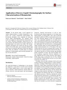
carried out with the cell extract of HEK293 cells transfected with pcDNA3.1-hCB2. Recombinant hCB2 protein was observed at the position of approximately 46 kDa, which is consistent with the value estimated from the deduced amino acid sequence of the recombinant protein (Fig. 2).
plate at 37°C for 16 h and then incubated with 500μM 3-isobutyl-1-methylxanthine (IBMX) in serum-free DMEM at 37 ℃ for 30 min. Cells were treated with 10X series dilution (10-9~10-5M) of CP55940 (Cayman Chemical Company; Ann Arbor, MI, USA) in DMEM plus 0.2% BSA, 10 mM HEPES, 100 μM IBMX, and 10 μM forskolin for 30 min at 37 ℃. The reaction was terminated by adding 300 μL of 0.1 M HCl and debris was removed after centrifugation. The concentration of intracellular cAMP was determined using the commercially available cAMP enzyme immunoassay kit (Assay Designs, Detroit, MI, USA) as described by the manufacturer’s published protocol. E. Analysis of ERK activity CB2 binding with a specific agonist, one of which is known to be CP55940, not only inhibits the activity of adenylyl cyclase, but it also activates the MAPK/ERK pathway. A clone of HEK293 cells stably expressing hCB2 receptors (5×10⁵ cells/mL) were seeded in a 6 cm plate at 37°C for 16 h. Cells were treated with 100 nM CP55,940 as a positive control or 5 μg/mL of ethanol extract of Noni fruit (Morinda citrifolia) (Kuang Chuan Biotech Co. Ltd.) for 0~4 h and then washed with cold PBS. After centrifugation, cells were treated with 200 μL lysis buffer (50 mM Tris– HCl, 150 mM NaCl, 1 mM EDTA, 1% Triton X-100, 1% protease inhibitor cocktail, pH 7.4) at 4 ℃ for 1 h and then the cell lysate was collected after centrifugation. Proteins in cell lysates were separated by subjection to sodium dodecyl sulfate–10% polyacrylamide gel electrophoresis (SDS-PAGE). The proteins on the gel were transfer onto PVDF membranes (Millipore; Billerica, MA, USA) with Semi-dry transfer unit (Hoefer Inc.; Holliston, MA, USA). Phosphorylated p42/44 ERK and total ERK proteins were then probed with the rabbit Phospho-p44/42 MAPK (Erk1/2) (Thr202/Tyr204) antibody and Erk antibody (Cell Signaling Technology; Danvers, MA, USA) at a dilution of 1:3,500 in 1× TPBS buffer (0.05% Tween20 in PBS buffer). After 1h incubation, anti-rabbit IgG-HRP antibody (Zymed; San Francisco, CA, USA) at a 1:15,000 dilution was added and the solution was incubated for another 1 h. Signals were developed by incubating the membrane with ECL Plus Western Blotting Detection Reagents (GE Healthcare, UK). III.
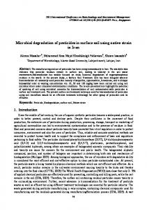
Figure 1. Electrophoretic analysis of KpnI and AgeI digested pcDNA3.1-hCB2 plasmid DNA.Fragment sizes were determined using a DNA ladder marker (lane 1). The upper band is the pcDNA™3.1/V5-His-TOPO® vector of 5.5 kb. The lower band corresponds to the hCB2 insert of approximate 1.1 kb (lane 3). Lane 2: pcDNA3.1- hCB2 plasmid.
Figure 2. Western immunoblot analysis of HEK293 cell expressing recombinant hCB2 protein. The total protein was detected with anti-hCB2 polyclonal serum and Rabbit anti-Goat IgG-HRP antibodies Lane 1: total protein extracted from HEK293; lane 2:total protein extracted from HEK293/pcDNA3.1; lane 3:total protein extracted from HEK293/pcDNA3.1-hCB2.
RESULTS
A. Cloning and expressions of hCB2 The amplified PCR products of hCB2 (1083 bp) were successfully ligated with the pcDNA3.1/V5-his TOPO expression vector (Fig. 1) and their nucleotide sequences were confirmed to be 100% homologous to that of hCB2 published with accession numbers of NM_001841.2 in GenBank. After transfection of the hCB2-expression plasmid into the HEK293 cells, the mRNA of hCB2 were detectable (data not shown); PCR product sequences of hCB2 (1083 bp) were confirmed to be 100% homologous to hCB2. Thus, the hCB2 were successfully transcribed into the mRNA of the transfected HEK293 cells. To further verify the synthesis of hCB2 protein, Western blotting was
B. Expression of hCB2 on HEK293 cell surface The surface localization of the recombinant hCB2 protein was confirmed by using the confocal laser microscope. HEK293 cells expressing hCB2 were labeled with rabbit antibody against CB2 and then fluorescently stained with Alexa Fluor® 488-conjugated goat anti-rabbit IgG. Results showed that the surface of the HEK293 cells was covered with the antibody-Alexa Fluor® 488 complex, which confirms that hCB2 can be successfully expressed on the cell surface of transfected HEK293 cells (Fig. 3A and 3B). In contrast, the control cells transfected with pcDNA were not immunostained (Fig. 3C and 3D).
50
Figure 5. Time-dependent effects of CP55,940 (A) and Noni fruit extract (B) on ERK signalling pathway in HEK293 cells transfected with hCB2 and pcDNA3.1 (control).
IV.
Figure 3. Phase micrographs and confocal immunofluorescence micrographs of HEK 293 cells transfected with pcDNA-hCB2 (panels A and B) and pcDNA (panels C and D).
DISCUSSION
Recently, new technologies based on mammalian cell-based display systems have been developed, and these technologies have been applied towards measurement of ligand-receptor interactions and towards screening natural products as candidate ligands for pharmacological therapy. A reference agonist CP55940 to the CB2 receptor has been employed in recombinant cell lines, as binding between CP55940 and the CB2 receptor triggers the impedance of cAMP accumulation and affects the potency ERK pathway activation. These parameters can be quantified using the well-established assays measuring cAMP and ERK phospholation levels [12]. Results of the present study revealed that cAMP accumulation in HEK293 cells expressing hCB2 was indeed significantly inhibited by agonist CP55,940 in a dose-dependent manner, and also that CP55,940 activated the ERK signaling pathways in a time-dependent manner; these effects were not present in the parental nontransfected cell line, confirming a specific, receptor-mediated response. Furthermore, these results demonstrate that indeed, hCB2-transfected cells bind to CP55940 leading to the activation of Gi/o, the inhibition of adenylyl cyclase, and ultimately resulting in the reduction of intracellular levels of cAMP. Noni has been used by Polynesians for over 2000 years for two main purposes, namely, as a dye for traditional and ceremonial clothes and a component of a variety of medicinally used recipes [13]. Binding affinity assay of noni extracts to CB2 receptors on stably transfected human recombinant CHO (Chinese Hamster Ovary) K1 cells confirmed that extracts of noni contains the component specific to the CB2 receptor [14]. Moreover, our results further showed that an ethanol extract of noni significantly increased ERK phosphorylation in hCB2-expressing cells. Therefore, the results of the present study not only establish the mammalian cell-based display system for CB2, but also allow for interpretation of part of the mechanism of action of the noni extracts on CB2-dependent physiological effects.
C. 3.3 Assays on hCB2 activation in HEK293 Results showed that Forskolin-stimulated cyclic AMP accumulation in the HEK293 cells expressing hCB2 was significantly inhibited by agonist CP55,940 in a dose-dependent manner (Fig. 4).
Figure 4. CP55940 induced inhibition of forskolin-stimulated cAMP accumulation in hCB2-expressing HEK293 cells.
Results from analysis of ERK activation within the CP55940-treated hCB2-expressing cells demonstrated that CP55940 activated the ERK signaling pathways in a time-dependent manner (Fig. 5A). Furthermore, ERK phosphorylation was significantly increased in hCB2-expressing cells after the cells were treated with 5 μg/mL of NE for 4 hrs (Fig. 5B).
51
ACKNOWLEDGMENT
[7]
This work was supported by research grants (NSC98-2313-B-020-012-MY3) from the National Science Council in Taiwan. We thank Kuang Chuan Biotech Co. Ltd. for kindly providing ethanol extract of Noni fruit.
[8]
[9]
REFERENCES [1] [2]
[3]
[4]
[5]
[6]
R.G. Pertwee, “Pharmacology of cannabinoid CB1 and CB2 receptors,” Pharmacol Ther, vol. 74, 1997, pp. 129-80. S. Galiegue, S. Mary, J. Marchand, D. Dussossoy, D. Carriere, P. Carayon, M. Bouaboula, D. Shire, G. Le Fur and P. Casellas, “Expression of central and peripheral cannabinoid receptors in human immune tissues and leukocyte subpopulations,” Eur J Biochem, vol. 232, Aug 1995, pp. 54-61. T.P. Jr Malan, M.M. Ibrahim, H. Deng, Q. Liu, H.P. Mata, T. Vanderah, F. Porreca and A. Makriyannis, “CB2 cannabinoid receptor-mediated peripheral antinociception,” Pain, vol. 93, 2001, pp. 239–45. M.D. Van Sickle, M. Duncan, P.J. Kingsley, A. Mouihate, P. Urbani, K. Mackie, N. Stella, A. Makriyannis, D. Piomelli, J.S. Davison, L.J. Marnett, V. Di Marzo, Q.J. Pittman, K.D. Patel and K.A. Sharkey, “Identification and functional characterization of brainstem cannabinoid CB2 receptors,”Science, vol. 310, 2005, pp. 329–32. E. Nunez, C. Benito, M.R. Pazos, A. Barbachano, O. Fajardo, S. Gonzalez, R.M. Tolon and J. Romero, “Cannabinoid CB2 receptors are expressed by perivascular microglial cells in the human brain: an immunohistochemical study,” Synapse, vol. 53, 2004, pp. 208–13. S.A. Golech, R.M. McCarron, Y. Chen, J. Bembry, F. Lenz, R. Mechoulam, E. Shohami and M. Spatz, “Human brain endothelium: coexpression and function of vanilloid and endocannabinoid receptors,” Brain Res Mol Brain Res, vol. 132, 2004, pp. 87–92.
[10]
[11]
[12]
[13]
[14]
52
C.J. LaBuda, M. Koblish and P.J. Little, “Cannabinoid CB2 receptor agonist activity in the hindpaw incision model of postoperative pain,” Eur J Pharmacol, vol. 527, 2005, pp. 172–174. M.D. Jhaveri, D.R. Sagar, S.J. Elmes, D.A. Kendall and V. Chapman, “Cannabinoid CB2 receptor-mediated anti-nociception in models of acute and chronic pain,” Mol Neurobiol, vol. 36, Aug 2007, pp. 26-35. P. Anand, G. Whiteside, C.J. Fowler and A.G. Hohmann, “Targeting CB2 receptors and the endocannabinoid system for the treatment of pain,” Brain Res Rev, vol. 60, Apr 2009, pp. 255-66. M.M. Ibrahim, H. Deng, A. Zvonok, D.A. Cockayne, J. Kwan, H.P. Mata, T.W. Vanderah, J. Lai, F. Porreca, A. Makriyannis and T.P. Jr. Malan, “Activation of CB2 cannabinoid receptors by AM1241 inhibits experimental neuropathic pain: pain inhibition by receptors not present in the CNS,” Proc Natl Acad Sci USA, vol. 100, 2003, pp. 10529–33. B. Bosier, G.G. Muccioli, E. Hermans and D.M. Lambert, “Functionally selective cannabinoid receptor signalling: therapeutic implications and opportunities,” Biochem Pharmacol, vol. 80, Jul 2010, pp. 1-12. P. Scandroglio, R. Brusa, G. Lozza, I. Mancini, R. Petro, A. Reggiani and M. Beltramo, “Evaluation of Cannabinoid Receptor 2 and Metabotropic Glutamate Receptor 1 Functional Responses Using a Cell Impedance-Based Technology,” J Biomol Screen, Sep 2010. [Epub ahead of print] M.Y. Wang, B.J. West, C.J. Jensen, D. Nowicki, C. Su, A.K. Palu and G. Anderson, “Morinda citrifolia (Noni): a literature review and recent advances in Noni research,” Acta Pharmacological Sinica, vol. 12, 2002, pp. 1127–41. A.K. Palu, A.H. Kim, B.J. West, S. Deng, J. Jensen and L. White, “The effects of Morinda citrifolia L. (noni) on the immune system: its molecular mechanisms of action,” J Ethnopharmacol, vol. 115, Feb 2008, pp. 502-6.