Segmentation of the organs near the pubic bone (prostate and bladder) is an important and .... rigid-body or affine transformations can be utilized. Deformable ...
Application of Automatic Image Registration in a Segmentation Framework of Pelvic CT Images� Attila Tan´ acs, E¨ ors M´ at´e, and Attila Kuba Department of Image Processing and Computer Graphics, University of Szeged, H–6701 Szeged P.O.Box 652, Hungary {tanacs, mate, kuba}@inf.u-szeged.hu
Abstract. In radiation treatment (RT) planning, clinicians must trace the outline of a few critical structures on a large number of images. Using automated image segmentation could save tremendous time and effort. Segmentation of the organs near the pubic bone (prostate and bladder) is an important and challenging task: Some of the neighboring organs have similar density values in the CT images and the border between the different organs is hardly visible. In a segmentation framework, transforming a CT study to a common reference frame is used in two tasks: For statistical atlas (model) generation, and in the clinical application, establishing the voxel-to-voxel correspondence between the study and the model. In these cases precise alignment of all anatomical structures is not crucial, the focus is on proper alignment of the pubic bone area and fast execution. Our proposed method solves this by a new, two step process based on a voxel similarity-based registration algorithm.
1
Introduction
During clinical diagnosis, the patient’s internal anatomy is imaged to determine how a disease has progressed. Several modalities are used to generate images of patient’s anatomy or functionality, suitable for diagnostic purposes or radiotherapy treatment, or for surgical planning. In case of radiation treatment (RT) planning, CT imaging is generally used because image voxel gray values (Hounsfield Units) are in direct function of radiation dose calculation. There are several regions of interest in radiation treatment planning either targets to radiation (e.g., tumor) or regions that should be avoided during radiation (e.g., healthy tissues and vital organs). Manually drawing the individual contours on a contiguous set of 2D slices then combining them is very time consuming and labor intensive. Using automated image segmentation could save tremendous time and effort that would otherwise be needed if using manual tracing. Also, automated segmentation could increase precision (intra-operator repeatability and inter-operator reproducibility) by eliminating subjectivity of the clinician. Segmentation of prostate and bladder is an important and challenging task. E.g., the contour of the prostate in CT images is very poor and its interface with �
This work was supported by GE Medical Systems.
A. Gagalowicz and W. Philips (Eds.): CAIP 2005, LNCS 3691, pp. 628–635, 2005. c Springer-Verlag Berlin Heidelberg 2005 �
Application of Automatic Image Registration in a Segmentation Framework
629
other structures, such as the bladder, seminal vesicles, rectum, and urethra is not always clearly defined. There are a few methods published about segmentation of pelvic organs. Philips already has a RT planning product containing modelbased segmentation of pelvic organs [1] based on deformable models. Their model building process starts from a representative training set of segmented organs delineated by clinical experts. The surface of an organ is triangulated and aligned to the shapes in the remaining segmented datasets by rigid and nonrigid registration. A point distribution model is generated by computing the mean shape and the shape variation modes. The segmentation is performed by interactively positioning the model and then the deformable model is adapted to the image data using energy minimization. In our proposed approach, the CT images of different patients are transformed to a common reference frame, thus besides the organ shapes, the variability of their positions in this frame can also be taken into account. The deformable model is described in this frame. In the clinical application, this model can be initialized automatically by applying the inverse of the transformation taking the study to be segmented to the reference frame. If necessary, this can be refined by the clinician interactively. By optimizing a cost function, the deformable model is adopted the image data. The scope of this paper is limited to the registration of the studies to a common reference frame and automatic initialization of the deformable segmentation process, which can be considered as a preprocessing step of a segmentation framework.
2
Methods
Registration of 3D medical data has been in focus of research for decades, several algorithms have been developed [2]. In our project manual and interactive registration algorithms are not convenient enough – the more automatic the method is the better. Fully automatic methods utilize geometric features, such as points, outlines or surfaces, or image intensities directly, computing statistical, information theory-based or correlation-based similarity of corresponding intensity values. Many of these methods can be used to register the pubic bone area. Methods based on voxel similarity measures provide fast and reliable results without any user interaction. Surface registration could also be used. The bone surface can be segmented automatically and the registration can be even faster than that of voxel based methods. Outliers (bone structures that can be found only in one of the studies) can cause problems though. Another important part of the registration algorithm is the type of transformation to consider. Here the anatomy non-rigidly differs from patient to patient, the goal of registration is to bring the anatomic structures “close” to each other. Since an approximate result is satisfactory and fast execution is required, rigid-body or affine transformations can be utilized. Deformable registration can take too much time, and it is hard, if not impossible, to adequately model the anatomical differences between patients. Furthermore, since both organ position and shape are taken into account during model creation, too much deformation of the organs is not welcome at this point.
630
A. Tan´ acs, E. M´ at´e, and A. Kuba
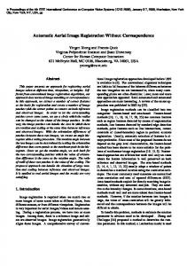
A previous work on pelvis registration focuses on precise bone alignment for bone atlas creation [3]. Such an application requires deformable registration due to the large differences between patient anatomies, since the size and shape of pelvises can vary widely. On the other hand, precise alignment of all bone structures is not crucial as a preliminary step in model-based segmentation of prostate and bladder, the focus is on acceptable alignment of the pubic bone. In addition, deformable registration requires more computing time, in our case fast registration is important. Furthermore, better bone alignment does not guarantee better soft tissue alignment. In model-based segmentation, transforming the CT studies to a common reference frame is a useful step before two tasks. For model making, when the studies are registered, the assumption is that the anatomic regions of different studies can be found nearly in the same voxel regions. By transforming the hand-segmented ground truth segmentations of the organs of interest to this common reference frame together with the CT studies a deformable organ model can be calculated in the reference frame. In the clinical program, automatic alignment of the organ model to the patient study — via applying the inverse of the transformation that takes the study to the reference frame — can reduce the necessary user input to initialize the model-based segmentation. Although the registration methods applied in these steps are almost the same, the requirements are different. The model generation is not part of the actual segmentation process thus the registration can be performed in “batch mode”, i.e., one after the other without user interaction. More precise alignment is preferred to fast execution time. On the other hand, in the clinical program, the execution must be as fast as possible, even sacrificing some precision. We propose an extension not addressed by previous algorithms. The idea is that after an affine transformation which gives global optimal alignment, a refinement step is performed. Global registration prefers alignment of body parts of big volume, like spine or pelvis causing slight or big differences in the pubic bone area (Fig. 1, top row). It is assumed that scale parameters are satisfactorily determined by this global part. During refinement, scale parameters are kept from the global part, then an optimal rigid-body transformation is searched in the pubic bone region only (Fig. 1, bottom row). Many methods could be extended this way [4]. In our actual implementation, we used a general registration method utilizing normalized mutual information [5].
3
Materials
Our test database consists of 33 pelvic CT images provided by General Electric Medical Systems Company. The chosen reference image has 83 2D slices of 512x512 voxels. The in-slice resolution is 0.936562 millimeters, while the slice distance is 3.00 millimeters. Most of the other studies have 60–100 slices, but in some extreme cases 33 or 189 slices are present. The spatial in-slice resolution is in the [0.60-0.98] interval, the slice distance is usually 2.5–3.00 mm. Studies are of varying quality, some are distorted by artificial (metallic) objects.
Application of Automatic Image Registration in a Segmentation Framework
631
Bladder
Bladder
Prostate
Prostate Bone
Pubic Bone
Bladder
Bladder
Prostate
Prostate Bone
Pubic Bone
Fig. 1. Top row: The optimal global registration of a study (dotted outline) against the reference volume (filled shape) — a coronal (left) and a sagittal cross section (right). It is well visible that the organs of the two studies are close to each other, but the overlapping region e.g., of the prostate, is small. Bottom row: The optimal local rigid-body refinement following the global registration of a study against the reference volume — a coronal (left) and a sagittal cross section (right). The result of the registration provides a good starting point for a segmentation algorithm. The figures are derived from real data.
Three clinical experts manually segmented prostate in 26 and bladder in all the 33 studies independently. The experts visually checked the result of the majority vote segmentation (if a voxel was identified as the given organ by at least two experts) together, and accepted it or made some modifications. This final segmentation is used as the gold standard. After visual inspection of the registration results, three studies were omitted from the database because of unacceptable misregistrations. In one of these cases the patient orientation was wrong (a preprocessing step is necessary to solve this), in another case the bladder was filled with contrast material. Note that there are some more studies of this kind and even heavily distorted ones where the registration gave good results. Table 1 summarizes the properties of the studies we use in this paper. Our proposed registration method requires the manual selection of the local neighborhood of the pubic bone in the reference study. This region of interest is used in the local refinement step of the algorithm. During local refinement, voxel intensity values inside this neighborhood only are taken into account. This region should contain the pubic bone, the lower part of ischium, and some soft
632
A. Tan´ acs, E. M´ at´e, and A. Kuba
Table 1. Study database. The last three columns show the size of the gold standard prostates. The volume is given in pixels and cm3 , the last column shows the diameter if a perfect spherical prostate shape is assumed. Study ID
Slices Slice spacing Slice Thick. Prostate Gold Standard (mm) (mm) (pixels) (cm3 ) Sp. diam. (cm) cd2pa2 (reference) 83 0.976562 3 7121 20.37 3.39 cd2pa3 70 0.976562 3 9927 28.40 3.79 cd2pa4 73 0.976562 3 6020 17.22 3.20 cd2pa5 68 0.976562 3 14267 40.82 4.27 cd2pa6 81 0.976562 3 11165 31.94 3.94 cd2pa7 64 0.976562 3 8936 25.57 3.66 cd1prostate1 86 0.9375 2.5 42501 93.39 5.63 cd1prostate2 149 0.820312 1.25 31181 26.23 3.69 cd1prostate3 81 0.9375 2.5 11269 24.76 3.62 cd3pa4st1se1 67 0.976562 2.5 13526 32.25 3.95 cd4pa5st1se1 112 0.976562 2.5 23520 56.08 4.75 cd4pa7st1se1 78 0.976562 2.5 8958 21.36 3.44 cd4pa8st1se1 83 0.976562 2.5 8402 20.03 3.37 cd4pa10st1se1 73 0.976562 2.5 15323 36.53 4.12 cd6pa5st1se2 84 0.976562 3 1570 4.49 2.05 cd6pa8st1se2 73 0.976562 3 15677 44.85 4.41 cd6pa9st1se2 55 0.976562 3 8642 24.72 3.61 cd6pa10st1se2 78 0.976562 3 13652 39.06 4.21 cd8pa8st1se1 39 0.9375 5 24620 108.19 5.91 cd8pa9st1se2 104 0.976562 2.5 33800 80.59 5.36 cd14anon10 49 0.98 5 5157 24.76 3.62 cd14anon14 46 0.976562 3 6362 18.20 3.26 cd14anon21 189 0.9375 5 8124 35.70 4.09
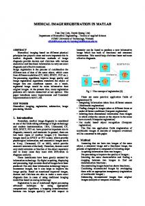
tissue region around them. This selection must be done only once and only for the reference study (Fig. 2).
4
Tests and Results
From the nature of the problem it is evident that ground truth information on the expected registration results is not available. What we have is the expert segmented gold standard database, and we expect that after spatial normalization, the organs will be “close” to each other. Since the sizes and shapes of the bladders vary too much and our database is too small to be able to select enough full, normal, and empty segmented bladders, only the prostate data is used for the evaluation of the global and local refinement methods. We conducted three different tests to find out whether the method utilizing local refinement is significantly better, i.e., brings organs, in this case prostates, closer to each other than the method using global registration only.
Application of Automatic Image Registration in a Segmentation Framework
633
Fig. 2. Surface model (left) and manually selected neighborhood in a transaxial slice (right) of the pubic bone selected for local refinement
Let N denote the number of the studies and P ROSTi ⊆ Z 3 (1 ≤ i ≤ N ) the voxel positions classified as prostate in the ith study after transforming to the common reference frame. In the first test we assume that the � prostates are spherical. For each study, the centroid of the prostate (COGi = x∈P ROSTi x/|P ROSTi |) is computed. The centroid of this set of centroids (COGglobal ), and for each study the Euclidean distance between COGi and COGglobal are computed. The second and third columns of Table 2 show these. The second and third tests utilize a so called probability atlas (P ROB) which is defined as follows. By transforming the hand-segmented ground truth segmentations of the organs of interest to the common reference frame together with the CT studies for each voxel, the probability that the given voxel belongs to a specific organ can be assigned. E.g., the value is 0 if that voxel was not classified as part of the organ in any of the studies, 0.5 indicates that this happened in half of the studies. The second test determines the average probability belonging to the voxels of the transformed gold standard prostates of each study, � P ROB(x) 1 . probi = x∈P ROSTi |P ROSTi | The third test shows the summed probability of the prostate region of a study relative to the whole probability map, � P ROB(x) 2 �ROSTi . probi = x∈P x P ROB(x) For each method and test, statistical parameters (mean, standard deviation and paired two-tailed t-Test) were computed. Table 2 shows the results of the three tests for the global registration only and global registration followed by the local refinement, respectively.
634
A. Tan´ acs, E. M´ at´e, and A. Kuba
Table 2. Results showing Euclidean distances of the centroids, and prob1i and prob2i values Study ID
Centroid Test Global Refined cd2pa3 8.08 2.92 cd2pa4 3.80 7.12 cd2pa5 9.81 7.14 cd2pa6 10.30 8.94 cd2pa7 4.75 4.95 cd1prostate1 9.90 11.19 cd1prostate2 3.73 5.51 cd1prostate3 3.64 6.64 cd3pa4st1se1 12.66 6.14 cd4pa5st1se1 11.40 14.82 cd4pa7st1se1 8.74 7.61 cd4pa8st1se1 22.95 18.07 cd4pa10st1se1 2.75 4.00 cd6pa5st1se2 8.77 8.18 cd6pa8st1se2 10.50 6.69 cd6pa9st1se2 17.95 8.65 cd6pa10st1se2 18.22 4.41 cd8pa8st1se1 22.49 18.99 cd8pa9st1se2 19.04 17.69 cd14anon10 2.57 3.39 cd14anon14 16.09 10.15 cd14anon21 13.19 7.04 Average 10.97 8.65 Std.Dev. 6.30 4.73 t-Test 0.0213
5
Probability Test 1 Global Refined 0.5635 0.6790 0.6439 0.6689 0.4937 0.5685 0.4853 0.5297 0.5934 0.6263 0.3188 0.3290 0.5576 0.5995 0.6274 0.6400 0.4028 0.6082 0.3350 0.3160 0.4867 0.5931 0.1870 0.3049 0.5299 0.5678 0.6395 0.8404 0.4588 0.5254 0.3148 0.4205 0.2792 0.4886 0.2164 0.2501 0.2661 0.4325 0.6212 0.6734 0.3897 0.6070 0.4180 0.5325 0.4468 0.5364 0.1435 0.1449 8.3513E-06
Probability Test 2 Global Refined 0.3837 0.4588 0.2970 0.3015 0.4890 0.5400 0.4143 0.4470 0.4268 0.4466 0.7591 0.7819 0.4752 0.5229 0.4220 0.4157 0.3012 0.4737 0.5391 0.5020 0.2622 0.3451 0.1112 0.1805 0.5515 0.5857 0.0811 0.1077 0.5451 0.6245 0.4175 0.5553 0.3284 0.5906 0.6947 0.7987 0.0165 0.0266 0.4101 0.4652 0.2036 0.3128 0.4290 0.5514 0.3890 0.4561 0.1836 0.1903 0.0001
Discussion
The results in Table 2 indicates that after the refinement step the prostate regions are significantly (P < 0.05) closer to each other compared to the global registration. Registrations were performed on a 3Ghz Pentium IV desktop PC. The running time is about two minutes for a study. It is acceptable for the model creation process since this must be done only once. In the clinical application, fast execution is crucial especially since the registration is only a preprocessing step in the segmentation process which is time consuming by itself. Utilizing several optimizations (e.g., by using only the coarser levels of a hierarchical representation), the running time is currently between 20–40 seconds. It can be further reduced to nearly its half by reducing the number of voxels in the reference volume removing the unnecessary parts (e.g., using the bounding box of the patient data). Visual inspection and preliminary statistical results show that although the precision is slightly reduced, the result is acceptable in most cases.
Application of Automatic Image Registration in a Segmentation Framework
6
635
Conclusions
In this paper we focused on a preprocessing step of a segmentation framework. Before model generation, transforming the studies of different patients to a common reference frame is useful. In the clinical application, initial organ model placement can be established automatically. Note that automatic registration does not guarantee acceptable results, so visual inspection is necessary. In our database the failure rate of registrations was low (three out of 26) even though the studies were “real-life”, many of them distorted by metallic objects, or not satisfying the assumed protocol (e.g., wrong patient position, contrast agent is visible in the images).
Acknowledgements This work has been supported and the image database was provided by GE Medical Systems. We thank our clinical experts, Dr. Katalin Gion, Dr. Istv´ an Csenkey-Sink´ o, and Dr. Endre Szab´ o from the Department of Radiology, University of Szeged for their indispensable work during the gold standard creation process.
References 1. Pekar, V., McNutt, T.R., Kaus, M.R.: Automated Model-based Organ Delineation for Radiotherapy Planning in Prostatic Region, Int. J. Radiation Oncology Biol. Phys. 60 No 3 (2004) 973–980 2. Maintz, J.B.A., Viergever, M.A.: A survey of medical image registration. Medical Image Analysis 2 (1998) 1–36 3. Yiqiang Zhan, Dinggang Shen, Russ Taylor: Deformable Registration of Male Pelvises in CT Images, IEEE International Symposium on Biomedical Imaging (ISBI), Arlington, VA, April 15-18, 2004. 4. West, J.B., Fitzpatrick, J.M., et al.: Retrospective Intermodality Registration Techniques for Images of the Head: Surface-Based Versus Volume-Based. IEEE Trans. on Medical Imaging 18 (1999) 144–150 5. Tan´ acs, A., Kuba, A.: Evaluation of a Fully Automatic Medical Image Registration Algorithm Based on Mutual Information, Acta Cybernetica 16 (2003) 327–336