Arrhythmia Classification in Long-Term Data Using Relative RR Intervals Marcus Vollmer Institute of Bioinformatics, University Medicine Greifswald, Germany Abstract Background: The automated heart beat detection and the classification of arrhythmic beats from ECG recordings are difficult tasks. Even thoroughly annotated standard databases have a wealth of misplaced heart beats and a visual identification of all affected ECG parts is time-consuming. The study of relative RR intervals can help in this regard. Methods & Results: Relative RR intervals are defined as the change of two successive RR intervals weighted by their mean. I have analyzed the return maps of relative RR intervals of the Normal Sinus Rhythm Database as well as the MIT-BIH Arrhythmia Database. Different structures have been investigated analytically and are reproducible due to modeling. I am able to distinguish structures regarding arrhythmia types and cases of insufficient heart beat detection. Upon that approach, I have developed filtering criteria of artifacts from RR interval sequences. Utilization: The return map of relative RR intervals can be used as visualization technique for the detection of arrhythmia types or failures in the automated beat detection. Further, it can be used to apply filtering criteria for the removal of irregular heart beats in RR interval data or to improve heart beat detection algorithms by applying a more sensitive method in the effected parts. This may help to distinguish irregular beats from false positive alarms caused by interference effects, possibly in postprocessing routines.
1.
Introduction
RR intervals are the differences of successive heart beats (R-peaks) in an ECG as the result of an automated heart beat detection. Besides the computation of the heart rate, RR intervals are mainly used for the quantification of heart rate variability (HRV) [1]. The geometry and the dynamics of the return map of RR intervals (Poincar´e plot) has been studied by [2] and [3] for example. Currently some conventional mobile monitoring systems (e.g. Polar sports watches) are able to save RR intervals or the heart rate as a processed information. Due to limited resources and the absence of the need, the storage of raw signals is not
Computing in Cardiology 2017; VOL 44
Page 1
practiced in general, in contrast to a clinical environment. In our digitized world, it is worth to study RR intervals more thoroughly, since the technical opportunity outside of clinics can assist remote diagnosis (telemedicine), but with problems of dirty data.
2.
Relative RR Intervals
In this context it is quite promising to analyze relative RR interval sequences. Relative RR intervals rri are defined as a weighted difference of successive RR intervals: rri :=
RRi − RRi−1 1 2 (RRi + RRi−1 )
for all i ∈ {2, . . . , n},
(1)
with the following properties: 1. 2. 3. 4.
−2 ≤ rri ≤ +2, rri = 0 if and only if RRi = RRi−1 , rri = −2 if and only if RRi = 0, rri = +2 if and only if RRi−1 = 0.
The first property implies that relative RR intervals are located between −200% and +200% as the result of weighting the difference by the mean. To prove this property we set RRi =c · RRi−1 , with c≥0. Then ( ) (c−1)RRi−1 c−1 2 rri = 1 =2· = 2 1− . (2) c+1 c+1 2 (c+1)RRi−1 2 c+1
is strictly monotonically decreasing for 0≤c≤∞. As a function of c, rri is strictly monotonically increasing and reaches its lowest value of −2 at c=0, that means for RRi =0. The limit of rri for c → ∞ is +2 that is reached for RRi−1 =0.
3.
Structures in the Return Map
The return map of relative RR intervals is the scatter plot of pairs of values (rri , rri+1 ) for i=1, . . . , n−1. In concrete terms, we use a standardized form of the return map of absolute RR intervals, due to the weighting of the difference of successive RR intervals. The multidimensional view has proven its reliability in methods of the chaos theory (e.g. [4] or [5]). To get practical experience, I calculated relative RR intervals from data of the “Normal Sinus Rhythm RR Interval Database” (nsr2db)[6]. The
ISSN: 2325-887X DOI:10.22489/CinC.2017.213-185
RR sequence 10
+4.0% +3.1% +3.7% -9.2% -3.3% +8.8% -0.8% -6.3% -5.9% +1.7% 0.95
0.99
1.02
1.06
0.97
0.94
1.02
1.02
0.95 0.90
Return map of relative RR intervals
8
0.91
6
3
10%
2 7
5%
1.05 1.00
1
0.95 0.90
5 10
8
rri+1
RRi+1
6
Return map of relative RR-Intervals
4
5
2 0 -2 -4
6 4
-5%
9
21
9
0%
4
rri+1 in %
Return map of RR-Intervals
8
-6
7 3
-10%
0.90 0.95 1.00 1.05
-10%
RRi
0%
-8 -10 -10 -8
10%
-6
-4
-2
0
2
4
Arrhythmia and artifacts (20000 random coordinates from nsr2db)
150
100
2"
2!rri 6+rri
rri+1 in %
50
0
-50
6!
16 2!rri
-100
-150
-200 -200
-150
-100
8
(b) Coordinates of 10000 random value pairs.
(a) Short sequence of absolute and relative RR intervals and the return maps.
200
6
rri in %
rri
-50
0
50
100
150
rri in % (c) Return map of 20000 random pairs of successive relative RR intervals lying outside ±20%.
Figure 1: Plots from intervals of the Normal Sinus Rhythm RR Interval Database
Page 2
200
10
RR sequences and relative RR intervals A
B
C
D
E
-56.6 % 0.73
+58.5% 0.41
+0.0% 0.75
0.74
0.75
0.66
+3.2%
-6.3% 0.77
+0.0% 0.66
+0.0%
+64.8% 0.38
+2.1%
+0.0%
0.63
-63.9 %
+0.0% 0.63
0.75
0.72 0.04
+63.9% 0.38
-179.4 + %180.0%
+1.0%
0.74
0.66
0.18
-42.4 % 0.63
0.48
0.41
-1.0%
-3.2%
0.83
0.74 +2.4%
-26.7 % 0.63
0.55
Extrasystole without compensatory pause or false positive detection
-1.2%
+2.6% 0.60
False positive heart beat detection
0.72
0.66
-5.1%
-180.3 %
0.71 0.73
0.55
0.64
Sequence of ventricular extrasystoles or misplaced annotations
+0.0%
0.74
-3.6%
0.66
+68.4%
-30.3 %
0.75
-114.8 %+91.8% +31.3%
+166.9%
A
-64.8 %
0.65 -39.4 % 0.62
Ventricular extrasystole or misplaced annotation
0.41
+53.1%-1.1% -1.1% +0.0% -1.1%
8.15
0.42 0.73 0.72 0.71 0.71 0.70
B
C
Asystole or false negative detection
D
E
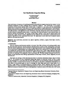
Figure 2: Arrhythmia types and annotation failures of heart beat detection correspond to special structures in the return map of relative RR intervals. heart beats of 54 long-term ECGs from test persons with a normal sinus rhythm were automatically annotated and checked visually. Figure 1a shows a sequence of 10 successive RR intervals from the database. For each pair of consecutive RR intervals, I computed the relative change. The detachment from the heart rate becomes apparent when comparing the return maps of absolute and relative RR intervals. Relative changes are centered around the origin (0, 0), whereas absolute RR intervals are basically stretched along the line of identity. Figure 1b shows the bundling of pairs near the coordinate origin. 97.6% of all pairs are located between −20% and +20%. With great certainty these are pairs of normal intervals (NN intervals). Interspaces between the coordinates arises as a result of the sampling frequency (fs =128 Hz for nsr2db). Impressive structures become visible when looking at the whole domain (see Figure 1c). Not all theoretical possible rr intervals occur. Typical patterns correspond to specific arrhythmia types or failures in heart beat detection (see Figure 2).
extrasystole (case C in Figure 2), which is sandwiched between two normal beats. The time between normal beats is RRi . Previous and following RR intervals varies only slightly with RRi−2 ≈ · · · ≈ RRi+2 . The extrasystole occurs after some coupling time that splits RRi into the intervals RRi1 =a·RRi and RRi2 =(1−a)·RRi with 020% and |rri+2 −rri+1 |>20%. This corresponds to extrasystoles or misplaced heart beats (cases B,C,D in 2). Next, restart the calculation of relative RR intervals and remove further irregularities: 3. Replace RRi with NaN if RRi−1 =NaN and |rri+1 |>15%. 4. Replace RRi and RRi−1 with NaN if |rri |>50% . Clifford et al. have used similar exclusion criteria, dealing with a mixture of absolute and relative intervals [9]. Instead of weighting with the mean of successive intervals, their relative rules are based on a reference interval (mean of the five last sinus intervals). Patient characterization A close look at the return map of relative RR intervals with its limited domain can assist cardiologists in order to characterize and classify patients. Recurring patterns in the return map (e.g. triangles of case C) are visible and refers to certain arrhythmia types. Detection problems I checked the return maps derived from annotations of different detection algorithms. One can clarify different and common detection problems and the robustness of several algorithms against different waveforms can be compared (not published). Reduce false alarms Further, it can be used to improve heart beat detection algorithms by applying a more sensitive method in the effected parts, e.g. to check additionally for P and T-waves in the ECG-complex. This could be
Page 4
done independently from an existing algorithm in a postprocessing routine. In my previous work [10] I have already used relative RR intervals to evaluate the signal quality of different biosignals and considered also relative intervals of higher grades. HRV Measurement Through its centered version of the classical Poincar´e plot, periodic orbits (caused by the vagal tone) become more visible than in standard reports. I proposed a heart rate variability measure based on relative RR intervals which is easy to understand and robust against artifacts and heart rate changes [11].
References [1]
Malik M, Bigger JT, Camm AJ, Kleiger RE, Malliani A, Moss AJ, Schwartz PJ. Heart rate variability. European Heart Journal 1996;17(3):354–381. [2] Piskorski J, Guzik P. Geometry of the poincare plot of rr intervals and its asymmetry in healthy adults. Physiological Measurement 2007;28(3):287. [3] Karmakar C, Khandoker A, Gubbi J, Palaniswami M. Complex correlation measure: a novel descriptor for poincare plot. BioMedical Engineering OnLine 2009;8(1):17. [4] Peng CK, Hausdorff JM, Goldberger A, Walleczek J. Fractal mechanisms in neuronal control: human heartbeat and gait dynamics in health and disease. Nonlinear Dynamics Self organization and Biomedicine 2000;66–96. [5] Fell J, Mann K, R¨oschke J, Gopinathan M. Nonlinear analysis of continuous ECG during sleep II. Dynamical measures. Biological cybernetics 2000;82(6):485–491. [6] Goldberger AL, Amaral LAN, Glass L, Hausdorff JM, Ivanov PC, Mark RG, Mietus JE, Moody GB, Peng CK, Stanley HE. PhysioBank, PhysioToolkit, and PhysioNet: Components of a New Research Resource for Complex Physiologic Signals. Circulation 2000;101(23):e215–e220. [7] Tsipouras M, Fotiadis D, Sideris D. An arrhythmia classification system based on the RR-interval signal. Artificial Intelligence in Medicine 2005;33(3):237–250. [8] Park J, Lee S, Jeon M. Atrial fibrillation detection by heart rate variability in Poincare plot. Biomedical engineering online 2009;8(1):38. [9] Clifford GD, Azuaje F, McSharry P. ECG Statistics, Noise, Artifacts, and Missing Data. Advanced Methods and Tools for ECG Data Analysis 2006;6:18. [10] Vollmer M. Robust detection of heart beats using dynamic thresholds and moving windows. In Computing in Cardiology. 2014; 569–572. [11] Vollmer M. A Robust, Simple and Reliable Measure of Heart Rate Variability using Relative RR Intervals. In Computing in Cardiology. 2015; 609–612.
Address for correspondence: Marcus Vollmer /
[email protected] Institute of Bioinformatics / University Medicine Greifswald Walther-Rathenau-Str. 48 / 17475 Greifswald / Germany


![RR` ]RR`](https://m.moam.info/img/260x300/rr-rr_59c1f6ef1723ddc052bf1ab1.jpg)
![RR` ]RR`](https://m.moam.info/img/260x300/rr-rr_599b63921723dd0f406edb17.jpg)