Jan 6, 2016 - Hwunjae Lee12, Seung-Hyun Yang12, Dan Heo12, Heyoung Son3, ..... A. Y. Kim, J. K. Han, C. K. Seong, T. K. Kim, and B. I. Choi, J. of.
Article Copyright © 2016 American Scientific Publishers All rights reserved Printed in the United States of America
Journal of Nanoscience and Nanotechnology Vol. 16, 196–202, 2016 www.aspbs.com/jnn
Molecular Imaging of CD44-Overexpressing Gastric Cancer in Mice Using T2 MR Imaging Hwunjae Lee1� 2 , Seung-Hyun Yang1� 2 , Dan Heo1� 2 , Heyoung Son3 , Seungjoo Haam2� 4 , Jin-Suck Suh1� 2� 3� 5 , Jaemoon Yang1� 5� ∗ , and Yong-Min Huh1� 2� 5� ∗ 1
2
Department of Radiology, College of Medicine, Yonsei University, Seoul, 120-752, Republic of Korea Nanomedical Interdisciplinary Program, National Core Research Center, Yonsei University, Seoul 120-749, Republic of Korea 3 Severance Biomedical Science Institute, College of Medicine, Yonsei University, Seoul, 120-752, Republic of Korea 4 Department of Chemical and Biomolecular Engineering, Yonsei University, Seoul 120-749, Republic of Korea 5 YUHS-KRIBB Medical Convergence Research Institute, Seoul 120-752, Republic of Korea Novel diagnostic techniques have been developed in many research area using targetable contrast agents with magnetic resonance imaging (MRI) for cancer diagnosis. For cancer diagnosis, the use of MRI with biocompatible targeting moieties and manganese ferrite nanoparticles (MFNPs) is preferred. Thus, we synthesized MFNPs using a thermal decomposition method which enables sensitive T2 or T2 Turbo Spin Echo (TSE) MRI and coated them with hyaluronic acid (HA). The high targeting ability of HA-MFNPs was observed at MKN-45 cells (gastric cancer cell line) which high-expressing CD44 in contrast with MKN-28 cells which to: low-expressing CD44. We also prepared Delivered by Publishing Technology Yonsei University the gastric cancer mice using MKN-45 which has the stem-like property was implanted IP: model 165.132.14.104 On:cells Wed, 06 Jan 2016 04:53:37 Copyright: American Scientific Publishers into BALB/c nude mice. And then HA-MFNPs of the T2 contrast enhancement effects and targeting ability were investigated by in vivo MR imaging. As a result of these studies, we conclude that HA coated MFNPs can be effectively used as a novel probes for visualizing gastric cancer stem cells.
Keywords: Contrast Agent, Gastric Cancer, CD44, Magnetic Nanoparticles, Magnetic Resonance Imaging.
1. INTRODUCTION Molecular imaging provides a tool to diagnose cancer at the cellular and molecular levels. It not only allows early and accurate tumor localization for diagnostic cancer imaging, but also has the potential to visualize the biological processes of tumor growth, metastasis, and response to treatment.1–10 Molecular magnetic resonance (MR) imaging has emerged as a key tool in the diagnosis of cancer,10–16 since it has advantages over noninvasive, anatomical imaging due to its high resolution, high contrast, and 3-dimensional information in real time, much more so than nuclear medicine (positron electron tomography and single-photon emission computed tomography) and optical imaging.17–21 In addition, molecular MR imaging is able to simultaneously detect the metabolism of cells and tissues, physiological and structural information, and biological processes occurring in deep tissues.22� 23 ∗
Authors to whom correspondence should be addressed.
196
J. Nanosci. Nanotechnol. 2016, Vol. 16, No. 1
Molecular MR imaging can be used to observe a variety of lesions in the diagnosis of gastric cancer but also has limitations.24� 25 In case of gastric cancer, it is difficult to diagnose using MR imaging because many foods and other digestive secretions exist inside the stomach.24� 26–29 Currently, many research groups are looking for a solution, but gastric cancer’s limitations and difficulties can be addressed using various MR imaging contrast agents and MR sequence. Many MR contrast agents have been used for good quality imaging.13� 30–34 However, the blood pool contrast agents cannot specifically reach their target goals. Thus, we aim to develop a targetable contrast agent using hyaluronic acid.35–41 In particular, hyaluronic acid is known to interact with the CD44 receptor, and gastric cancer is known to overexpress the CD44 receptor, a biomarker of cancer stem cells.42–47 It is crucial for MR probes to target early gastric cancer from a diagnostic point of view. CD44 is important as a gastric cancer stem cell marker as it interacts with hyaluronic acid. hyaluronic 1533-4880/2016/16/196/007
doi:10.1166/jnn.2016.11782
Lee et al.
Molecular Imaging of CD44-Overexpressing Gastric Cancer in Mice Using T2 MR Imaging
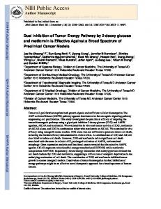
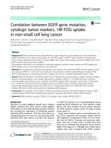
Scheme 1. Schematic illustration of hyaluronic acid-conjugated manganese ferrite nanoparticles (HA-MFNPs) for CD44-expressing gastric cancer cell-specific MR imaging
acid is a nontoxic, biocompatible polymer, with tandem 2.2. Synthesis of Manganese Ferrite Nanoparticles (MFNPs) disaccharide repeats of ß-1,4-D-glucuronic acid-ß-1,3-DN-acetylglucosamine; components of the glycosaminoglyThe monodispersed MFNPs were prepared by thercan family have been used in various areas as targeting mal decomposition method.54 Briefly, 2 mmol iron (III) 41� 48–53 Technology to: Yonsei University Delivered by Publishing moieties for MR probes as well as antibodies. acetylacetonate, 1 mmol manganese (II) acetylacetonate, In IP: 165.132.14.104 On: Wed, Jan 2016 04:53:37 1006 mmol 1,2-hexadecanediol, 6 mmol dodecanoic acid, addition, MRI sequence is also important in cancer diagnoCopyright: American Scientific Publishers and 6 mmol dodecylamine were dissolved in 20 mL bensis, In this study, molecular MR imaging was investigated zyl ether under an ambient nitrogen atmosphere. The mixto find biological processes that occur in gastric cancer. � ture was then preheated to 200 C for 2 h and refluxed at A T2 TSE sequence was used to confirm better diagnos� 300 C for 30 min. After the reactants were cooled to room tic possibilities and targeting effects were demonstrated temperature, the products were purified with an excess of using hyaluronic acid-conjugated MFNPs (HA-MFNPs) in pure ethanol. Approximately 11 nm MFNPs were synthea heterotopic xenograft gastric cancer model (Scheme 1). 55 sized using the seed-mediated growth method. Various experiments were conducted to evaluate specific binding affinity and diagnostic effectiveness both in vivo 2.3. Preparation of HA-MFNPs and in vitro. Aminated MFNPs were fabricated using the nanoemulsion method. First, 30 mg of MFNPs was dissolved 2. EXPERIMENTAL DETAILS in 4 ml of n-haxane (organic phase). The organic phase 2.1. Materials was injected into 30 ml of de-ionized water (aqueous Polysorbate 80 (P80), ethylenediamine, 1,4-dioxane phase) containing 100 mg of aminated P80. After mutual (99.8%), and 1,1� -carbonyldiimidazole (CDI) were pursaturation, the solution was emulsified for 20 min under chased from Sigma Aldrich Chemical Co. Phosphate ultrasonification (ULH700S, Ulssohitech, Cheonwon-gum, buffered saline (PBS: 10 mM, pH 7.4), Roswell Park South Korea) at 450 W. The mixture was kept overnight at Memorial Institute-1640 (RPMI-1640), fetal bovine serum room temperature to remove the volatile organic solvent. (FBS) and antibiotic-antimycotic solution were purchased The product were purified using a centrifugal filter (Cenfrom Gibco and dialysis membrane (1 kDa MWCO) triprep YM-3, 3-kDa molecular weight cutoff (MWCO), from Spectrum laboratory. Hyaluronic acid (1 MDa) was Amicon, Millipore Corporation, Billerica, MA, USA) in supplied from Yuhan Phrmaceutical Corporation (Seoul, three times at 3,000 rpm for 20 min. HA-MFNPs were Korea). MKN-45 and MKN-28 (American Tissue Type fabricated by EDC-NHS chemistry. First, the pH of the Culture) cell lines were grown in medium containing 10% aminated MFNP solution was adjusted to neutral condiFBS and 1% antibiotic antimycotic at 37 � C and a humidtion by the addition of 0.1 N HCl solution. Then, HA ified 5% CO2 atmosphere. Ultrapure deionized water was (9.63 �mol) were dissolved in the 40 ml of de-ionized water followed by the addition of EDC and sulfo-NHS. used for all of the syntheses. J. Nanosci. Nanotechnol. 16, 196–202, 2016
197
Molecular Imaging of CD44-Overexpressing Gastric Cancer in Mice Using T2 MR Imaging
Each HA solution was added to aminated MFNPs containing 50 mg of MFNPs. The HA and aminated MFNPs were reached for 2 h at room temperature. Finally, EDC, sulfo-NHS, and unbound HA were removed using dialysis (MWCO, 25,000) against excess de-ionized water. The hydrodynamic diameter and zeta potential of HA-MFNPs were analyzed by laser scattering (ELS-Z, Otsuka Electronics) and the relaxivity (R2) data of the HA-MFNPs was obtained by MR imaging analysis.
Lee et al.
The working solution was prepared by mixing 10% potassium ferrocyanide and 20% HCl in equal amounts. The working solution was added to the cells and incubated for 30 min at room temperature. The cells were washed with PBS and stained with nuclear fast red solution (Sigma, USA) for 30 min at room temperature. The cells were observed using an optical system microscope (Olympus BX51, Japan).
2.7. Heterotopic Animal Model and Experimental Procedure 2.4. Biocompatibility Tests for HA-MFNPs All animal experiments were conducted with the approval The cytotoxic effects of HA-MFNPs in MKN-45 cells and of the Association for Assessment and Accreditation of MKN-28 was evaluated by 3-(4,5- dimethylthiazol-2-yl)Laboratory Animal Care (AAALAC) International. Female 2,5-diphenyltetrazolium bromide (MTT) assay. MKN-45 BALB/c nude mice at 7–8 weeks of age were anesthetized and MKN-28 cells were maintained in RPMI-1640 conby intraperitoneal injection of a Zoletil/Rompun mixture taining 10% FBS and 1% antibiotics at 37 � C in a humidand 1.0 × 10⁷ MKN-45 cells which suspended in 200 �L ified atmosphere with 5% CO2 . MKN-45 and MKN-28 saline were implanted into the femoral region. After cancer cells (1.0 × 10⁴ cells/well) were seeded into a 96-well � cell implantation, MR imaging was performed between 2 plate at 37 C overnight and the cells were incubated with and 3 weeks. After in vivo MR imaging, MR imaging of various concentrations of HA-MFNPs for 4 h. The cells the harvested organ was also performed. In addition, the were washed with 100 �L PBS (pH 7.4, 1 mM), and extracted tumor tissues from tumor-bearing mice treated 100 �L phenol red free RPMI-1640 was added. Subsewith HA-MFNPs were frozen, sectioned, and stained using quently, the cells were treated with MTT assay solution Prussian blue. All stained tissue sections were analyzed according to the manufacturer’s instructions. Cell viabilusing a virtual microscope (Olympus BX51, Japan) and ity was evaluated using a microplate reader (Synergy H4 Olyvia software. hybrid reader, BioTek) at an absorbance wavelength of Delivered by Publishing Technology to: Yonsei University 575 nm (reference wavelength of 650 nm). Cell viabilIP: 165.132.14.104 On: Wed, 06MR Jan Imaging 2016 04:53:37 2.8. ity was represented by normalization against HA-MFNPsCopyright: American Scientific Publishers We performed solution and in vitro MR imaging experinon-treated cells (which were considered as having 100% ments with a 1.5 T clinical MRI instrument with a microcell viability). 47 surface coil (Intera, Philips Medical Systems, Best, The Netherlands). The R2 (T2 relaxation rate, 1/T2, s−1 ) 2.5. Darkfield Microscopy of the HA-MFNPs solution and HA-MFNPs-treated cells MKN-45 and MKN-28 cells (2.0 × 10⁵ cells/well) were (1 × 10⁷) were measured by using the Carr-Purcellseeded onto cover glass in 4 well plates and incubated Meiboom-Gill (CPMG) sequence at room temperature at for 4 h at 37 � C. Prepared various concentrations of with the following parameters: TR = 10 sec, 32 echoes HA-MFNPs were added to RPMI. After incubation for with 12 msec even echo space, number of acquisitions = 1, � 48 h at 37 C, the cells were washed with PBS and fixed point resolution of 156 × 156 �m, and section thickness of with 4% paraformaldehyde. To observe HA-MFNPs in the 0.6 mm. For the acquisition of T2-weighted MR images of cells, the light scattering images were recorded using an MFNPs solution and HA-MFNPs or HA-MFNPs-treated inverted microscope (Olympus BX51, Japan) with a highly cells, the following parameters were adopted: resolution numerical darkfield condenser (U-DCW, Olympus), which of 234 × 234 �m, section thickness of 2.0 mm, TE = delivers a very narrow beam of white light from a tung15 msec, TR = 400 msec, and number of acquisitions = 1. sten lamp to the surface of the sample. Immersion oil (nd: The r2 (mM−1 s−1 ) is equal to the ratio of the R2 to 1.516, Olympus) was used to narrow the gap between the the HA-MFNPs concentration. And in vivo MR imaging condenser and the glass slide, and to balance the refractive experiments were performed with a 3T Siemens clinical index. MR imaging instrument using a human wrist coil with T2 TSE sequence (TR: 4,000 ms, TE: 114 ms, slice thickness: 2.6. Prussian Blue Staining 1.0 mm, FOV read: 180 mm). MKN-45 and MKN-28 cells (1.0 × 10⁶ cells/well) were seeded into 6-well plates and incubated at 37 � C. Pre3. RESULTS AND DISCUSSION pared various concentrations of HA-MFNPs were mixed 3.1. Preparation of MFNPs and HA-MFNPs with RPMI-1640 and these mixtures were added to the cells. After incubation for 4 h at 37 � C, the cells were To detect the target cancer cells in MR images using low washed with PBS and fixed with 4% paraformaldehyde. dose of contrast agent, the contrast agent should have the 198
J. Nanosci. Nanotechnol. 16, 196–202, 2016
Lee et al.
Molecular Imaging of CD44-Overexpressing Gastric Cancer in Mice Using T2 MR Imaging
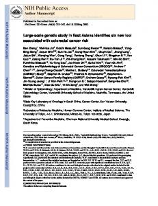
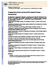
high magnetic susceptibility. Thus, we synthesized high magnetic properties at the appropriate size to avoid reticuloendothelial system detection and prolong retention in crystalline MFNPs (MnFe2 O4 ) by the thermal decompothe circulation. In Figure 1(d), the T2-weighted MR image sition method as previously reported.54 It was reported exhibited a strong black color, which signified a decrease that the relaxivity coefficient of MnFe2 O4 is approximately in signal intensity for the thicker HA-MFNPs solution. 150% larger than Fe3 O4 .57 The targeting moiety is also necessary for target specific MR imaging, thus we selected hyaluronic acid to targeting CD44-overexpressed gastric 3.2. In Vitro Analysis of HA-MFNPs cancer cells. For the conjugation with hyaluronic acid, The MTT assay was then performed, in which yellow the MFNPs were enveloped by aminated P80 using nanotetrazolium salt is reduced to purple formazan crystals in emulsion method. HA-MFNPs were synthesized using metabolically active cells. The relative percentage of cell EDC/sulfo-NHS cross-linker. The hydrodynamic diameter viability was determined as the ratio of formazan intensity of the aminated MFNPs and HA-MFNPs were determined in viable cells which treated with HA-MFNPs to the intento be 76.0 ± 18.5 nm and 132.4 ± 33.4 nm, respectively sity in non-treated (control) cells. As shown in Figure 1(c), (Fig. 1(a)). In addition, the surface charge of the aminated the in vitro cytotoxicity measured by MTT assay showed MFNPs also changed from 21.2 ± 1.1 mV to −15�9 ± that the viability of MKN-45 and MKN-28 cells was 80% 1�2 mV after conjugation of hyaluronic acid due to the at a concentration of 4.1 �g Fe + Mn/mL in HA-MFNPs. negative charge of carboxyl group of hyaluronic acid For verifying specific targeting efficacy of HA-MFNPs, (Fig. 1(a)). After the conjugation of hyaluronic acid and darkfield microscopy and Prussian blue staining analythe aminated MFNPs, the size slightly increased due to sis were carried out and MKN-45 (CD44 +) and MKNthe large molecular weight of hyaluronic acid (1 MDa). 28 (CD44 −) cell lines were selected due to difference The zeta potential was changed from the positive to negof their CD44-expressing level. In Figure 2(a), darkfield ative after conjugation of hyaluronic acid because of the microscopy images demonstrated that HA-MFNPs disnegative charge of carboxyl structure of hyaluronic acid. played excellent binding to MKN-45 cells in comparison As shown in Figure 1(b), the characteristic band of with MKN-28 cells. HA-MFNPs conjugates were verified by FT-IR specMKN-45 cells treated with HA-MFNPs exhibited bright tra, which exhibits O–H stretching at 3200–3400 cm−1 , spots. Furthermore, MKN-28 cells treated with HAC O stretching at 1100–1300 cm−1 ,byCO–NH(amide) MFNPsto:exhibited a low number of bright spots, indiDelivered Publishing Technology Yonsei University −1 bonds at 1630–1680 cm andIP: CH165.132.14.104 bending in HA at cating that cellular binding affinity was low. Darkfield On: Wed, 06 Jan 2016 04:53:37 2 Copyright: Publishers 1430–1470 cm−1 . To assess the potential use American of HA- Scientific microscopy method is excluding the unscattered beam MFNPs as MR imaging agents, we performed MR imaging from image. As a result, the field around the specianalysis using HA-MFNPs, which exhibited the highest men is generally dark. However, nanoparticles under same
Figure 1. Characterization of HA-MFNPs for molecular MR imaging. (a) Hydrodynamic diameter (gray bar) and zeta potential (black circle) of aminated manganese ferrite nanoparticles (aminated MFNPs) and hyaluronic acid-conjugated MFNPs (HA-MFNPs), respectively. (b) Fourier transform infrared spectra of HA (green line), MFNPs (red line) and HA-MFNPs (black line). (c) Cell proliferation assay of MKN-28 (CD44 −), MKN-45 (CD44 +) cells after treatment of HA-MFNPs at several concentrations. (d) Table of R2 values and MR images for each Fe concentration of HA-MFNPs.
J. Nanosci. Nanotechnol. 16, 196–202, 2016
199
Molecular Imaging of CD44-Overexpressing Gastric Cancer in Mice Using T2 MR Imaging
Lee et al.
Figure 2. In vitro microscopic analysis of MKN-28 (CD44 −) and MKN-45 (CD44 +) cells after treatment of HA-MFNPs. (a) darkfield microscopic images of MKN-28 (CD44 −), MKN-45 (CD44 +) cells were incubated with 10 �g/mL of HA-MFNPs in 4 h. (b) prussian blue staining images of MKN-28 (CD44 −), MKN-45 (CD44 +) cells were treated with 40 �g/mL of HA-MFNPs in 4 h. All scale bars are 5 �m.
brightness were scattered more than cellular matrix, thus they shine brightly like a white spots. In Figure 2(b), the extent of intracellular uptake of HA-MFNP in both MKN-45 and MKN-28 cells was confirmed using microscopic images after Prussian blue
staining. Prussian blue staining is a commonly used in histopathology to detect the presence of iron in specimens. Any ferric ion present in the specimens combines with the ferrocyanide and results in the formation of a bright blue color. As a result, one can confirm the presence of a small
Delivered by Publishing Technology to: Yonsei University IP: 165.132.14.104 On: Wed, 06 Jan 2016 04:53:37 Copyright: American Scientific Publishers
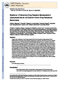
Figure 3. In vivo MR imaging of MKN-45 xenograft mouse model and histological analysis. (a) T2 TSE MR images of xenograft mouse model after intravenous injection of HA-MFNPs. TR: 4,000 ms, TE: 114 ms, Slice thickness: 1.0 mm, FOV read: 180 mm, coil elements: wrist coil. (b) �R2/R2Pre-injection (%) graph verses time after intravenous injection of HA-MFNP respectively. (c) Prussian blue staining image of tumor in mouse xenograft model. Right pictures are magnified image of 80 times from left.
200
J. Nanosci. Nanotechnol. 16, 196–202, 2016
Lee et al.
Molecular Imaging of CD44-Overexpressing Gastric Cancer in Mice Using T2 MR Imaging
amount of iron contained in the nanoparticles in cells or tissues. The transport of HA-MFNPs into MKN-45 cells took place efficiently. In contrast, MKN-45 and MKN-28 cells treated with HA-MFNPs exhibited insignificant cellular uptake. The small fractions of observed iron were represented the nonspecific cellular binding of MFNPs.
Research Foundation of Korea (NRF) grant funded by the Korea government (MEST) (NRF-2014R1A1A2059806).
References and Notes
1. J. K. Kim, K. J. Choi, M. Lee, M. H. Jo, and S. Kim, Biomaterials 33, 207 (2012). 2. M. F. Kircher, H. Hricak, and S. M. Larson, Molecular Oncology 6, 182 (2012). 3.3. In Vivo MR Imaging Analysis of HA-MFNPs 3. X. Meng, B. W. Loo, Jr., L. Ma, J. D. Murphy, X. Sun, and J. Yu, The xenograft mice model were prepared to heterotopic The J. of Nuclear Medicine: Official Publication, Society of Nuclear tumor model, thus MKN-45 cells were implanted into Medicine 52, 1573 (2011). 4. P. Gong, B. Shi, M. Zheng, B. Wang, P. Zhang, D. Hu, D. Gao, right thigh intrasubcutanously because it has some advanZ. Sheng, C. Zheng, Y. Ma, and L. Cai, Biomaterials 33, 7810 tages to observe the volume or morphology of the tumor (2012). rather than orthotopic gastric cancer model. In Figure 3, 5. R. G. Blasberg, Molecular Cancer Therapeutics 2, 335 (2003). contrast enhancement in MR imaging was identified after 6. J. M. Hoffman and A. E. Menkens, Academic Radiology 7, 905 (2000). 7. J. Bzyl, W. Lederle, A. Rix, C. Grouls, I. Tardy, S. Pochon, HA-MFNPs injection into xenograft mice model. In T2 M. Siepmann, T. Penzkofer, M. Schneider, F. Kiessling, and TSE MR images, clear anatomic details were observed, M. Palmowski, European Radiology 21, 1988 (2011). and there was no artifact due to a difference in suscep8. M. Nishino, D. M. Jackman, H. Hatabu, P. A. Janne, B. E. Johnson, tibility. Initially, the center of the tumor instantly darkand A. D. Van den Abbeele, Academic Radiology 18, 424 (2011). ened, and contrast enhancement at surrounding vessels 9. F. Kiessling, Radiology 256, 331 (2010). 10. K. Pinker, A. Stadlbauer, W. Bogner, S. Gruber, and T. H. Helbich, was simultaneously observed (Fig. 3(a)). Four hours after European Journal of Radiology 81, 566 (2012). intravenous injection of HA-MFNPs into the lateral tail 11. D. Artemov, N. Mori, B. Okollie, and Z. M. Bhujwalla, Magnetic vein of the mice model, the T2-contrast enhancement Resonance in Medicine: Official Journal of the Society of Magnetic was observed at the tumor site. In contrast, in the MR Resonance in Medicine/Society of Magnetic Resonance in Medicine 49, 403 (2003). image of post-injection 30 min, the change of T2-signal 12. A. Gossmann, Y. Okuhata, D. M. Shames, T. H. Helbich, T. P. L. was not shown significantly. This result was re-confirmed Roberts, M. F. Wendland, S. Huber, and R. C. Brasch, Radiology �R2/R2Pre-injection (%) graph. We think this data demonstrate 213, 265 (1999). Publishing Technology Yonsei that the HA-MFNPs have theDelivered ability ofbylong circulation 13. M. to: Q. Tan, S. M.University Burden-Gulley, W. Li, X. M. Wu, D. Lindner, IP: 165.132.14.104 On: Wed, 06 2016 04:53:37 S. Jan M. Brady-Kalnay, V. Gulani, and Z. R. Lu, Pharm Res-Dordr against of other T1 contrast agent. Copyright: American Scientific 29, 953Publishers (2012). 14. H. T. Song and J. S. Suh, J. Korean Med. Assoc. 52, 121 (2009). 3.4. Histological Analysis 15. N. Grenier, B. Quesson, B. D. de Senneville, H. Trillaud, In Figure 3(c), the histological morphology of tumor was F. Couillaud, and C. Moonen, Jbr-Btr. 92, 8 (2009). 16. A. Gossmann, Y. Okuhata, D. M. Shames, T. H. Helbich, T. P. confirmed using H&E staining. We noticed that each of Roberts, M. F. Wendland, S. Huber, and R. C. Brasch, Radiology the cancer cells could be observed through hematoxylin 213, 265 (1999). (nucleus: blue) and eosin Y (cytoplasm: pink) staining. 17. D. Thomas, H. Bal, J. Arkles, J. Horowitz, L. Araujo, P. D. Acton, Figure 3(c) shows that the iron content (black arrows) of and V. A. Ferrari, Magnetic Resonance in Medicine: Official Journal of the Society of Magnetic Resonance in Medicine/Society of accumulated HA-MFNPs in the tumors was also observed Magnetic Resonance in Medicine 59, 252 (2008). by Prussian blue staining. We thus confirmed that our 18. A. Coimbra, D. S. Williams, and E. D. Hostetler, Current Topics in developed HA-MFNPs had an adequate capability for tarMedicinal Chemistry 6, 629 (2006). geting CD44-overexpressing gastric cancer. 19. S. S. Spencer, W. H. Theodore, and S. F. Berkovic, Magnetic Resonance Imaging 13, 1119 (1995). 20. A. Y. Kim, J. K. Han, C. K. Seong, T. K. Kim, and B. I. Choi, J. of 4. CONCLUSIONS Computer Assisted Tomography 24, 389 (2000). 21. L. M. Portnoi, L. B. Denisova, G. A. Stashuk, and V. O. Nefedova, In summary, we synthesized HA-MFNPs as MR imagVestnik Rentgenologii I Radiologii 1, 26 (2000). ing agents for effective diagnoses of CD44-overexpressing 22. M. Bradbury and H. Hricak, Magnetic Resonance Imaging Clinics gastric cancer. Gastric cancer is difficult to diagnose using of North America 13, 225 (2005). MR imaging, but we tried to solve this problem by using 23. E. J. Delikatny and H. Poptani, Radiologic Clinics of North America 43, 205 (2005). an HA-MNFP contrast agent. We plan to evaluate addi24. I. M. de Zwart and A. de Roos, European Radiology 20, 2609 tional imaging tools using variable MR sequences for a (2010). better imaging technique. 25. M. Takeda, Y. Amano, T. Machida, S. Kato, Z. Naito, and S. Kumita, Japanese Journal of Radiology 30, 602 (2012). 26. T. Motohara and R. C. Semelka, Abdominal Imaging 27, 376 (2002) Acknowledgments: This work was supported by the 27. M. E. Spieth and B. S. Gauger, AJR. American Journal of Nation Research Foundation grant (2006-2004652) funded Roentgenology 182, 259 (2004). by the Korea government (MEST)/grant from the National 28. C. J. Das, J. Debnath, and S. Mukhopadhyay, Indian Journal of R&D Program for Cancer Control, Ministry for Health Gastroenterology: Official Journal of the Indian Society of Gastroenand Welfare, Republic of Korea (1220100)/the National terology 25, 81 (2006).
J. Nanosci. Nanotechnol. 16, 196–202, 2016
201
Molecular Imaging of CD44-Overexpressing Gastric Cancer in Mice Using T2 MR Imaging
Lee et al.
29. I. Y. Kim, S. W. Kim, H. C. Shin, M. S. Lee, D. J. Jeong, C. J. Kim, 42. B. I. Jang, Y. Li, D. Y. Graham, and P. Cen, Gut, and Liver 5, 397 and Y. T. Kim, World Journal of Gastroenterology: WJG 15, 3992 (2011). (2009). 43. C. S. Yong, C. M. Ou Yang, Y. H. Chou, C. S. Liao, C. W. Lee, and 30. M. Buijs, I. R. Kamel, J. A. Vossen, C. S. Georgiades, K. Hong, and C. C. Lee, BMC Gastroenterology 12, 95 (2012). 44. C. Zhang, C. Li, F. He, Y. Cai, and H. Yang, Journal of Cancer J. F. Geschwind, Journal of Vascular, and Interventional Radiology Research, and Clinical Oncology 137, 1679 (2011). 18, 957 (2007). 45. N. Matsuura, H. Waki, A. Tsukiyama, and M. Tsujimoto, Nihon 31. J. Rydland, A. BjOrnerud, O. Haugen, G. Torheim, C. Torres, K. A. Rinsho. Japanese Journal of Clinical Medicine 59, 101 (2001). Kvistad, and O. Haraldseth, Acta Radiol. 44, 275 (2003). 46. H. F. Hsieh, J. C. Yu, L. I. Ho, S. C. Chiu, and H. J. Harn, Molecular 32. K. Nikolaou, H. Kramer, C. Grosse, D. Clevert, O. Dietrich, Pathology: MP 52, 25 (1999). M. Hartmann, P. Chamberlin, S. Assmann, M. F. Reiser, and S. O. 47. A. Yamaguchi, M. Saito, T. Gio, A. Iida, K. Takeuchi, K. Hirose, Schoenberg, Radiology 241, 861 (2006). G. Nakagawara, T. Urano, K. Furukawa, and H. Shiku, Japanese 33. C. P. Stracke, M. Katoh, A. J. Wiethoff, E. C. Parsons, Journal of Cancer Research: Gann 86, 1166 (1995). P. Spangenberg, and E. Spuntrup, Stroke; A Journal of Cerebral Cir48. R. W. Moskowitz, Current Rheumatology Reports 2, 466 (2000). culation 38, 1476 (2007). 49. M. Wiig and S. O. Abrahamsson, J. Hand Surg. Br. 25, 183 (2000). 34. N. McDannold, S. L. Fossheim, H. Rasmussen, H. Martin, 50. R. N. Rosier and R. J. O’Keefe, Instructional Course Lectures N. Vykhodtseva, and K. Hynynen, Radiology 230, 743 (2004). 49, 495 (2000). 35. E. K. Lim, H. O. Kim, E. Jang, J. Park, K. Lee, J. S. Suh, Y. M. 51. D. M. Schwartz, R. Equi, and M. Jumper, Archives of Ophthalmology Huh, and S. Haam, Biomaterials 32, 7941 (2011). 118, 445 (2000). 36. Y. He, G. D. Wu, T. Sadahiro, S. I. Noh, H. Wang, D. Talavera, 52. T. Kaneko, H. Saito, M. Toya, T. Satio, K. Nakahara, and M. Hiroi, J. M. Vierling, and A. S. Klein, American Journal of Physiology. J. of Assisted Reproduction, and Genetics 17, 162 (2000). Gastrointestinal, and Liver Physiology 295, G305 (2008). 53. V. B. Lokeshwar, C. Obek, H. T. Pham, D. Wei, M. J. Young, R. C. 37. M. M. Knupfer, H. Poppenborg, M. Hotfilder, K. Kuhnel, J. E. Duncan, M. S. Soloway, and N. L. Block, The J. of Urology 163, 348 Wolff, and M. Domula, Clinical and Experimental Metastasis 17, 71 (2000) (1999). 54. J. Yang, L. C. H., K. H. J., S. J. S., Y. H. G., L. K., H. Y. M., and 38. H. Miyake, I. Hara, I. Okamoto, K. Gohji, K. Yamanaka, S. Haam, Angew. Chem. Int. Edn 46, 8836 (2007). S. Arakawa, H. Saya, and S. Kamidono, The J. of Urology 160, 1562 55. S. H. Yang, D. Heo, J. Park, S. Na, J. S. Suh, S. Haam, S. W. Park, (1998). Y. M. Huh, and J. Yang, Nanotechnology 23, 505702 (2012). 39. J. Lesley and R. Hyman, European Journal of Immunology 22, 2719 56. E.-K. Lim, J. Yang, J.-S. Suh, Y.-M. Huh, and S. Haam, J. Mater. (1992). Chem. 19, 8958 (2009). 40. R. Hyman, J. Lesley, and R. Schulte, Immunogenetics 33, 392 57. J. H. Lee, Y. M. Huh, Y. W. Jun, J. W. Seo, J. T. Jang, H. T. Song, (1991). S. Kim, E. J. Cho, H. G. Yoon, J. S. Suh, and J. Cheon, Nature 41. W. Zhang, L. Gao, S. Qi, D. Liu, D. Xu, J. Peng, P. Daloze, H. Chen, by Publishing Technology to: Yonsei University Medicine 13, 95 (2007). and R. Buelow, TransplantationDelivered 69, 665 (2000).
IP: 165.132.14.104 On: Wed, 06 Jan 2016 04:53:37 Copyright: American Scientific Publishers Received: 19 January 2015. Accepted: 24 April 2015.
202
J. Nanosci. Nanotechnol. 16, 196–202, 2016