COGNITIVE NEUROPSYCHOLOGY, 2006, 23 (3), 448 – 462
Attending to space within and between objects: Implications from a patient with Balint’s syndrome Lynn C. Robertson Veterans Administration, Martinez, California, and University of California, Berkeley, CA, USA
Anne Treisman Princeton University, Princeton, NJ, USA
Neuropsychological conditions such as Balint’s syndrome have shown that perceptual organization of parts into a perceptual unit can be dissociated from the ability to localize objects relative to each other. Neural mechanisms that code the spatial structure within individual objects or words may seem to be intact, while between-object structure is compromised. Here we investigate the nature of withinobject spatial processing in a patient with Balint’s syndrome (RM). We suggest that within-object spatial structure can be determined (a) directly by explicit spatial processing of between-part relations, mediated by the same dorsal pathway as between-object spatial relations; or (b) indirectly by the discrimination of object identities, which may involve implicit processing of between-part relations and which is probably mediated by the ventral system. When this route is ruled out, by testing discrimination of differences in part location that do not change the identity of the object, we find no evidence of explicit within-object spatial coding in a patient without functioning parietal lobes.
Objects are usually defined by their internal spatial structure and individuated by their different locations in space (Biederman, 1987; Palmer, 1983; Rock, 1973; Treisman & Kanwisher, 1998). It was long assumed that the spatial representations that relate objects to one another are also involved in relating parts of a single object to one another. However, neuropsychological cases have suggested that this need not be so. Humphreys and Riddoch (1995) have shown that in a single patient visual neglect can occur for one side of individual objects in a scene but
for the opposite side of the scene itself. They suggested that the ventral system directs attention within objects, while the dorsal system directs attention in the overall visual field. A closely related distinction plays a central role in many theories of attention (Duncan, 1984; Egly, Driver, & Rafal, 1994; Kahneman & Treisman, 1984; Kramer & Jacobson, 1991; Robertson, Egly, Lamb, & Kerth, 1993). Is attention directed to objects, to the space they occupy, or to both? Again, the neuropsychological literature gives some clues. In cases diagnosed with Balint’s
Correspondence should be addressed to Lynn C. Robertson, Department of Psychology, Tolman Hall, University of California, Berkeley, CA, USA (Email:
[email protected]). This research was supported by a Senior Research Career Scientist award to LCR by the Office of Veterans Affairs and by funding from the Veterans Administration Merit Review Board as well as a grant from NIMH (MH62331) to LCR and from NIH (MH58383) to AT. This manuscript is submitted for publication with the understanding that the US government is authorized to reproduce and distribute reprints for governmental purposes.
448 http://www.psypress.com/cogneuropsychology
# 2005 Psychology Press Ltd DOI:10.1080/02643290500180324
ATTENDING TO SPACE AND OBJECTS
syndrome patients are able to perceive single objects but are unable to perceive other objects even in the same line of sight (i.e., they exhibit simultanagnosia). In addition, they cannot accurately reach for the object they see or verbally report its location, although they can locate their own body parts through touch or proprioception (e.g., obey the command “touch your left ear”). They have lost the ability to know where things are in the visually experienced external world (Balint, 1909; Holmes & Horax, 1919). The symptoms of Balint’s syndrome are not due to “tunnel vision” or to perceptual oversimplification. The objects these patients see may be large or small, simple or complex. Furthermore, features of other objects in the field are clearly available. Illusory conjunctions (the miscombination of features such as colour, motion, size, and shape) are common (Bernstein & Robertson, 1998; Friedman-Hill, Robertson, & Treisman, 1995; Humphreys, Cinel, Wolfe, Olseon, & Klampen, 2000; Robertson, Treisman, Friedman-Hill, & Grabowecky, 1997). What appears to be missing is an explicit representation of external space. Lesions in such patients are almost always bilateral in occipital-parietal lobes (Rafal, 2001). Damage affects the dorsal “where” pathway while sparing the ventral “what” pathway associated with the temporal lobes (Ungerleider & Mishkin, 1982). The fact that these patients can accurately report what they see (for one object) but not where they see it is consistent with this dual-pathway theory. One exception to this rule is the case of GK, who has been extensively studied by Glyn Humphreys and his colleagues. GK’s lesion on the left is in occipital-parietal areas that have been indicated in both neglect and Balint’s syndrome. However, his lesion on the right is most probably due to posterior cerebral artery infarct that appears to have affected much of extrastriate (and possibly striate) visual cortex as well as deep white matter underlying these areas (see images in Humphreys et al., 2000). GK showed the classic signs of Balint’s syndrome, but one deficit he did not seem to have is difficulty in perceiving the correct orientations of objects that entered his awareness. Although his
ability to report location was poor, his ability to report orientation appeared to be quite good. Other Balint’s patients and patient RM reported here have both location and orientation deficits. One question that has been investigated before is whether patients with Balint’s syndrome can identify the locations of parts within an object (see also Cooper & Humphreys, 2000). This question can be further refined: Identifying an object sometimes entails discriminating the locations of parts within the object. For example, the letters “L” and “T” differ in the relative locations of the vertical and horizontal lines. Thus discrimination of the two identities implies localization of their parts. The same logic can apply to orientation. For instance, the letters “d” and “p” differ only in their orientation. Here, discrimination of the two identities implies the correct localization and orientation of their parts “o” and “l”. Nevertheless, a form of template matching without separately representing the parts and their relative locations could support both discriminations, but only if the template maintained the canonical orientation of the object. For several years we tested a patient (RM) with a rare and debilitating bilateral loss of parietal function resulting in severe spatial deficits. We reported some evidence that spatial information coded to discriminate object identities was better preserved than spatial information coded as relations between objects (Robertson et al., 1997). For example, we showed RM three dots on a page and asked him to tell us whether the line they formed was straight or bent. If he reported it was bent, we asked him whether the middle dot was above or below the other two. He was correct on all 15 trials in saying whether the virtual line formed by the three dots was straight or bent. However, he made four out of eight errors in judging in which direction the line was bent (a “roof top” or a “V”). Also, he could often tell that a schematic face with jumbled features differed from a normal face, but not whether the normal face was upright or upside down. However, with nonmeaningful objects he was apparently not helped by the presence of topological distinctions like COGNITIVE NEUROPSYCHOLOGY, 2006, 23 (3)
449
ROBERTSON AND TREISMAN
inside/outside or touching/not touching, which one might have thought would be coded as part of the perceptual routines underlying object perception. In the present paper, we report some further exploration of his spatial abilities relevant to object recognition. Specifically, in Experiment 1 we test whether, like GK, he is able to localize a part within a unitary perceptual object any better than between two separate objects in conditions where he is unable to use the object identification route.
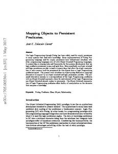
EXPERIMENT 1 In Experiment 1 we presented a simple rectangular figure, oriented horizontally in some blocks and vertically in others. The sides of the rectangle were either connected to form a single closed object, or separated by gaps in the centre of the longer sides (see Figure 1), which converted the rectangle into two separate perceptual units. On 67% of trials, one end of the rectangle was curved inward. RM was given two tasks: first, to report whether he saw a curve or not and, second, if he reported a curve, he was to report its location (“right” or “left” in several blocks with the long axis of the
rectangle oriented horizontally, “up” or “down” in other blocks with the long axis oriented vertically). The question of interest was the effect of the gap in the rectangle on the two different tasks— detection and localization of the curve. We predicted that detection would be impaired by the gap, consistent with evidence collected with other Balint’s patients showing that connected objects are more likely to be perceived than disconnected objects. For instance, Luria (1962) reported that one of his patients perceived only one of two circles placed horizontally side by side, but when a line connected the two circles, the patient reported seeing “spectacles”. Humphreys and Riddoch (1993) reported a similar effect in two patients with Balint’s syndrome. These patients were more likely to detect the presence of two different colours in a display of connected dot pairs if the lines connected pairs of dots of different colours than if they connected pairs of the same colour. Would a similar benefit for connectedness accrue in a within-object part localization task? Are parts more likely to be correctly located within a single object than between two different objects? We compared the effect of the gap on detection and on localization of the curve within the same displays on each trial. If the localization of parts within an object is mediated by identifying
Figure 1. Examples of stimuli used in Experiment 1: connected rectangle (A), separated rectangle (B), connected rectangular stimulus with curve (target) (C), separated rectangular stimulus with curve (D).
450
COGNITIVE NEUROPSYCHOLOGY, 2006, 23 (3)
ATTENDING TO SPACE AND OBJECTS
the whole object as one of two alternatives that differ only in the relative locations of their parts, RM should fail on the curve localization task. This is because the two locations of the curve left the identity of the object unchanged, changing only its orientation through rotation and/or reflection. On the other hand, detecting the presence of the curve could rely on identifying the shape, since, when present, the curve converted a rectangle into a different shape. Here RM’s intact ventral pathways could allow him to sense the presence of the curve as long as he saw the object at all.
Method Patient RM A thorough description of RM’s medical history, mental and neurological status, and visual abilities has been provided elsewhere (Robertson et al., 1997), as have 3-D reconstructed MRIs of his brain (Friedman-Hill et al., 1995; FriedmanHill, Robertson, Ungerleider, & Desimone, 2003). For this reason, the general description here is brief and highlights the most relevant information for the present study. In 1991– 92 RM suffered two separate stokes, probably due to an embolic source, a few months apart (the first affecting the occipital-parietal area of his right hemisphere and the second affecting a nearly symmetrical part of his left hemisphere). RM’s lesions included both occipital-parietal lobes and underlying white matter and were centred in Brodmann’s areas 7 and 39 with limited extension into areas 5 and 19 in both hemispheres and area 6 in the right hemisphere. These lesions left him with a classical case of Balint’s syndrome. In late 1999 RM suffered another stroke,
resulting in right-sided weakness. The radiological report of the initial MRI scan indicated no new lesions, and an MRI taken over a year later did not reveal any new lesions, although some cortical atrophy was noted (Friedman-Hill et al., 2003). A neurologist (Dr Robert Knight) determined that the most probable cause of the new motor deficits was a small lacunar infarct in the internal capsule of the left hemisphere that was undetectable on the T1 weighted image. The location of his original bilateral occipital-parietal lesions were unchanged, and his vision was unaffected. In October 1999, before the present series of studies could be completed, RM was moved to a residential facility, and his health deteriorated further after that time. This prevented us from running as many trials as we would have liked in Experiment 2 and from following up the findings with further experiments. However, a large amount of data was collected in Experiment 1 and, in concert with the data from Experiment 2, provides preliminary support for the hypotheses. We therefore felt that they should be included. Formal visual examination performed by the UC Davis ophthalmologic department in 1994 showed normal visual acuity, colour perception, and full visual fields. Another examination in August, 1999 by the optometry clinic at UC Berkeley showed no substantial change. RM could recognize shapes, single objects, and faces and could read single words. Other mental abilities, including language, were within normal range, although mild dysarthria was present. RM could not localize the items he saw, nor could he accurately reach for them or move his eyes to them when testing began in late 1992 (about 7 months after his second stroke).1 It is important to note that RM clearly understood the meanings
1 Perenin and Vighetto (1988) described ten patients with unilateral parietal lesions who showed optic ataxia but could still locate items verbally. There has been a great deal of evidence that optic ataxia and visual spatial deficits can be dissociated, and so reaching problems need not imply visual spatial problems. However, visual spatial problems do necessarily result in problems with volitionally reaching and pointing, so these problems are consistent with a visual spatial loss. The fact that reaching or pointing in the direction of a target was equally difficult with RM’s right and left hands and equally manifested in right and left fields, like his verbal reports, suggests a common origin in spatial perception deficits. Whether or not this is the case for RM, it does not detract from the differences reported here between feature detection and feature localization. RM had both reaching and verbal localization problems, and whether his dorsal spatial deficits were due to two separate functional systems or to one is an interesting question, but one that does not detract from our conclusions regarding ventral stream processing.
COGNITIVE NEUROPSYCHOLOGY, 2006, 23 (3)
451
ROBERTSON AND TREISMAN
of the words “left”, “right”, “up”, and “down”. We tested him in advance to make sure by touching him lightly to the right or left of his back and asking him on which side he felt the touch. He was very accurate, making only one error out of 27 trials. Any difficulty must arise in mapping the meanings of directional words onto visual space. RM’s spatial abilities improved slowly but significantly over the years but never reached anywhere near normal levels. Experiment 1 was run at a time when RM was able to occasionally locate items in his everyday life, but he was still well below normal and continued to require daily help due to his spatial problems.
Stimuli The stimuli were created from dark grey lines on a light grey background, forming a connected or disconnected rectangular figure (Figure 1A and 1B), subtending 5, 6, or 7 cm by 2.5 cm. The disconnected figure was the same as the connected figure except that a gap replaced the central 1.5, 2.5, or 4.5 cm of each of the two long sides. On 67% of trials, one of the shorter sides was replaced by a line that curved inwards relative to the rectangle, as shown in Figures 1C and 1D.
Procedure The computer monitor was placed approximately 60 cm in front of RM’s face. The rectangle was oriented horizontally in 11 testing blocks and vertically in 9 blocks. RM was tested on 4 different days. There were approximately 6 weeks between 2 earlier and 2 later sessions. Seven blocks of the horizontal display were first tested, followed by 6 vertical blocks. Three vertical and 4 horizontal blocks were tested at the end. Table 1 presents the block order and display parameters in each block. The stimuli were individually presented, centred in the middle of a computer monitor. The curve, when present, was equally often at the left and the right of the rectangle (in horizontal displays, or at the top and bottom in vertical displays). Presentation time, horizontal or vertical visual angle, and gap width (for separated figures) were all parameters within the program that could be varied across blocks of trials. There were 528 trials with horizontal displays and 432 trials with vertical displays; 28 trials were eliminated due to equipment error such as early noise detection by the VOR used to record RM’s verbal responses. Half the trials showed the connected rectangle, and half showed the separated parts. For each of these types of trials, a curved line was present 2/3 of the time, half
Table 1. Block order and parameters Display time Display
Order
Blocks
1 2 3 4 5
1 2 1 2 1
6 7 8 9 10
1 1 1 2 1
Vertical
11
Horizontal
12 13
Horizontal
Vertical
452
Sec
Ms
Dimensions (cm) Height
Gap
Usable trials
5 5 7 7 6
2.5 2.5 2.5 2.5 2.5
1.5 2.5 4.5 4.5 2.5
47 93 47 96 48
197 197 197
2.5 2.5 2.5 2.5 2.5
5 5 5 7 7
1.5 1.5 2.5 4.5 4.5
48 47 47 93 48
3
197
2.5
5
2.5
143
2 2
197 197
5 7
2.5 2.5
2.5 4.5
80 95
197 197 197 5 197 10
5
COGNITIVE NEUROPSYCHOLOGY, 2006, 23 (3)
Width
ATTENDING TO SPACE AND OBJECTS
on the left and half on the right, or half at the top and half at the bottom. The stimuli on the remaining trials contained no curve. Closure (connected vs. separated), curvature (curve or no curve), and side of curve (left vs. right, or top vs. bottom) were randomly varied within each block. RM was instructed that on each trial he should first report whether or not a curved line was present. The computer recorded his voice onset time to respond “yes” or “no”, although reaction time was not stressed. If RM said “yes”, he was then to report whether the curved line was on the left or the right (or at the top or the bottom) of the screen. His location responses were keyed in by the experimenter. Errors for both detection and localization of the curve were recorded by computer in the data file. Each trial began with a 500-ms central fixation followed 500 ms later by a stimulus that was displayed for 197 ms in all but 3 blocks. In these 3 blocks the stimuli were shown for 5 to 10 sec in order to determine whether time limiting the displays was critical.
Results Curve detection As shown in Table 2, curve detection was well above chance (87.6%) and quite accurate overall (89.5% for the horizontally oriented rectangle and 85% for the vertically oriented rectangle). There were only 53 errors in a total of 506 usable trials (10.5%) with the horizontal displays and 64 in the 426 usable vertical displays (15.0%). Misses accounted for the majority of errors (13.7% of the curve present trials, 4 false alarms or 2.5% of the curve absent trials). More importantly, chi square analyses showed that curve detection overall was better for connected (92.7%) than for separated (82.5) displays, x2 ¼ 21.33, p , .001. However, the advantage for connected displays did not appear with all parameter settings.
Increasing the gap size for separated displays or increasing the length of the sides by the same amount for connected displays had equal and opposite effects on the two types of figures (– 7.4% and þ 5.1%, respectively; compare B and D, Table 2). On the other hand, increasing the width of the figure without increasing the gap size had no effect for separated displays (compare D and E, Table 2), and both widths for the separated figures produced significantly worse curve detection than did the connected display (x2 ¼ 4.36, p , .05). Increasing both the gap size (4.5 cm) and the overall width of the display (7 cm) impaired curve detection accuracy (Table 2, F and G), but the advantage of connected over separated displays remained, x2 ¼ 5.81, p , .02 (for brief presentations) and was in the expected direction, x2 ¼ 2.74, p , .10 (for longer presentations).2 In sum, gap distance did have an effect, with gaps of 1.5 cm producing no significant advantage for connected over separated displays, while gaps of 2.5 cm or more did do so. RM apparently perceived the display with the smaller gap (1.5 cm) more often as a unitary whole than as two separate figures. Finally, practice seemed to have little effect on the detection advantage for connected displays. Detection accuracy was better for connected than for separated displays in the last sessions both when the overall width was 5 cm with a 2.5-cm gap, x2 ¼ 3.35, p , .01, and when it was 7 cm with a 4.5-cm gap, x2 ¼ 7.03, p , .01 (Table 2, H and I). Remarkably, exposure time had no effect on the detection advantage for connected over separated displays for a range of durations from 197 ms to 10 seconds (compare B with C and F with G, Table 2). It seems that RM’s problems in detecting the curve in two shapes rather than one shape were not remedied by giving him ample time to switch attention or to process more information. The curve was simply not accessible on some proportion of trials, and this was more often the case when the gap created two shapes rather than one.
2
For reasons that are not clear, increasing presentation time up to 5 seconds for displays that were 7 cm overall with a 4.5-cm gap produced a significant connected advantage only for horizontal displays, x2 ¼ 6.16, p , .02, but not for vertical displays. COGNITIVE NEUROPSYCHOLOGY, 2006, 23 (3)
453
ROBERTSON AND TREISMAN
Table 2. Detection accuracy Curve detection % A. Overall mean B. Base (197ms presentation time)
C. Increase presentation time (10 sec or to response)
D. Inrease gap (197 ms presentation time)
E. Increase width (197 ms presentation time)
F. Increase width and gap (197 ms presentation time)
G. Increase presentation time (5 sec or to response)
H. Last Session w/increased gap
N
87.6 92.6 89.4 95.7
42/47 45/47
84/93 44/47
37/47 79/93
96.3 77.6
I. Last Session w/increased width and gap
89.5
(197 ms presentation time)
89.5
83.0 87.0 83.3 60.9 82.2 79.2 83.3 95.3 70.8
39/47 20/23
81.3
Orientation
Widtha
Gapa
91.3 95.7
21/23 22/23
H V
5 5
1.5 1.5
95.8
23/24
V
5
1.5
97.8 100
45/46 24/24
H V
5 5
2.5 2.5
24/24
H
6
2.5
23/24 42/48
H V
7 7
4.5 4.5
46/48 19/24
H V
7 7
4.5 4.5
36/37 60/71
H V
5 5
2.5 2.5
46/47
H
7
4.5
100 20/24
100
90.3 14/23 37/45
95.8 87.5
90.3 38/48 20/24
95.8 79.2
88.9 41/42 51/72
81.3 85/95
N
98.6
80.0 77/80 111/143
%
95.8 21/24
80.5 84/96 39/48
84.3
(197 ms presentation time)
87.5
75.0
85.5 87.5 81.3
21/24 23/24
83.3 44/48
82.9 78.7 84.9
87.5 95.8
Connected
92.7 93.5
84.3
91.7 91.7
N
87.5 44/48
91.4 90.3 93.6
% 82.5 91.7
91.7 91.7
Separated
97.3 84.5
97.9 39/48
97.9
Overall percentages in bold. ain cm.
Localization In contrast to curve detection, localization was poor (63.1% overall), although significantly better than chance, x2 ¼ 6.45 p , .02. In the horizontal conditions there was a clear response bias to report the curve as on the right, although RM was no more likely to correctly detect a curve on the right (48.3%) than on the left (51.7%). However, in the vertical condition he was more likely to accurately detect a curve on the top (62.7%) than on the bottom (37.3%), and he was also more likely to report it as being on the top. The most important finding concerns the effects of connectedness, which made no difference to RM’s ability to localize the curve (Table 3). None of the comparisons between connected and separate displays for localization accuracy showed any significant difference, all x2 , 1 except for one (means shown in Table 3E), which did
454
COGNITIVE NEUROPSYCHOLOGY, 2006, 23 (3)
not reach significant levels, x2 ¼ 2.96, ns, and this was in the opposite direction. In direct contrast to curve detection, RM’s localization was no better for unitary rectangles than for rectangles with gaps.
Discussion Curve detection was easier for RM when the curve was part of a single closed figure than when it could appear in either of two separated parts, although the physical separation of the two possible locations in the separated and connected conditions was the same. These results are consistent with his simultanagnosia and suggest that a connected figure made the display appear to him as a single perceptual object. The detection of a curved end could then be made by discriminating the shape as a normal or distorted rectangle. When the curve was part of
ATTENDING TO SPACE AND OBJECTS
Table 3. Location accuracy Location detection % A. Overall mean B. Base (197ms presentation time)
C. Increase presentation time (10 sec or to response)
D. Increase gap (197 ms presentation time)
E. Increase width (197 ms presentation time)
F. Increase width and gap (197 ms presentation time)
G. Increase presentation time (5 sec or to response)
H. Last Session w/increased gap (197 ms presentation time)
I. Last Session w/increased with and gap (197 ms presentation time)
N
63.1 60.7 51.9 69.0
14/27 20/29
30/54 18/28
13/23 31/51
12/23 7/12
69.2
32/55 51/66
17/29 24/31
45.8 27/55
45.8
Gapa
57.1 69.2
8/14 9/13
H V
5 5
1.5 1.5
60.0
9/15
V
5
1.5
58.1 68.8
18/31 11/16
H V
5 5
2.5 2.5
37.5
6/16
H
6
2.5
53.8 57.1
7/13 16/28
H V
7 7
4.5 4.5
24/31 10/11
H V
7 7
4.5
15/26 27/35
H V
5 5
2.5 2.5
16/31
H
7
4.5
81.0 17/22 10/12
58.6 77.4
Widtha
56.1 6/10 15/23
77.3 83.3
Orientation
37.5 9/13
60.0 65.2
N
61.7
68.3
49.1 40.1
52.2 58.3
%
60.0 8/13
79.4 41/53 20/23
68.6 58.2 77.3
61.5
63.6
80.3 77.4 87.0
6/13 11/16
69.2 15/29
59.5 56.5 60.8
46.2 68.8
Connected
62.9 63.0
54.3
51.7 51.7
N
61.5 17/28
58.5 55.5 64.3
% 63.5 58.6
60.7 60.7
Separated
77.4
90.9 68.9 57.7 77.1
4.5
51.6 11/24
51.6
Overall percentages in bold. ain cm.
one of two separate figures, RM may on some trials simply not have seen both parts of the display, although this was less likely when the gap was small. Since a smaller gap did make a difference, it appears that it was not closure per se that was important, but how closely the stimulus features approximated closure and thus suggested a unitary figure. In contrast, RM found it no easier to localize the curve he had just detected when it was embedded in a single object than when it was in two objects. We should note that the curve was always concave relative to the rectangle. If the location of the curve had changed the global shape and identity of the object for RM (by being concave relative to the rectangle in one location and convex in the other), the connected shape might have invoked an object identification system in RM’s intact ventral pathway and resulted in better performance. Instead the changed
locations of the curve could be interpreted as differences in the orientation of the same shape rather than differences in its identity. RM had very poor orientation discrimination. We previously reported evidence showing that he had great difficulty in judging the orientation of single letters that he had no difficulty in identifying (Robertson et al., 1997). He could recognize an A or a T with almost no errors, but he was unable to say any better than chance whether the letter was upright or inverted. This was also true for faces. His orientation abilities were again tested in 2003 in a more systematic study. Although he was not totally at chance in judging the orientation, his error rate was close to 40% (Friedman-Hill et al., 2003). The absence of any benefit from the unitary object in the localization task suggests that within-object locations are not explicitly available as such to Balint’s patients with bilateral parietal damage. COGNITIVE NEUROPSYCHOLOGY, 2006, 23 (3)
455
ROBERTSON AND TREISMAN
EXPERIMENT 2
point halfway between the two letters. The letters were shown for 3 seconds, although RM was given as long as he needed to respond.
In Experiment 2 we tested the hypothesis that within-object localization could be mediated by object identification. We directly compared RM’s ability to explicitly localize components relative to each other with his ability to identify the pair of components as a unit. Experiment 2a used the words “ON” and “NO”, while Experiment 2b used the letters “b”, “d”, “p”, and “q”, which are built of the same components in different relative locations but for which the differences can be interpreted as differences in the orientation of the same physical object. In both cases localizing the components and identifying the whole required the same information but framed the question differently. Localization treats parts as separate objects and asks whether one is to the right or the left of the other. Identification treats the pair of letters as a single object to be identified, either a word or a letter, where the relative location of their parts just happens to be what differentiates the two alternatives.
Procedure In the localization task, RM was asked to report whether the “N” was to the right or to the left of the “O”. He was run in one block of 69 trials, with random selection of the separation and the side of the “N”, giving unequal numbers in the different categories. In the word identification task he was asked to report which word he saw—“NO” or “ON” or both—in a total of 197 trials. We gave him the option of saying “both” because he spontaneously told us on many trials that he could see both words, often alternating or fading into one another. This was the case for both binocular and monocular presentations. In a follow-up experiment several months later, we repeated the study using a forced choice identification test, asking him to report which word he saw first, “ON” or “NO” in a block of 30 trials. In another block of 30 trials, we also tested him with “OZ” and “ZO”, which are presumably less familiar and perhaps less unitized. We had planned to return for more data, but RM’s health suffered a further deterioration, and we were unable to test him further.
EXPERIMENT 2A: WORDS Method Stimuli The displays consisted of the two upper-case letters, “N” and “O”, presented side by side at one of three different separations: 18, 38, or 58 centre to centre. Each letter subtended approximately 18. The letters were preceded for 500 ms by a fixation
Results Localization The mean percentages of correct reports are shown in Table 4. RM was at chance in locating
Table 4. Percentage of correct localization reports and number of correct trials Separation 18
38
58
%
N
%
N
%
N
Mean
N on left N on right
92 17
11/12 2/12
67 50
8/12 6/12
70 9
7/10 1/11
76 26
Mean
54
13/24
58
14/24
39
8/21
51
456
COGNITIVE NEUROPSYCHOLOGY, 2006, 23 (3)
ATTENDING TO SPACE AND OBJECTS
the “N” relative to the “O”, averaging 51% overall. He showed a strong bias to report the “N” to the left of the “O”. He clearly had no information at all about the relative locations of the two letters when asked the localization question, but, as the next section demonstrates, he did have some information about the identity of the word.3 Word identification Table 5 shows the results of the word identification task using the same displays. RM found this a difficult task, in part because he often saw both words in the single display. He said things like “It did switch on me. I saw ‘on’ and ‘no’. One switches to the other one. Sometimes I see them at the same time like the one is in front of the other. I saw them both—‘no’ and ‘on’.” We decided to let him report both when that was what he saw because we were interested in how frequently this occurred and whether seeing both correlated more with one display than with the other. Again RM showed a large bias to report the word “NO”, consistent with his bias to locate the “N” to the left of the “O”. However, he did convey a considerable amount of information about the display in performing the word identification task. He reported seeing a single word on 63.5% of the trials and both words on the remaining trials.4 Looking at “Both” responses, he was much more likely to say “Both” when “ON” was presented than when “NO” was presented.
Counting “Both” and “On” responses as indications that he saw the word “ON”, even if he also saw “NO”, the mean correct responses were 63, 73 and 71% for the separations of 18, 38, and 58, respectively, and 69% overall, or well above chance (x2 ¼ 11.4, p , .001). In a forced choice word identification test almost a year later RM was asked to respond only with the word he saw first, “NO” or “ON”, and he averaged 50%, 80%, and 90% correct at the separations of 18, 38, and 58, respectively, or 73% overall (22 out of 30). Although he still had a slight bias to report the word “NO”, his correct responses were now more evenly divided between the two alternatives (9 correct reports for “ON” and 13 for “NO”). Thus, the word he saw first was more likely to be the word that was displayed. At this time we also tested the less familiar letter pairs “OZ” and “ZO”, and again he was asked to report which word he saw first. In reading “OZ” and “ZO”, he was correct on 8, 6, and 4 out of 10 trials for each of the separations of 18, 38, and 58, respectively, or 60% overall, which is not significantly better than chance. In localizing the “O” relative to the “Z”, he averaged 40% correct (4, 3, and 6 out of 13 trials for each of the separations of 18, 38, and 58, respectively), which is again at chance levels. Unlike the familiar words “ON” and “NO”, an unfamiliar word did not produce above-chance performance in reading. It appears that he was more likely to
3
This was not due to differential difficulty with the localization and naming tasks. We also ran two experiments with groups of 8 normal participants in each experiment using a threshold measure (Kaernbach, 1990) in which the stimuli (“ON”, “NO”, “OZ”, “ZO”) were varied in the time of presentation in a staircase method that depended on the subjects’ performance and were immediately masked by a set of random lines. (The only difference between the experiments was that we increased the mask complexity in the second experiment, making the stimuli more difficult to detect.) In each experiment participants named the word in one block of trials and located the “O” in another (block order was counterbalanced between participants). Stimuli were randomly presented, and a block ended after 10 reversals in the staircase with the mean of the last 8 reversals used as the estimate of threshold presentation time to reach chance performance. In addition, the location of the whole word was jittered around the centre of the screen so that if participants fixated or attended to one location on the screen and only noted whether there was an “O” there or not, it would be uninformative. In the first experiment the mean presentation threshold was 34.2 ms for identification and 33.4 ms for localization (F , 1). In the second both thresholds increased, but there was still no difference between the two conditions (65.6 ms for naming and 58.2 ms for localization), again F , 1. 4 The identity of the words “ON” and “NO” also depends on their perceived orientation. If the display is rotated 1808, the identity is reversed. It may be that the ventral word recognition system has its own orientation analysis, independently of the parietal system. Note that RM was far from perfect in identifying the words, perhaps because recognition of word orientation by the ventral system is somewhat crude and inaccurate. COGNITIVE NEUROPSYCHOLOGY, 2006, 23 (3)
457
ROBERTSON AND TREISMAN
Table 5. Percentage of “Both”, “No”, and “On” responses for each stimulus condition Separation 18
38
58
Mean
Display
Reported Both Reported No Reported On Counting “Both” as correct for ON and “No” as correct for NO
NO
ON
NO
ON
NO
ON
NO
ON
21 76 3
47 50 3
22 75 3
66 28 6
13 84 3
50 41 9
19 78 3
54 40 6
63
treat familiar words as units than unfamiliar words.5 The evidence, though sparse due to RM’s deteriorating health, suggests that reading the familiar words “ON” and “NO” benefited from better unitization than reading unfamiliar syllables. This finding is consistent with the proposal of Shalev and Humphreys (2002) that Balint’s patients perform better when guided by stored representations of the display.
73
71
69
Procedure In the localization task, RM was asked to report which side of the line the “o” was presented, left or right. In the identification task, he was asked which of the four letters he saw, “b”, “d”, “p”, or “q”. Each consisted of 16 trials. Another block of 16 trials with the letters “e”, “o”, “c” and “d” was shown as a control to examine his ability to discriminate fine detail based on differences of only a pixel or two.
Results EXPERIMENT 2B: LETTERS Stimuli In Experiment 2b, the displays used for the localization task consisted of a circle and a vertical line, subtending approximately 0.68 and 18, at the same three separations of 18, 38, and 58, presented until RM responded. The displays used for the letter identification task consisted of a single letter—“b”, “d”, “p”, or “q”—presented foveally. The letters “e”, “o”, “c” and “d” were shown in a separate block of control trials.
Although the data were sparse due to RM’s health condition, he was again at chance in localizing the “o” relative to the “l”, getting 7 out of 16 correct (43.8%). He did better on letter identification, getting 23 out of 32 correct (71.9%), although he did make a substantial number of errors (9), mostly confusing mirror-image pairs (which he did 5 times). There was a trend toward a significant difference, x2 ¼ 3.12, p , .08. We compared his performance on a block of identification trials with the perceptually confusable letter set “o”, “e”, “c”, and “d”, where he made only one error in 16 trials (93.8% correct), and that was to call
5 One might expect RM to be better at reading the words when the letters were closer together, as this is the more familiar form. However, he could not estimate the length of lines or distances between two objects on a page accurately (Robertson et al., 1997), consistent with his explicit lack of space. We do not know the distance he perceived between the “N” and “O”. All the letters may have looked about equally close in his experience, regardless of their spatial separation in the stimulus. If so, this would also conform to the more familiar spatial relation that was provided by the stored representation, and not to the space within the stimulus itself.
458
COGNITIVE NEUROPSYCHOLOGY, 2006, 23 (3)
ATTENDING TO SPACE AND OBJECTS
the “d” a “b”. His ability to discriminate between letters that required perceiving small differences in terms of pixel count was better than discriminating between letters on the basis of orientation, x2 ¼ 9.18, p , .01. His difficulty seemed to be primarily with judging spatial orientations of the letters, and this, in turn, was less damaged than his ability to localize one letter relative to another (“o” and “l”).
GENERAL DISCUSSION It is generally assumed that the ventral pathway plays the major role in the perception of objects, whereas the dorsal pathway is involved in spatial localization and action. By testing patients with bilateral parietal lesions, it is possible to demonstrate additional contributions to object perception made by the dorsal pathway. These appear to be substantial. Our earlier research (Robertson et al., 1997) showed that (a) the parietal lobes are essential in binding features such as colour, shape, and motion for perception when more than one object is present; (b) they are necessary for the individuation of objects through their separate spatial locations, and therefore for the perception of more than one object at a time; and (c) they also play a role in perceiving object orientations. The present paper continued this exploration, contrasting detection, identification, and the localization of parts within an object. In Experiment 1 we compared detection of a feature with localization, using the same pairs of displays. These consisted of either a single object or the same object divided in two by a spatial gap. The idea was first to confirm earlier findings that Balint’s patients are more likely to see the parts of a single object than two separate objects even when the figures are exactly matched except for the presence of the spatial gap in the centre. RM was indeed better at detecting a feature (a curved line) when the display was seen as a single object than when the same object was divided by a spatial gap large enough to clearly separate the display into two parts. Second, we tested the hypothesis that spatial localization might also
be better within an object than between two parts, which might be seen as separate objects. We found no evidence to support this view. The single connected object provided no benefit when the task was to localize the feature. RM’s ability to localize the curve he had just detected was no better within an object than between two parts. In Experiment 2, we used familiar stimuli for which switching the locations of the components would change the perceived identity of the whole. Using the words “ON” and “NO”, we compared two ways of defining what was essentially the same task: In one version RM was asked to localize one of the letters relative to the other, and in the other version he was asked to identify the pair of letters by reading the word. The displays were identical for the two tasks; all that differed was the description of the task and the definition of the displays as two letters or as one word. We found a clear dissociation between RM’s ability to use the relative locations of the letters to identify familiar words and his ability to judge the relative locations of the letters in the same words. The former was well above chance, while the latter was not. The better performance in word identification of “ON” and “NO” suggests that the letter localization used in familiar word recognition may depend at least in part on pattern analysis involving areas other than the parietal lobes. We found this pattern only with familiar words. When he was asked to read the words “ZO” and “OZ”, both localization and naming were at chance levels (although the number of trials was necessarily small). We also found an effect of word familiarity in a previous report (Robertson et al., 1997) where RM rapidly and confidently read relatively long words, such as the city in which he lived and his name, but missed shorter, less familiar words. Familiar words may be more likely to function as units rather than individual letters. Humphreys (1998; Cooper & Humphreys, 2000; Humphreys & Riddoch, 1993) suggested that the dorsal and ventral systems code spatial relations between objects and within objects, respectively. More recently Shalev and Humphreys (2002) studied another Balint’s COGNITIVE NEUROPSYCHOLOGY, 2006, 23 (3)
459
ROBERTSON AND TREISMAN
patient, GK. They showed that with arbitrary nonmeaningful objects, his between-object localization was better than within-object localization. For instance, when asked to discriminate the relative locations of two small rectangles and a large oval, he did better when the rectangles were outside the oval. However, when the rectangles were replaced with a pair of horizontally aligned circles and the whole configuration was described as eyes in a face, he did better with the circles inside the oval. Shalev and Humphreys suggested that when GK was told the stimulus was a face, he used an internal template and deduced the part locations by reference to this face template. If differences in the location of a part change the identity of the whole, they will be seen, but otherwise not. Note that two assumptions must hold under these conditions (a) the template must include the canonical orientation of a face, and (b) the orientation of the stimulus must be explicitly accessible. Unlike RM, GK retained the explicit ability to discriminate orientation and was able to use this information when he accessed stored representations.6 RM, too, was influenced by stored representations, but due to his explicit orientation difficulties (see Friedman-Hill et al., 2003; Robertson et al., 1997) the canonical orientation had little influence. In an earlier study, we presented RM with scrambled and normal faces that were either upright or inverted and were either complete or missing a feature (such as an eye). He was able to discriminate the complete faces from the scrambled or missing feature faces with 100% accuracy, but he was unable to report the orientation of the faces above chance levels. His performance was similar with letters, where he only made one identification error in a series of upright and inverted letter stimuli but was able to report the correct orientation of the letters only 61% of the time (Robertson et al., 1997). 6
The word and letter stimuli in the present experiments were more ambiguous than faces in the sense that they had different stored representations for each possible orientation. Both “ON” and “NO” correspond to English words, and “d”, “b”, “q”, and “p” are all letters of the alphabet. The identities depend on the relative locations of the parts in their canonical orientation. Without information about the current orientation and relative location of object parts, neither the locations nor the identities can be correctly discriminated. In fact, in the “ON”/“NO” experiment, RM reported he saw both representations on about one third of the trials. This was true even under monocular viewing conditions, so it could not be an effect of diplopia. This double perception is consistent with the notion that the ventral system derives part locations from the identity of the whole rather than the reverse. Where both patterns activate a familiar word, RM tended to see both, giving priority to “NO”, which may be semantically more salient than the preposition “ON”. These observations suggest that the withinobject system is not really a spatial system as such. It codes the locations only indirectly, if and when the features and/or the orientations can unambiguously differentiate the identities of two objects. This is consistent with Shalev and Humphrey’s (2002) conclusion that the ventral pathway allows the identification of objects, including the discrimination of objects that are implicitly defined only by the relative locations of their parts, but that explicit localization and, as we have shown here, the orientation of parts within objects requires the participation of the parietal lobes in the same way as they are required in discriminating locations between objects. The data we collected with the letters “b”, “d”, “p”, and “q” were unfortunately too sparse to support strong conclusions without further evidence, but the implications of the results were consistent with those from the words, thus lending
This difference was probably due to the different distributions of the lesions on the right for RM and GK. Whereas RM had two separate embolic strokes creating lesions in the territory of the middle cerebral artery (MCA) on the right and left, GK’s lesion on the right is more consistent with a posterior artery infarct. Scans published in Humphreys et al. (2000) show a lesion that includes the right calcarine cortex with lesion extension deep into white matter but sparing areas in the distribution of the MCA.
460
COGNITIVE NEUROPSYCHOLOGY, 2006, 23 (3)
ATTENDING TO SPACE AND OBJECTS
converging support. RM was considerably better at identifying the unitized letters than at localizing the component parts. On the other hand, he did have trouble in judging the orientations of the letters, whereas he had no trouble identifying a set of letters (“e”, “c”, and “o”) that should have been more confusable in terms of overlapping pixels but the identities of which are independent of their orientations. In sum, we tested RM in four different perceptual tasks: (a) detection of the presence of a part (a curved edge) in one versus two objects (a rectangle or the same rectangle split in two by a gap), (b) explicit localization of that part when it left the identity of the object unchanged, (c) identification of objects the identities of which depend on the locations and/or orientations of their parts (“ON” vs “NO”, and “b” vs “d” vs “q” vs “p”), and (d) localization of a part when the task requires seeing the part as one of two separate objects (“O” and “N”, “o” and “l”). We found clear dissociations between part detection and object identification, (a and c) giving much better performance than explicit localization (b and d). Curve detection benefited from the presence of a single object the shape of which was changed by the presence of the part, presumably a ventral process. An explicit word identification task (“NO” versus “ON”) mediated the discrimination of two different spatial arrangements, again a task for the ventral system. On the other hand, explicit localization and orientation within an object and explicit localization between objects both appear to depend on parietal function. Since determining the correct orientation of an object depends on a relative spatial judgement and thus a comparison to some other intact spatial reference frame, it might be expected that it too would be affected by parietal lesions. This was what we observed both here and in previous studies with RM. The present results add to those of Shalev and Humphreys (2002) by demonstrating an influence of stored representations in a patient with an even more severe spatial deficit: clear bilateral dorsal damage but with posterior ventral areas anatomically intact in both hemispheres. The differences between our results and
previous studies can be attributed to the fact that GK had at least one dimension of space available to him (orientation), while RM did not. Manuscript received 24 December 2004 Revised manuscript received 29 March 2005 Revised manuscript accepted 19 May 2005 PrEview proof published online January 2006
REFERENCES Biederman, I. (1987). Recognition by components: A theory of human image understanding. Psychological Review, 94, 115– 147. Balint, R. (1909). Seelenla¨hmung des “Schauens”, optische Ataxie, ra¨umliche Sto¨rung der Aufmerksamkeit. Monatschrift fu¨r Psychiatrie und Neurologie, 25, 5 – 81 (translated in Cognitive Neuropsychology, 12, 1995, 265– 281). Bernstein, L. J., & Robertson, L. C. (1998). Independence between illusory conjunctions of color and motion with shape following bilateral parietal lesions. Psychological Science, 9, 167–175. Cooper, A. C. G., & Humphreys, B. W. (2000). Coding space within but not between objects: Evidence from Balint’s syndrome. Neuropsychologia, 38, 723– 733. Duncan, J. (1984). Selective attention and the organization of visual information. Journal of Experimental Psychology: General, 113, 501– 517. Egly, R., Driver, J., & Rafal, R. D. (1994). Shifting visual attention between objects and locations: Evidence for normal and parietal-lesion subjects. Journal of Experimental Psychology: General, 123, 161– 172. Friedman-Hill, S., Robertson, L. C., & Treisman, A. (1995). Parietal contributions to visual feature binding: Evidence from a patient with bilateral lesions. Science, 269, 853– 855. Friedman-Hill, S. R., Robertson, L. C., Ungerleider, L. G., & Desimone, R. (2003). Posterior parietal cortex and the filtering of distractors. Proceedings of the National Academy of Sciences, 7, 4263– 4268. Holmes, G., & Horax, G. (1919). Disturbances of spatial orientation and visual attention with loss of stereoscopic vision. Archives of Neurology and Psychiatry, 1, 385– 407. Humphreys, G. W. (1998). Neural coding of objects in space: A dual coding account. Philosophical Transactions of the Royal Society, B, 353, 1341– 1351. COGNITIVE NEUROPSYCHOLOGY, 2006, 23 (3)
461
ROBERTSON AND TREISMAN
Humphreys, G. W., Cinel, C., Wolfe, J., Olseon, A., & Klampen, N. (2000). Fractioning the binding process: Neuropsychological evidence distinguishing binding of form from binding of surface features. Vision Research, 40, 1569– 1596. Humphreys, G. W., & Riddoch, M. J. (1993). Interactions between objects and space systems revealed through neuropsychology. In S. K. David & E. Meyer (Eds.), Attention and performance, XIV. Cambridge, MA: MIT Press. Humphreys, G. W., & Riddoch, M. J. (1995). Separate coding of space within and between perceptual objects: Evidence from unilateral visual neglect. Cognitive Neuropsychology, 12, 283– 311. Kaernbach, C. (1990). A single-interval adjustmentmatrix (SIAM) procedure for unbiased adaptive testing. Journal of the Acoustical Society of America, 88, 2645– 2655. Kahneman, D., & Treisman, A. (1984). Changing views of attention and automaticity. In R. Parasuraman & D. R. Davies (Eds.), Varieties of attention. New York: Academic Press. Kramer, A. F., & Jacobson, A. (1991). Perceptual organization and focused attention: The role of objects and proximity in visual processing. Perception and Psychophysics, 50, 267– 284. Luria, A. R. (1962). Higher cortical functions in man. Moscow: Moscow University Press. Palmer, S. E. (1983). The psychology of perceptual organization: A transformational approach. In J. Beck, B. Hope, & A. Baddeley (Eds.), Human and machine vision. New York: Academic Press.
462
COGNITIVE NEUROPSYCHOLOGY, 2006, 23 (3)
Perenin, M. T., & Vighetto, A. (1988). Optic ataxia: A specific disruption in visuomotor mechanisms. I. Different aspects of the deficit in reaching for objects. Brain, 111, 643–674. Rafal, R. (2001). Balint’s syndrome. In M. Behrmann (Ed.), Disorders of visual behavior (Vol. 4). Amsterdam: Elsevier Science. Robertson, L. C., Egly, R., Lamb, M. R., & Kerth, L. (1993). Spatial attention and cueing to global and local levels of hierarchical structure. Journal of Experimental Psychology: Human Perception and Performance, 19, 471– 487. Robertson, L. C., Treisman, A., Friedman-Hill, S., & Grabowecky, M. (1997). The interaction of spatial and object pathways: Evidence from Balint’s syndrome. Journal of Cognitive Neuroscience, 9, 295– 317. Rock, I. (1973). Orientation and form. New York: Academic Press. Shalev, L., & Humphreys, G. W. (2002). Implicit location encoding via stored representations of familiar objects: Neuropsychological evidence. Cognitive Neuropsychology, 19, 721– 744. Treisman, A., & Kanwisher, N. K. (1998). Perceiving visually-presented objects: Recognition, awareness, and modularity. Current Opinion in Neurobiology, 8, 218– 226. Ungerleider, L. G., & Mishkin, M. (1982). Two cortical visual systems. In D. J. Ingle, J. W. Mansfield, & M. A. Goodale (Eds.), Advances in the analysis of visual behavior. Cambridge, MA: MIT Press.