NeuroImage 55 (2011) 1200–1207
Contents lists available at ScienceDirect
NeuroImage j o u r n a l h o m e p a g e : w w w. e l s e v i e r. c o m / l o c a t e / y n i m g
Auditory cortex activation is modulated by emotion: A functional near-infrared spectroscopy (fNIRS) study M.M. Plichta a,1, A.B.M. Gerdes b,c,⁎,1, G.W. Alpers b,c,d, W. Harnisch e, S. Brill e, M.J. Wieser b, A.J. Fallgatter f a
Central Institute of Mental Health (CIMH), Mannheim, Germany Department of Psychology, Biological Psychology, Clinical Psychology, and Psychotherapy, University of Würzburg, Germany Department of Clinical and Biological Psychology, University of Mannheim, Germany d Otto-Selz-Institute, University of Mannheim, Germany e Department of Otorhinolaryngology, Plastic, Aesthetic and Reconstructive Head and Neck Surgery, University of Würzburg, Germany f Department of Psychiatry and Psychotherapy, University of Tübingen, Germany b c
a r t i c l e
i n f o
Article history: Received 13 December 2010 Accepted 6 January 2011 Available online 12 January 2011 Keywords: Emotion Auditory cortex Enhanced sensory processing Early processing Functional near-infrared spectroscopy (fNIRS)
a b s t r a c t Visual emotional stimuli evoke enhanced activation in early visual cortex areas which may help organisms to quickly detect biologically salient cues and initiate appropriate approach or avoidance behavior. Functional neuroimaging evidence for the modulation of other sensory modalities by emotion is scarce. Therefore, the aim of the present study was to test whether sensory facilitation by emotional cues can also be found in the auditory domain. We recorded auditory brain activation with functional near-infrared-spectroscopy (fNIRS), a non-invasive and silent neuroimaging technique, while participants were listening to standardized pleasant, unpleasant, and neutral sounds selected from the International Affective Digitized Sound System (IADS). Pleasant and unpleasant sounds led to increased auditory cortex activation as compared to neutral sounds. This is the first study to suggest that the enhanced activation of sensory areas in response to complex emotional stimuli is apparently not restricted to the visual domain but is also evident in the auditory domain. © 2011 Elsevier Inc. All rights reserved.
Introduction An organisms' capacity to detect and efficiently process emotional information in the environment can be crucial for its survival, e.g. it can be life-saving to quickly respond to subtle visual or auditory cues that may indicate the approach of a potential predator. Human studies on the neural underpinnings of such processing capacities revealed that emotionally arousing visual stimuli evoke enhanced activation in early visual cortex, including areas which are responsible for basic visual feature analyses (V1 and V2). This phenomenon has been consistently observed across a wide range of methods including electroencephalography (EEG) (Junghofer et al., 2006; Keil et al., 2005; Leutgeb et al., 2009; Schupp et al., 2003, 2004), functional magnetic resonance imaging (fMRI) (Alpers et al., 2009; Lane et al., 1999; Lang et al., 1998), and optical imaging (Alpers et al., 2005; Herrmann et al., 2008; Minati et al., 2009). Investigating the underlying mechanisms, neuro-anatomical (Amaral et al., 2003; Catani et al., 2003) and functional imaging studies (e.g. Davis and Whalen, 2001; LeDoux, 1998, 2000; Sabatinelli et al., 2005, 2009; Vuilleumier et al., 2004) suggest that core structures of emotional processing, in particular the amygdala, prime these early visual processing ⁎ Corresponding author. University of Mannheim, Department of Clinical and Biological Psychology, 68131 Mannheim, Germany. Fax: + 49 621 181 2107. E-mail address:
[email protected] (A.B.M. Gerdes). 1 Both authors contributed equally to this work. 1053-8119/$ – see front matter © 2011 Elsevier Inc. All rights reserved. doi:10.1016/j.neuroimage.2011.01.011
circuits through re-entrant projections. Advantageous consequences of enhanced early visual cortex activation comprise enhanced perception (Alpers and Gerdes, 2007, 2008), improved sensory discrimination (Phelps et al., 2006), and facilitated detection of potentially relevant environmental stimuli (Öhman and Mineka, 2001; Padmala and Pessoa, 2008), which may support the organism to quickly initiate appropriate approach or avoidance behavior in response to salient cues (Lang, 1995; Lang et al., 1997). Thus, the modulating impact of emotional arousal on neural processing at basic steps within the visual information pathway may be part of an evolutionary adaptive mechanism. While the literature on emotional modulation in sensory areas has been well advanced with respect to the visual domain, the picture is less than complete for other sensory modalities. Salient information, however, is often also provided by auditory cues. Different from vision, auditory sensitivity allows organisms to receive information at long distances (e.g. thunder or a growling predator) and in any direction without the need to be focused on it. Furthermore, compared to visual cues, auditory stimuli lead to slightly faster motor reaction times (e.g. Hovland and Bradshaw, 1937) making audition interesting for emotional processing. Therefore, it is particularly important to examine whether emotion also enhances activity of basic sensory areas in the auditory domain. There are several arguments why enhanced sensory cortex activity may be independent of sensory modality: from an anatomical perspective afferent and efferent connections between the amygdala and cortical areas have also been documented for the auditory
M.M. Plichta et al. / NeuroImage 55 (2011) 1200–1207
1201
system (Doron and LeDoux, 1999; Woodson et al., 2000; Yukie, 2002). Furthermore, auditory signals of emotion activate emotional core structures in the human brain such as the amygdala (Anders et al., 2008; Belin et al., 2008; Ethofer et al., 2006; Fecteau et al., 2007; Johnstone et al., 2006; Royet et al., 2000; Sander and Scheich, 2005; Wiethoff et al., 2009; Wildgruber et al., 2006). Finally, emotional sounds elicit behavioral (e.g., reaction times) and psychophysiological responses (e.g., modulated startle) which are very similar to those evoked by emotional pictures (e.g., Bradley and Lang, 2000; Czigler et al., 2007; Magnée et al., 2007; Partalaa and Surakka, 2003; Royet et al., 2000). Nevertheless, studies on hemodynamic changes in response to auditory emotional stimuli in humans are relatively rare. Moreover, the existing studies did not investigate neural responses to unpleasant and pleasant stimuli (Blood and Zatorre, 2001; Frey et al., 2000; Maddock and Buonocore, 1997) or did not specifically focus on emotional modulation of the auditory cortex (Anders et al., 2008; Fecteau et al., 2007; for an exception see Wiethoff et al., 2008). Finally, the existing studies with auditory cues mainly used restricted subsets of auditory stimuli such as emotional prosody or human vocalization (Ethofer et al., 2006, 2009; Johnstone et al., 2006; Morris et al., 1999; Wiethoff et al., 2009, 2008; Wildgruber et al., 2006), so that direct comparisons with results from the visual domain are difficult to achieve. The relatively sparse information on auditory cortex activation during emotion processing may be partially due to methodological issues. It can be difficult to study the effects of emotional sounds in conventional fMRI scanners where MR gradient noise often exceeds 100 dB sound pressure level (SPL). Gradient noise has been shown to interfere with auditory stimulation (Bandettini et al., 1998), potentially even in a non-additive manner (Gaab et al., 2007). Furthermore, it can induce stress and annoyance (e.g. Pripfl et al., 2006) which is prone to interact with affective stimulation. While sophisticated fMRI protocols have been developed to reduce the impact of acoustic scanner noise (Amaro et al., 2002; Blackman and Hall, in press; Griffiths et al., 2001; Hall et al., 1999; Hennel et al., 1999; Marcar et al., 2002; Obleser et al., 2007; Yetkin et al., 2003), the fMRI scanner environment (strict movement restrictions; claustrophobic situation) may still interfere with emotional processing at least in some humans. Alternative functional imaging methods may complement existing approaches to investigate the auditory system. One such alternative is functional near-infrared spectroscopy (fNIRS), a silent and non-invasive optical imaging method where movement restrictions are less rigorous (no head fixation necessary) and the subject can sit in an upright position. Like fMRI, fNIRS measures hemodynamic changes (Ferrari, 2004; Hoshi, 2003; Obrig and Villringer, 2003) and enables to reliably detect task-specific brain activation (Plichta et al., 2006a, 2007c, 2006b). Recently, fNIRS has been successfully applied to investigate emotional modulation of the visual cortex (Herrmann et al., 2008; Minati et al., 2009) but an investigation of the auditory domain is pending. Therefore, the aim of the present study is to investigate whether (complex) emotional sounds modulate activation within auditory sensory areas, an effect that has been consistently observed in the visual domain. Standardized pleasant, unpleasant, and neutral sounds (including both human and nonhuman sources) were selected from the International Affective Digitized Sound System (IADS, Bradley and Lang, 1999) and presented to healthy participants while evoked auditory cortex activation was recorded with a silent hemodynamic imaging technique (fNIRS).
about the nature of the experiment as well as the operating mode of the fNIRS instrument, before giving written informed consent. A brief instruction to remain relaxed and to avoid major body movement was given. The fNIRS-investigation of healthy participants and the whole experimental procedure was in accordance with the Declaration of Helsinki and was approved by the Ethics Committee of the medical faculty of the University of Würzburg.
Methods
The continuous wave system (ETG-4000, Hitachi Medical Co., Japan) is described in detail elsewhere (Plichta et al., 2006b). The inter-optode distance was 30 mm and the sampling rate was set to 10 Hz. The two 22-channel probe-sets were placed on the scalp above the left and right temporal lobes based on the results of a pilot study (see Supplementary material) where the identical fNIRS system, probe-
Participants A total of 17 participants were examined (mean age = 25.94 ± 4.59 years; 7 males). All participants had normal hearing and no history of any neurological disorder. All participants were informed
Auditory stimulation The stimulus material consisted of 20 unpleasant (e.g., screaming adult, crying child and humming wasps), 20 pleasant (e.g., laughter, erotic sighs, and applause) and 20 neutral sounds (e.g. clacking type writer, cackling chickens, and ticking clock) selected from the International Affective Digitized Sounds (IADS) (Bradley and Lang, 1999) — see Appendix A. Pleasant and unpleasant sounds were matched for levels of arousal. Due to the heterogeneity of the sound material, two raters categorized the IADS sounds into two classes (human vocal sounds and non-vocal material). Subsequently, the three emotional categories were balanced for these classes of sounds (see Appendix A for details of the selected IADS sounds) preventing biased results due to dominance of human vocalizations within the categories (Belin et al., 2000). Prior to testing, all IADS sounds were adjusted to equal peak sound pressure level (dB SPL). Furthermore, it was assured that the mean intensity contours and power spectra were comparable across the sound categories. Standard insert earphones for audiometry (E-A-RTONE® 3A 10 Ω, Aearo Company, Indianapolis, IN) were used for acoustic stimulation. A detailed description of earphone characteristics, pre-processing of the IADS sound material and tests on differences between the sound categories concerning physical parameters (including spectro-temporal modulation analyses according to Kumar et al., 2008) can be found in the Supplementary material. Procedure The presentation of instructions and stimuli as well as the recording of behavioral responses was controlled by the high-precision software Presentation© running on an Intel® Celeron Processor (500 MHz) with a 19″ monitor (resolution of 1280 × 1024). After the two 22-channels fNIRS probe-sets were adjusted and the in-ear headphones inserted, three example sounds were presented (which were not used in the subsequent experimental run) in order to familiarize the participants with the stimulation set-up. The following experiment consisted of 5 blocks with unpleasant, 5 blocks with pleasant, and 5 blocks with neutral sounds which were presented in pseudo-randomized order. Each block had a duration of 24 s with four different, randomly chosen sounds (each of 6 s duration in maximum — see Table 1 for exact sound durations) of the same valence category presented every 6th second. No inter-stimulus-interval appeared between the sounds except for minimal time gaps (0.05 s on average) between the actual ending of a sound and the 6th stimulation second. Each block was followed by a quiet resting phase of 24 s. The entire fNIRS recording took about 15 min. After this, all sounds were presented again and the participant had to rate each one of them on valence (1 to 9, anchored as “very unpleasant” and “very pleasant”) and arousal (1 to 9, anchored as “not at all intense” and “very intense”). fNIRS measurements and analyses
1202
M.M. Plichta et al. / NeuroImage 55 (2011) 1200–1207
Table 1 HHb amplitude estimates (mean and standard errors)a.
Table 2 O2Hb amplitude estimates (mean and standard errors)a.
Region
Hemisphere
Unpleasant
Neutral
Pleasant
Region
Hemisphere
Unpleasant
Neutral
ROI
Left Right Pooled Left Right Pooled
0.147 0.177 0.162 0.068 0.055 0.061
0.094 0.088 0.091 0.068 0.022 0.045
0.150 0.170 0.160 0.048 0.033 0.041
ROI
Left Right Pooled Left Right Pooled
0.263 0.360 0.311 − 0.026 − 0.106 − 0.066
0.202 0.269 0.236 0.034 − 0.060 − 0.013
Non-ROI
a
(0.030) (0.025) (0.024) (0.029) (0.021) (0.022)
(0.024) (0.025) (0.023) (0.029) (0.012) (0.016)
(0.030) (0.030) (0.026) (0.011) (0.013) (0.010)
Scale has been inverted and transformed by ⁎103.
sets and a neutral sound stimulation (1000 Hz tone) were applied. Accordingly, channel #7 was placed above EEG marker position T3/T4 (10–20 system) and the probe-sets were then rotated in such a way that the vertical mid optode column (between channels #7 and #16) paralleled a line between T3/T4 and C3/C4. Channels covering and directly surrounding T3/T4 which indicate significant activation in the pilot study (i.e., channel #2, #3, #7 and #11 (left) or #12 (right), respectively) were defined as a-priori region-of-interest (ROI). We defined non-ROI channels as outside ROI minus the directly adjacent channels of the ROI (Fig. 1). The fNIRS data were analyzed according to the general linear model (GLM) with custom software programmed in Matlab® (MathWorks, Natick, MA) as described in Plichta et al. (2007b). For detrending of the functional data, a discrete cosine transform (DCT) based high-pass filter (156 s) was applied. The design matrix included three boxcar regressors (one for each emotion category), which were convolved with a Gaussian hemodynamic response function (HRF) to predict brain activation (HRF peak-time = 6.5 s; FWHM = 5.89). Beta-weights, scaling the predictors, served as the fNIRS amplitude estimates. All analyses were corrected for serial correlated errors by fitting a first-order autoregressive process (AR[1]) to the error term by the Cochrane–Orcutt procedure (Cochrane and Orcutt, 1949). In order to ensure that the statistical comparison of emotional vs. neutral sounds is restricted to the auditory cortex we identified channels that indicate activation evoked by all three sound categories separately (conjunction null hypothesis according to Nichols et al., 2005) within the a-priori ROI (alpha = 0.05). The beta-estimates from those channels were then averaged and subsequently tested by a 2 × 2 × 3 repeated-measures ANOVA comprising the three withinsubject factors hemisphere (2: left and right), region (2: ROI and nonROI), and emotion (3: neutral, pleasant, and unpleasant sounds). Significant interaction effects (simple or second order) were analyzed by post-hoc analyses on the individual factor-levels. Greenhouse– Geisser correction was used where appropriate. For directed hypotheses, one-tailed post-hoc t-tests were used (pleasant sounds N neutral sounds and unpleasant sounds N neutral sounds; Bonferroni corrected alpha = 0.05). The difference between pleasant vs. unpleasant sounds was tested by means of a two-tailed t-test at an uncorrected alphalevel of 0.25, which is associated with a statistical power of 0.80 at a moderate effect size of dz = 0.5 (Faul et al., 2007). In addition to p-values, effect size parameters are always reported as η2p for ANOVAs and dz for dependent t-tests. While both fNIRS parameters (O2Hb and HHb) were fully analyzed, the focus of the results section is on the HHb parameter because O2Hb have lead to ambiguous results in previous studies while HHb results are more closely in line with fMRI data on the emotional modulation of brain activation (see Alpers et al., 2005; Herrmann et al., 2008; Plichta et al., 2007a). Results Self-report As expected, the ratings differed between the sound categories, with a main effect of sound category for valence (pleasant: M=5.72, SD=
Non-ROI
a
(0.124) (0.118) (0.119) (0.050) (0.051) (0.046)
Pleasant (0.059) (0.068) (0.060) (0.093) (0.048) (0.068)
0.268 0.309 0.289 − 0.036 − 0.113 − 0.074
(0.088) (0.095) (0.087) (0.057) (0.051) (0.052)
Scale has been transformed by ⁎103.
0.93; neutral: M=3.81, SD=0.72; unpleasant: M=2.94, SD=0.69), F(2, 32)=137.61, pb 0.001, η2p =0.90; and arousal (pleasant: M=5.31, SD=1.16; neutral: M=4.01, SD=1.53; unpleasant: M=5.24, SD= 1.10), F(2, 32)=27.51, pb 0.001, η2p =0.63. Post-hoc t-tests showed that pleasant sounds were rated as more pleasant than neutral (t(16)=10.11, pb 0.001, dz=2.45) or unpleasant ones (t(16)=15.38, pb 0.001, dz=3.73). Unpleasant sounds were rated as more unpleasant than neutral ones (t(16)=6.18, pb 0.001, dz =1.50). Arousal ratings of pleasant and unpleasant sounds did not differ significantly (t(16)=0.35, p=0.731, dz=0.08), whereas both categories were rated as more arousing than neutral sounds (pleasant-neutral: t(16) = 6.01, p b 0.001, dz = 1.46; unpleasant-neutral: t(16) = 6.52, pb 0.001, dz=1.58). fNIRS results A complete list of the mean fNIRS amplitude estimates including standard errors resulting from the second level analyses can be found in Tables 1 and 2. For the HHb data, sound stimulation led to highly significant bilateral activation within the auditory cortex (Fregion(1, 16)= 17.27, pb 0.001, η2p =0.52). Testing the main hypothesis of the study, the interaction of region⁎emotion was highly significant (F(2, 32)=6.50; p=0.007; η2p =0.29): Post-hoc analyses on the separate factor-levels of region revealed an emotional modulation of auditory cortex activation (F(2, 32)=5.83; p=0.012; η2p =0.27), while no modulating effect was evident outside the auditory cortex (F(2, 32)=0.49; p=0.56; η2p =0.03). Final post-hoc t-tests within the ROI indicated that pleasant as well as unpleasant sounds led to significantly higher auditory cortex activation than neutral sounds (pleasant vs. neutral: t(16) = 3.79; p b 0.001; dz=0.92; unpleasant vs. neutral: t(16)=3.00; p=0.004; dz=0.73). No evidence appeared for an amplitude difference between pleasant and unpleasant sounds (t(16)=0.06; p=0.95; dz =0.01). Activation maps and signal time courses are shown in Figs. 2 and 3. All other main or interaction effects were not significant (all psN 0.20). For the O2Hb data, sound stimulation led to highly significant bilateral activation within auditory cortex (Fregion(1,16)=23.61, pb 0.001, η2p = 0.60). However, there was an unexpected region⁎hemisphere interaction effect (Fregion ⁎ hemisphere(1,16)=8.54; p=0.01, η2p =0.35) indicating that right hemispheric ROI vs. non-ROI signal differences were more pronounced compared to left hemisphere differences. More crucially, but in line with previous results, no main effect or interaction effect with the factor emotion was evident for O2Hb data (all psN 0.10). However, descriptively, the pattern of O2Hb results mirrored the HHb results reported above, i.e., the ROI amplitudes for pleasant and unpleasant sounds were increased as compared to neutral sounds (see Supplementary material). Discussion This is the first fNIRS study to examine whether auditory cortex activation is modulated by emotionally arousing complex auditory stimuli. Consistent with our hypothesis, we found that pleasant as well as unpleasant sounds led to higher auditory cortex activation as compared to neutral sounds. This finding is in line with previous functional imaging studies on visual stimuli, where occipital regions
M.M. Plichta et al. / NeuroImage 55 (2011) 1200–1207
1203
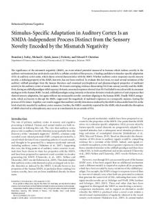
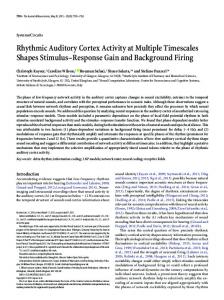
Fig. 1. Schematic representation of the 22-channel fNIRS probe sets placed on the participants' scalps, separated for left and right hemisphere. White squares represent light emitters; black squares represent detectors and numbers represent the measurement channels. The ROI is indicated by the dark grey shaded areas of the probe sets (channels: 2, 3, 7, 11 (left) or 12 (right)). Non-ROI channels are shown within light grey shaded areas. (b) Cranial optode positions in relation to the underlying anatomical structures are shown for one representative subject. ROI channels as described above are labeled. White circles represent light emitters; black circles represent detectors.
were found to be more activated by emotionally arousing compared to neutral stimuli (Alpers et al., 2009, 2005; Bradley et al., 2003; Herrmann et al., 2008; Lane et al., 1997; Lang et al., 1998; Minati et al., 2009). Thus, the present results clearly demonstrate that human auditory cortex activation is comparably modulated by complex emotional sounds. This adds to the growing evidence that the enhanced activation of sensory areas in response to emotional stimuli is apparently not restricted to a specific sensory modality, but that it is a more general principle of sensory processing (see Wiethoff et al., 2008). Most importantly, our results support the idea that emotional enhancement of basic sensory activation may represent an important adaptive mechanism to facilitate sensory encoding of emotionally significant stimuli. This may help the organism to quickly discriminate between relevant and irrelevant information and to prepare for adequate behavioral responses. Ultimately, successful survival could depend on enhanced sensory processing associated with facilitated identification of crucial information coming from the surroundings (Öhman et al., 2000; Öhman and Mineka, 2001). The finding that both pleasant and unpleasant sounds evoked enhanced activation in auditory cortex is also in line with results from the visual domain. Functional imaging and EEG studies
typically document that pleasant and unpleasant pictures are both processed more intensively in occipital regions (e.g. Bradley et al., 2003; Herrmann et al., 2008; Lang et al., 1998; Schupp et al., 2003). It is generally assumed that this is induced by re-entrant projections from the amygdala (e.g. Keil et al., 2009), which does not respond to valence of emotional stimuli per se (see meta-analyses from Murphy et al., 2003; Sergerie et al., 2008; Wager et al., 2003), but rather encodes the emotional salience of highly arousing stimuli (D. Sander et al., 2003), and is, thus, involved in the processing of both unpleasant and pleasant stimuli (but see also e.g. Gerdes et al., 2010). Our findings support the view that this mechanism also applies for auditory cues (Baxter and Murray, 2002; Fecteau et al., 2007; K. Sander et al., 2003; Sander and Scheich, 2001; Zald, 2003). It is plausible, that activation of the amygdala in response to auditory emotional stimuli (Fecteau et al., 2007) may be the driving force of the enhanced processing in auditory cortex via direct or indirect modulating ascending neural pathways (Doron and LeDoux, 1999; LeDoux, 1996; Woodson et al., 2000). However, there may also exist top-down influences on sensory areas due to re-entrant processing not only from subcortical areas like the amygdala, but also from the higher-order cortical areas as observed for emotional
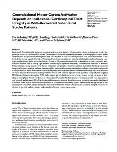
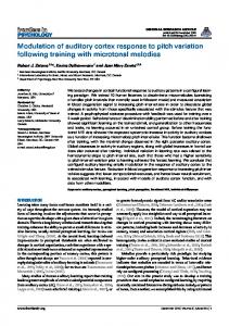
Fig. 2. Left and right auditory cortex activation evoked by sounds of the three emotional categories. Note that HHb results are shown and that scale has been inverted — see supplements for O2Hb results.
1204
M.M. Plichta et al. / NeuroImage 55 (2011) 1200–1207
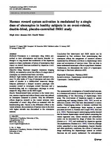
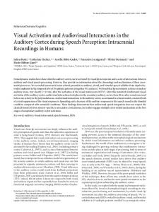
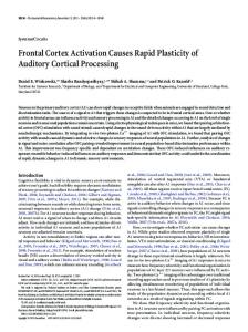
Fig. 3. Averaged activation and standard errors (SE) within and outside auditory cortex areas evoked by emotional sound categories (UNP = unpleasant; NEU = neutral; PLE = pleasant) is shown on the left side (a). The averaged signal time courses and SE separated for emotional sound categories are shown on the right side (b). Note that HHb results are shown and that the scale has been inverted. Results separated for left and right hemisphere are shown in the Supplementary material.
pictures in the visual modality (Keil et al., 2009; Sabatinelli et al., 2009). To this end, one may speculate that analogue to the visual domain (Martínez et al., 1999), an initial bottom-up auditory analysis is followed-up by an re-entrant modulation stemming from feedback signals originating in higher auditory cortex, prefrontal areas, or subcortical structures. Thus, prefrontal feedback in order to enhance sensitivity of auditory neurons to attended relevant stimulus features is highly likely (Jäncke et al., 1999; Jäncke and Shah, 2002) and does not weaken the implication of our findings. However, the involvement of the amygdala could not be tested in our study since fNIRS measures are limited to cortical areas. Activation of the prefrontal cortex, which was only partially accessible with the current setting, may have remained unrevealed due to methodological issues (Singh et al., 2005). Emotional stimuli: inherent physical properties or not? The investigation of perceptual and cognitive functions with noninvasive brain imaging methods critically depends on the careful selection of stimuli in experiments. Comparing human vocal and nonvocal sounds, Belin et al. (2000) showed that large parts of the auditory cortex respond stronger to human vocalizations than to other non-vocal sounds. Furthermore, Ethofer et al. (2009) demonstrated that emotional modulation effects also occurred within these regions. Therefore, in the present study the stimuli of the three categories are matched according to the ratio of human vocal sounds in the present study. A further important aspect is the characterization of the stimuli's physical properties and its comparability across the categories (see also Wiethoff et al., 2008). Focusing on the perceived unpleasantness of sounds, Kumar et al. (2008) noted that two classes of explanation for the perceived unpleasantness can be distinguished: first, sounds may contain features that inherently evoke negative (or positive) emotional responses or, second, associative learning. As shown in the visual research domain, visual perception and associated brain activation is strongly influenced by physical properties of the pictures, such as size, luminance, contrast or spatial frequency (Baas et al., 2002; Plichta et al., 2007b; Yue et al., 2011). This has been often neglected when emotional vs. neutral stimulus material was compared. Delplanque et al. (2007) showed that some of the negative stimuli contained in the international affective picture system (IAPS) have a higher fraction of lower spatial frequency as compared to neutral ones. Meanwhile, however, it has been shown that the emotional effect is not solely explained by physical properties (Bradley et al., 2007; De Cesarei and
Codispoti, 2006; Sánchez-Navarro et al., 2006) and studies which carefully controlled their visual stimulus material for physical characteristics still report strong emotional effects (Junghofer et al., 2001, 2006; Keil et al., 2003; Sabatinelli et al., 2007). However, in principle an infinite number of possible physical properties exists which may have an impact on neural sensory processing. In the case of sound material, it might well be that besides very basal parameters that were explicitly held constant across the sound categories in the current study (mean amplitude; frequency composition), other features e.g. the spectro-temporal modulation (Shamma et al., 1993) content of the sound material has an impact. Kumar et al. (2008) showed spectral frequencies between 2500 and 5500 Hz temporally modulated in a range of 1–16 Hz to be associated with the perceived unpleasantness. Therefore, we analyzed our selected sound material post-hoc according to the procedure described in Kumar et al. (2008). Analyses indeed showed subtle differences between emotional and neutral sounds. Re-analyses of our fNIRS data, however, demonstrated that these differences in spectrotemporal modulation do not solely explain our findings at the neural level (the complete results can be found in supplementary material). fNIRS related issues We used fNIRS as a brain imaging technique to shed light on emotional modulation in the auditory domain because it has inherent advantages to study activation evoked by emotional sounds: fNIRS is non-invasive, silent and the neuroimaging setting may be ecologically more valid for the examination of human cortical emotional processing (Hoshi et al., in press). The results obtained in the present experiment confirm that fNIRS is capable of detecting auditory activation. However, fNIRS specific shortcomings had to be critically considered and accounted for during experimental planning and data analyses: because fNIRS does not provide an anatomical image, we carefully planned the ROI definitions. A pilot study with emotionally neutral 1000 Hz tone stimulation was performed to identify channels that reflect auditory cortex activation. In addition, all tests of the emotional modulation were performed on amplitude estimates from channels which were identified as being significantly active during all acoustic stimulations, irrespective of the emotion condition (conjunction hypothesis). This is particularly important because it ensures that genuine auditory cortex activation was compared across conditions. This strategy can be described as datadriven and relying on functional criteria rather than probabilistic projection methods (Okamoto et al., 2004). In addition, we analyzed
M.M. Plichta et al. / NeuroImage 55 (2011) 1200–1207
non-ROI data to inspect the specificity of the results. Indeed, the results showed that the identified emotional modulation of activation was limited to auditory cortex and did not extend to areas surrounding the ROI. Nevertheless, the interpretation that we found emotional modulation in a single and homogeneous neural system can be challenged because spatial resolution of our fNIRS measure is rather low. With the higher spatial resolution provided by fMRI it can be demonstrated that complex sounds engage distinct areas within the auditory cortex (Leaver and Rauschecker, 2010). It might be argued that the applied pleasant and/or unpleasant sounds in our study do engage distinct areas as compared to neutral sounds and therefore mimic higher amplitudes as measured with fNIRS putatively because these distinct areas are larger or more surface prone. However, careful selection and matching of sounds across the categories according to different classes and physical properties prevents (as far as possible) this potentially confounding effect. Moreover, we did not consider discrete emotional categories as e.g. happiness, sadness, anger, fear, or disgust (Stevenson and James, 2008), which were shown to activate distinct behavioral and neural patterns for visual and auditory stimuli (Bradley et al., 2001; Grandjean et al., 2005; Murphy et al., 2003; K. Sander et al., 2003). Thus, specific contents may also account for activation in distinct areas of the auditory sensory system. Finally, with the current spatial resolution it is not possible to reliably disentangle primary auditory cortex and non-primary auditory cortex activation. While clear-cut primary auditory cortex modulation would definitely strengthen the parallel of the current findings to results in the visual domain, the unambiguous identification of primary auditory cortex is not a prerequisite for the conclusion that basal sensory processing is modulated by emotion and that this may reflect an important evolutionary principle. Importantly, it is rather unlikely that we only captured activation in areas which are unspecific to audition (multimodal areas or higher cortical associations): the location of our detected activation is nearly identical to the results obtained in our event-related 1000 Hz tone stimulation study (compare Fig. 2 and Supplementary material) for which primary auditory cortex sensitivity is high. Furthermore, the detected activation declines rather abruptly outside of the pre-defined ROI at channels #4, #8, #12, #16 (left hemisphere) and channels #1, #6, #11, #16 (right hemisphere) which parallels the presumed position of the sylvian fissure. This boundary is useful to render more precisely the position of our activation cluster in relation to the underlying cortex: accordingly, relative to channel #7 (i.e. T3/T4) for which Brodmann area (BA) 21 may be most likely (Okamoto et al., 2004) posterior–superior channel #11 (left hemisphere) and #12 (right hemisphere), positioned at the upper border of the activation cluster, are most likely located above BA 41/42. Separate analyses for these channels did not change the reported results. In line with existing fNIRS studies on emotional modulation of brain activation (Alpers et al., 2005; Herrmann et al., 2008), we found statistically more robust results for HHb compared to O2Hb (but see also Minati et al., 2009). However, in most other research domains, O2Hb appears to be more robust than HHb results (Holper et al., 2009; Plichta et al., 2006b; Schecklmann et al., 2007, 2008; Wolf et al., 2002) and diverging results are often interpreted as a lack of statistical power for the HHb parameter. On the other hand, we have previously suggested that HHb may have a higher spatial specificity and a higher local statistical power as compared to O2Hb (Plichta et al., 2007c). A possible explanation for the differences we observed here may be the impact of emotional arousal on systemic variables. For example, it has been shown that unpleasant pictures decrease heart rate compared to neutral and pleasant pictures, and that pleasant pictures increase blood pressure compared to neutral and unpleasant stimuli (Hempel et al., 2005; Sarlo et al., 2005). Because heart rate and blood pressure correlate with signal changes in O2Hb but not in HHb (Mehagnoul-Schipper et al., 2000), such systemic changes may have reduced the robustness of O2Hb as compared to the HHb effects. Because we did not measure heart rate or blood pressure in this study,
1205
this interpretation remains speculative and needs to be considered in further research.
Conclusion The present study demonstrated that auditory emotional cues modulate the activity of sensory areas as it has been shown for the visual domain. Our results underscore the idea that enhanced sensory processing of emotional stimuli may be an adaptive mechanism that enables the organisms to quickly and effectively respond to meaningful stimuli in the environment.
Acknowledgments This work was supported by the Research Group “Emotion and Behavior” which is sponsored by the German Research Society (DFG; FOR 605) and the SFB-TRR 58, subproject C4. The authors would like to thank Hitachi Medical Corporation for the ETG-4000 equipment. Furthermore, we thank Ramona Baur and Katharina Spacek for their help with data collection and Edda Bilek for proof reading the manuscript.
Appendix A. Stimulus list
Description
IADS number
Emotional Category
Human vocal soundsa
Sound duration (sec)b
DogGrowl Bees MaleSnore BabyCry FemScream3 ChildAbuse Funeral Attack2 Victim CardiacArrest Fight Prowler MaleScream JackHammer CarHorns Wind EngineFailure War Radio GlassBreak TomCat Rooster NoseBlow Yawn Crowd1 Crowd2 Office1 TypeWriter Traffic Restaurant Football Helicopter1 Helicopter2 Countdown Injury CboyIndians Belch ClockTick Cuckoo Walking Carousel BabyLaugh
106 115 252 261 277 278 280 285 286 287 290 291 292 380 420 500 502 706 723 730 100 120 251 262 310 311 320 322 325 361 362 403 410 415 423 610 702 708 710 722 109 110
Unpleasant Unpleasant Unpleasant Unpleasant Unpleasant Unpleasant Unpleasant Unpleasant Unpleasant Unpleasant Unpleasant Unpleasant Unpleasant Unpleasant Unpleasant Unpleasant Unpleasant Unpleasant Unpleasant Unpleasant Neutral Neutral Neutral Neutral Neutral Neutral Neutral Neutral Neutral Neutral Neutral Neutral Neutral Neutral Neutral Neutral Neutral Neutral Neutral Neutral Pleasant Pleasant
Non vocal Non vocal Vocal Vocal Vocal Vocal Vocal Vocal Vocal Non vocal Vocal Vocal Vocal Non vocal Non vocal Non vocal Non vocal Non vocal Non vocal Non vocal Non vocal Non vocal Vocal Vocal Vocal Vocal Non vocal Non vocal Non vocal Vocal Vocal Non vocal Non vocal Vocal Vocal Vocal Vocal Non vocal Non vocal Non vocal Vocal Vocal
5.99 5.99 5.99 5.99 5.99 5.98 5.69 5.99 5.57 5.75 5.99 6.00 5.99 5.99 5.99 5.99 5.99 5.94 5.99 5.99 5.99 5.99 5.99 5.99 5.99 6.00 5.99 5.99 5.99 5.99 5.64 5.99 5.99 6.00 6.00 5.99 5.41 5.99 5.99 5.99 5.99 5.99 (continued on next page)
1206
M.M. Plichta et al. / NeuroImage 55 (2011) 1200–1207
Appendix A (continued) Description
IADS number
Emotional Category
Human vocal soundsa
Sound duration (sec)b
MusicBox KidsInPark EroticFem1 EroticFem3 EroticMale1 EroticCouple2 BoyLaugh MaleLaugh ClapGame Laughing Giggling VideoGame SportsCrowd RollerCoaster CourtSport Beethoven RockNRoll FunkMusic
111 112 201 205 210 215 220 221 225 226 230 254 352 360 370 810 815 820
Pleasant Pleasant Pleasant Pleasant Pleasant Pleasant Pleasant Pleasant Pleasant Pleasant Pleasant Pleasant Pleasant Pleasant Pleasant Pleasant Pleasant Pleasant
Non vocal Vocal Vocal Vocal Vocal Vocal Vocal Vocal Non vocal Vocal Vocal Non vocal Vocal Non vocal Non vocal Non vocal Non vocal Non vocal
5.99 5.99 5.99 5.99 6.00 5.98 5.99 5.99 5.99 5.99 5.99 5.98 5.99 5.99 5.99 5.99 6.00 5.86
a
Sound categories did not statistically differ with regard to the ratio of vocal/non-vocal sounds (Chi2 (2) = 0.536, p = 0.765). b Mean duration across all sounds was 5.95 s. Separated for the emotional categories the mean duration was 5.94 s (unpleasant), 5.94 s (neutral) and 5.98 s (pleasant) — there was no significant difference of duration between the sound categories (F(2,59) = 0.92; p N 0.20).
Appendix B. Supplementary materials Supplementary data to this article can be found online at doi:10.1016/j.neuroimage.2011.01.011. References Alpers, G.W., Gerdes, A.B.M., 2007. Here is looking at you: emotional faces predominate in binocular rivalry. Emotion 7, 495–506. Alpers, G.W., Gerdes, A.B.M., 2008. Emotional pictures in binocular rivalry. In: Columbus, F. (Ed.), Visual Perception: New Research. Nova Science Publishers, Hauppauge, NY, pp. 227–247. Alpers, G.W., Herrmann, M.J., Pauli, P., Fallgatter, A.J., 2005. Emotional arousal and activation of the visual cortex: a near infrared spectroscopy analysis [abstract]. J. Psychophysiol. 19, 106. Alpers, G.W., Gerdes, A.B.M., Lagarie, B., Tabbert, K., Vaitl, D., Stark, R., 2009. Attention and amygdala activity: an fMRI study with spider pictures in spider phobia. J. Neural Transm. 116, 747–757. Amaral, D.G., Behniea, H., Kelly, J.L., 2003. Topographic organization of projections from the amygdala to the visual cortex in the macaque monkey. Neuroscience 118, 1099–1120. Amaro Jr., E., Williams, S.C., Shergill, S.S., Fu, C.H., MacSweeney, M., Picchioni, M.M., Brammer, M.J., McGuire, P.K., 2002. Acoustic noise and functional magnetic resonance imaging: current strategies and future prospects. J. Magn. Reson. Imaging 16, 497–510. Anders, S., Eippert, F., Weiskopf, N., Veit, R., 2008. The human amygdala is sensitive to the valence of pictures and sounds irrespective of arousal: an fMRI study. Soc. Cogn. Affect. Neurosci. 3, 233–243. Baas, J.M.P., Kenemans, J.L., Mangun, G.R., 2002. Selective attention to spatial frequency: an ERP and source localization analysis. Clin. Neurophysiol. 113, 1840–1854. Bandettini, P.A., Jesmanowicz, A., Van Kylen, J., Birn, R.M., Hyde, J.S., 1998. Functional MRI of brain activation induced by scanner acoustic noise. Magn. Reson. Med. 39, 410–416. Baxter, M.G., Murray, E.A., 2002. The amygdala and reward. Nat. Rev. Neurosci. 3, 563–573. Belin, P., Zatorre, R.J., Lafaille, P., Ahad, P., Pike, B., 2000. Voice-selective areas in human auditory cortex. Nature 403, 309–312. Belin, P., Fecteau, S., Charest, I., Nicastro, N., Hauser, M.D., Armony, J.L., 2008. Human cerebral response to animal affective vocalizations. Proc. Biol. Sci. 275, 473–481. Blackman, G.A., Hall, D.A., in press. Reducing the effects of background noise during auditory functional magnetic resonance imaging of speech processing: Qualitative and quantitative comparisons between two image acquisition schemes and noise cancellation. J. Speech Lang. Hear. Res. doi:10.1044/1092-4388(2010/10-0143). Blood, A.J., Zatorre, R.J., 2001. Intensely pleasurable responses to music correlate with activity in brain regions implicated in reward and emotion. Proc. Natl Acad. Sci. USA 98, 11818–11823. Bradley, M.M., Lang, P.J., 1999. International affective digitized sounds (IADS): stimuli, instruction manual and affective ratings (Tech. Rep. No. B-2). The Center for Research in Psychophysiology. University of Florida, Gainesville, FL. Bradley, B.P., Lang, P.J., 2000. Affective reactions to acoustic stimuli. Psychophysiology 37, 204–215. Bradley, M.M., Codispoti, M., Sabatinelli, D., Lang, P.J., 2001. Emotion and motivation II: sex differences in picture processing. Emotion 1, 300–319.
Bradley, M.M., Sabatinelli, D., Lang, P.J., Fitzsimmons, J.R., King, W.M., Desai, P., 2003. Activation of the visual cortex in motivated attention. Behav. Neurosci. 117, 369–380. Bradley, M.M., Hamby, S., Löw, A., Lang, P.J., 2007. Brain potentials in perception: picture complexity and emotional arousal. Psychophysiology 44, 364–373. Catani, M., Jones, D.K., Donato, R., Ffytche, D.H., 2003. Occipito-temporal connections in the human brain. Brain 126, 2093–2107. Cochrane, D., Orcutt, G.H., 1949. Application of least squares regression to relationships containing auto-correlated error terms. J. Am. Stat. Assoc. 44, 32–61. Czigler, I., Cox, T.J., Gyimesi, K., Horvath, J., 2007. Event-related potential study to aversive auditory stimuli. Neurosci. Lett. 420, 251–256. Davis, M., Whalen, P.J., 2001. The amygdala: vigilance and emotion. Mol. Psychiatry 6, 13–34. De Cesarei, A., Codispoti, M., 2006. When does size matter? Effects of stimulus size on affective modulation. Psychophysiology 43, 207–215. Delplanque, S., N'Diaye, K., Scherer, K., Grandjean, D., 2007. Spatial frequencies or emotional effects? A systematic measure of spatial frequencies for IAPS pictures by a discrete wavelet analysis. J. Neurosci. Meth. 165, 144–150. Doron, N.N., LeDoux, J.E., 1999. Organization of projections to the lateral amygdala from auditory and visual areas of the thalamus in the rat. J. Comp. Neurol. 412, 383–409. Ethofer, T., Anders, S., Wiethoff, S., Erb, M., Herbert, C., Saur, R., Grodd, W., Wildgruber, D., 2006. Effects of prosodic emotional intensity on activation of associative auditory cortex. NeuroReport 17, 249–253. Ethofer, T., Van De Ville, D., Scherer, K., Vuilleumier, P., 2009. Decoding of emotional information in voice-sensitive cortices. Curr. Biol. 19, 1028–1033. Faul, F., Erdfelder, E., Lang, A.G., Buchner, A., 2007. G*Power 3: a flexible statistical power analysis program for the social, behavioral, and biomedical sciences. Behav. Res. Meth. 39, 175–191. Fecteau, S., Belin, P., Joanette, Y., Armony, J.L., 2007. Amygdala responses to nonlinguistic emotional vocalizations. Neuroimage 36, 480–487. Ferrari, M., Mottola, L., Quaresima, V., 2004. Principles, techniques, and limitations of near infrared spectroscopy. Can. J. Appl. Physiol. 29, 463–487. Frey, S., Kostopoulos, P., Petrides, M., 2000. Orbitofrontal involvement in the processing of unpleasant auditory information. Eur. J. Neurosci. 12, 3709–3712. Gaab, N., Gabrieli, J., Glover, G., 2007. Assessing the influence of scanner background noise on auditory processing-II: an fMRI study comparing auditory processing in the absence and presence of recorded scanner noise using a sparse temporal sampling design. Hum. Brain Mapp. 28, 721–732. Gerdes, A.B., Wieser, M.J., Muhlberger, A., Weyers, P., Alpers, G.W., Plichta, M.M., Breuer, F., Pauli, P., 2010. Brain activations to emotional pictures are differentially associated with valence and arousal ratings. Front. Hum. Neurosci. 4, 175. Grandjean, D., Sander, D., Pourtois, G., Schwartz, S., Seghier, M.L., Scherer, K.R., Vuilleumier, P., 2005. The voices of wrath: Brain responses to angry prosody in meaningless speech. Nat. Neurosci. 8, 145–146. Griffiths, T.D., Uppenkamp, S., Johnsrude, I., Josephs, O., Patterson, R.D., 2001. Encoding of the temporal regularity of sound in the human brainstem. Nat. Neurosci. 4, 633–637. Hall, D.A., Haggard, M.P., Akeroyd, M.A., Palmer, A.R., Summerfield, A.Q., Elliott, M.R., Gurney, E.M., Bowtell, R.W., 1999. “Sparse” temporal sampling in auditory fMRI. Hum. Brain Mapp. 7, 213–223. Hempel, R.J., Tulen, J.H., van Beveren, N.J., van Steenis, H.G., Mulder, P.G., Hengeveld, M. W., 2005. Physiological responsivity to emotional pictures in schizophrenia. J. Psychiatr. Res. 39, 509–518. Hennel, F., Girard, F., Loenneker, T., 1999. “Silent” MRI with soft gradient pulses. Magn. Reson. Med. 42, 6–10. Herrmann, M.J., Huter, T.J., Plichta, M.M., Ehlis, A.-C., Alpers, G.W., Mühlberger, A., Lesch, K.-P., Fallgatter, A.J., 2008. Enhancement of neural activity of the primary visual cortex for emotional stimuli measured with event-related functional near infrared spectroscopy (NIRS). Hum. Brain Mapp. 29, 28–35. Holper, L., Biallas, M., Wolf, M., 2009. Task complexity relates to activation of cortical motor areas during uni- and bimanual performance: a functional NIRS study. Neuroimage 46, 1105–1113. Hoshi, Y., 2003. Functional near-infrared optical imaging: utility and limitations in human brain mapping. Psychophysiology 40, 511–520. Hoshi, Y., Huang, J., Kohri, S., Iguchi, Y., Naya, M., Okamoto, T., Ono, S., in press. Recognition of Human Emotions from Cerebral Blood Flow Changes in the Frontal Region: A Study with Event-Related Near-Infrared Spectroscopy. J. Neuroimaging. doi:10.1111/j.1552-6569.2009.00454.x. Hovland, C.I., Bradshaw, D.A., 1937. Visual reaction time as a function of stimulusbackground contrast. Psychol. Res. 21, 50–55. Jäncke, L., Shah, N.J., 2002. Does dichotic listening probe temporal lobe functions? Neurology 58, 736–743. Jäncke, L., Mirzazade, S., Shah, N.J., 1999. Attention modulates activity in the primary and the secondary auditory cortex: a functional magnetic resonance imaging study in human subjects. Neurosci. Lett. 266, 125–128. Johnstone, T., van Reekum, C.M., Oakes, T.R., Davidson, R.J., 2006. The voice of emotion: an FMRI study of neural responses to angry and happy vocal expressions. Soc. Cogn. Affect. Neurosci. 1, 242–249. Junghofer, M., Bradley, M.M., Elbert, T.R., Lang, P.J., 2001. Fleeting images: a new look at early emotion discrimination. Psychophysiology 38, 175–178. Junghofer, M., Sabatinelli, D., Bradley, M.M., Schupp, H.T., Elbert, T.R., Lang, P.J., 2006. Fleeting images: rapid affect discrimination in the visual cortex. NeuroReport 17, 225–229. Keil, A., Gruber, T., Müller, M.M., Moratti, S., Stolarova, M., Bradley, M.M., Lang, P.J., 2003. Early modulation of visual perception by emotional arousal: evidence from steady-state visual evoked brain potentials. Cogn. Affect. Behav. Neurosci. 3, 195–206. Keil, A., Moratti, S., Sabatinelli, D., Bradley, M.M., Lang, P.J., 2005. Additive effects of emotional content and spatial selective attention on electrocortical facilitation. Cereb. Cortex 15, 1187–1197.
M.M. Plichta et al. / NeuroImage 55 (2011) 1200–1207 Keil, A., Sabatinelli, D., Ding, M., Lang, P.J., Ihssen, N., Heim, S., 2009. Re-entrant projections modulate visual cortex in affective perception: evidence from Granger causality analysis. Hum. Brain Mapp. 30, 532–540. Kumar, S., Forster, H.M., Bailey, P., Griffiths, T.D., 2008. Mapping unpleasantness of sounds to their auditory representation. J. Acoust. Soc. Am. 124, 3810–3817. Lane, R.D., Reiman, E.M., Bradley, M.M., Lang, P.J., Ahern, G.L., Davidson, R.J., Schwartz, G.E., 1997. Neuroanatomical correlates of pleasant and unpleasant emotion. Neuropsychologia 35, 1437–1444. Lane, R.D., Chua, P.M.-L., Dolan, R.J., 1999. Common effects of emotional valence, arousal and attention on neural activation during visual processing of pictures. Neuropsychologia 37, 989–997. Lang, P.J., 1995. The emotion probe: studies of motivation and attention. Am. Psychol. 50, 372–385. Lang, P.J., Bradley, M.M., Cuthbert, M.M., 1997. Motivated attention: affect, activation and action. In: Lang, P.J., Simons, R.F., Balaban, M.T. (Eds.), Attention and Orienting: Sensory and Motivational Processes. Lawrence Erlbaum Associates, Hillsdale, New Jersey. Lang, P.J., Bradley, M.M., Fitzsimmons, J.R., Cuthbert, B.N., Scott, J.D., Moulder, B., Nangia, V., 1998. Emotional arousal and activation of the visual cortex: an fMRI analysis. Psychophysiology 35, 199–210. Leaver, A.M., Rauschecker, J.P., 2010. Cortical representation of natural complex sounds: effects of acoustic features and auditory object category. J. Neurosci. 30, 7604–7612. LeDoux, J., 1996. The Emotional Brain: The Mysterious Underpinnings of Emotional Life. Simon & Schuster, New York. LeDoux, J.E., 1998. Fear and the brain: where have we been, and where are we going? Biol. Psychiatry 44, 1229–1238. LeDoux, J.E., 2000. Emotion circuits in the brain. Annu. Rev. Neurosci. 23, 155–184. Leutgeb, V., Schafer, A., Schienle, A., 2009. An event-related potential study on exposure therapy for patients suffering from spider phobia. Biol. Psychol. 82, 293–300. Maddock, R.J., Buonocore, M.H., 1997. Activation of left posterior cingulate gyrus by the auditory presentation of threat-related words: an fMRI study. Psychiatry Res. 75, 1–14. Magnée, M.J., Stekelenburg, J.J., Kemner, C., de Gelder, B., 2007. Similar facial electromyographic responses to faces, voices, and body expressions. NeuroReport 18, 369–372. Marcar, V.L., Girard, F., Rinkel, Y., Schneider, J.F., Martin, E., 2002. Inaudible functional MRI using a truly mute gradient echo sequence. Neuroradiology 44, 893–899. Martínez, A., Anllo-Vento, L., Sereno, M.I., Frank, L.R., Buxton, R.B., Dubowitz, D.J., Wong, E.C., Hinrichs, H., Heinze, H.J., Hillyard, S.A., 1999. Involvement of striate and extrastriate visual cortical areas in spatial attention. Nat. Neurosci. 2, 364–369. Mehagnoul-Schipper, D.J., Vloet, L.C., Colier, W.N., Hoefnagels, W.H., Jansen, R.W., 2000. Cerebral oxygenation declines in healthy elderly subjects in response to assuming the upright position. Stroke 31, 1615–1620. Minati, L., Jones, C.L., Gray, M.A., Medford, N., Harrison, N.A., Critchley, H.D., 2009. Emotional modulation of visual cortex activity: a functional near-infrared spectroscopy study. NeuroReport 20, 1344–1350. Morris, J.S., Scott, S.K., Dolan, R.J., 1999. Saying it with feeling: neural responses to emotional vocalizations. Neuropsychologia 37, 1155–1163. Murphy, F.C., Nimmo-Smith, I., Lawrence, A.D., 2003. Functional neuroanatomy of emotions: a meta-analysis. Cogn. Affect. Behav. Neurosci. 3, 207–233. Nichols, T., Brett, M., Andersson, J., Wager, T., Poline, J.-B., 2005. Valid conjunction inference with the minimum statistic. Neuroimage 25, 653–660. Obleser, J., Wise, R.J.S., Alex Dresner, M., Scott, S.K., 2007. Functional integration across brain regions improves speech perception under adverse listening conditions. J. Neurosci. 27, 2283–2289. Obrig, H., Villringer, A., 2003. Beyond the visible-imaging the human brain with light. J. Cereb. Blood Flow Metab. 23, 1–18. Öhman, A., Mineka, S., 2001. Fears, phobias, and preparedness: toward an evolved of fear and fear learning. Psychol. Rev. 108, 483–522. Öhman, A., Flykt, A., Lundqvist, D., 2000. Unconscious emotion: evolutionary perspectives, psychophysiological data and neuropsychological mechanisms. In: Lane, R.D.R., Nadel, L. (Eds.), Cognitive Neuroscience of Emotion. Oxford University Press, New York, pp. 296–327. Okamoto, M., Dan, H., Sakamoto, K., Takeo, K., Shimizu, K., Kohno, S., Oda, I., Isobe, S., Suzuki, T., Kohyama, K., Dan, I., 2004. Three-dimensional probabilistic anatomical cranio-cerebral correlation via the international 10–20 system oriented for transcranial functional brain mapping. Neuroimage 21, 99–111. Padmala, S., Pessoa, L., 2008. Affective Learning Enhances Visual Detection and Responses in Primary Visual Cortex. J. Neurosci. 28, 6202–6210. Partalaa, T., Surakka, V., 2003. Pupil size variation as an indication of affective processing. Int. J. Hum. Comput. Stud. 59, 185–198. Phelps, E.A., Ling, S., Carrasco, M., 2006. Emotion facilitates perception and potentiates the perceptual benefits of attention. Psychol. Sci. 17, 292–299. Plichta, M.M., Herrmann, M.J., Baehne, C.G., Ehlis, A.C., Richter, M.M., Pauli, P., Fallgatter, A.J., 2006a. Event-related functional near-infrared spectroscopy (fNIRS): are the measurements reliable? Neuroimage 31, 116–124. Plichta, M.M., Herrmann, M.J., Ehlis, A.-C., Baehne, C.G., Richter, M.M., Fallgatter, A.J., 2006b. Event-related visual versus blocked motor task: detection of specific cortical activation patterns with functional near-infrared spectroscopy. Neuropsychobiology 53, 77–82. Plichta, M.M., Gerdes, A.B.M., Alpers, G.W., Fallgatter, A.J., 2007a. The sound of emotion: An fNIRS study of the processing of emotional sounds. Brain Topogr. 20, 48. Plichta, M.M., Heinzel, S., Ehlis, A.C., Pauli, P., Fallgatter, A.J., 2007b. Model-based analysis of rapid event-related functional near-infrared spectroscopy (NIRS) data: a parametric validation study. Neuroimage 35, 625–634. Plichta, M.M., Herrmann, M.J., Baehne, C.G., Ehlis, A.C., Richter, M.M., Pauli, P., Fallgatter, A.J., 2007c. Event-related functional near-infrared spectroscopy (fNIRS) based on
1207
craniocerebral correlations: reproducibility of activation? Hum. Brain Mapp. 28, 733–741. Pripfl, J., Robinson, S., Leodolter, U., Moser, E., Bauer, H., 2006. EEG reveals the effect of fMRI scanner noise on noise-sensitive subjects. Neuroimage 31, 332–341. Royet, J.P., Zald, D., Versace, R., Costes, N., Lavenne, F., Koenig, O., Gervais, R., 2000. Emotional responses to pleasant and unpleasant olfactory, visual, and auditory stimuli: a positron emission tomography study. J. Neurosci. 20, 7752–7759. Sabatinelli, D., Bradley, M.M., Fitzsimmons, J.R., Lang, P.J., 2005. Parallel amygdala and inferotemporal activation reflect emotional intensity and fear relevance. Neuroimage 24, 1265–1270. Sabatinelli, D., Lang, P.J., Keil, A., Bradley, M.M., 2007. Emotional perception: correlation of functional MRI and event-related potentials. Cereb. Cortex 17, 1085–1091. Sabatinelli, D., Lang, P.J., Bradley, M.M., Costa, V.D., Keil, A., 2009. The timing of emotional discrimination in human amygdala and ventral visual cortex. J. Neurosci. 29, 14864–14868. Sánchez-Navarro, J.P., Martínez-Selva, J.M., Román, F., Torrente, G., 2006. The effect of content and physical properties of affective pictures on emotional responses. Span. J. Psychol. 9, 145–153. Sander, K., Scheich, H., 2001. Auditory perception of laughing and crying activates human amygdala regardless of attentional state. Cogn. Brain Res. 12, 181–198. Sander, K., Scheich, H., 2005. Left auditory cortex and amygdala, but right insula dominance for human laughing and crying. J. Cogn. Neurosci. 17, 1519–1531. Sander, D., Grafman, J., Zalla, T., 2003a. The human amygdala: an evolved system for relevance detection. Rev. Neurosci. 14, 303–316. Sander, K., Brechmann, A., Scheich, H., 2003b. Audition of laughing and crying leads to right amygdala activation in a low-noise fMRI setting. Brain Res. Protoc. 81–91. Sarlo, M., Palomba, D., Buodo, G., Minghetti, R., Stegagno, L., 2005. Blood pressure changes highlight gender differences in emotional reactivity to arousing pictures. Biol. Psychol. 70, 188–196. Schecklmann, M., Ehlis, A.C., Plichta, M.M., Boutter, H.K., Metzger, F.G., Fallgatter, A.J., 2007. Altered frontal brain oxygenation in detoxified alcohol dependent patients with unaffected verbal fluency performance. Psychiatry Res. Neuroimaging 152, 129–138. Schecklmann, M., Ehlis, A.C., Plichta, M.M., Fallgatter, A.J., 2008. Functional nearinfrared spectroscopy: a long-term reliable tool for measuring brain. Neuroimage 43, 147–155. Schupp, H.T., Junghöfer, M., Weike, A.I., Hamm, A.O., 2003. Emotional facilitation of sensory processing in the visual cortex. Psychol. Sci. 14, 7–13. Schupp, H.T., Junghöfer, M., Weike, A.I., Hamm, A.O., 2004. The selective processing of briefly presented affective pictures: an ERP analysis. Psychophysiology 41, 441–449. Sergerie, K., Chochol, C., Armony, J.L., 2008. The role of the amygdala in emotional processing: a quantitative meta-analysis of functional neuroimaging studies. Neurosci. Biobehav. Rev. 32, 811–830. Shamma, S.A., Fleshman, J.W., Wiser, P.R., Versnel, H., 1993. Organization of response areas in ferret primary auditory cortex. J. Neurophysiol. 69, 367–383. Singh, A.K., Okamoto, M., Dan, H., Jurcak, V., Dan, I., 2005. Spatial registration of multichannel multi-subject fNIRS data to MNI space without MRI. Neuroimage 27, 842–851. Stevenson, R.A., James, T.W., 2008. Affective auditory stimuli: characterization of the International Affective Digitized Sounds (IADS) by discrete emotional categories. Behav. Res. Meth. 40, 315–321. Vuilleumier, P., Richardson, M.P., Armony, J.L., Driver, J., Dolan, R.J., 2004. Distant influences of amygdala lesion on visual cortical activation during emotional face processing. Nat. Neurosci. 7, 1271–1278. Wager, T.D., Phan, K.L., Liberzon, I., Taylor, S.F., 2003. Valence, gender, and lateralization of functional brain anatomy in emotion: a meta-analysis of findings from neuroimaging. Neuroimage 19, 513–531. Wiethoff, S., Wildgruber, D., Kreifelts, B., Becker, H., Herbert, C., Grodd, W., Ethofer, T., 2008. Cerebral processing of emotional prosody — influence of acoustic parameters and arousal. Neuroimage 39, 885–893. Wiethoff, S., Wildgruber, D., Grodd, W., Ethofer, T., 2009. Response and habituation of the amygdala during processing of emotional prosody. NeuroReport 20, 1356–1360. Wildgruber, D., Ackermann, H., Kreifelts, B., Ethofer, T., 2006. Cerebral processing of linguistic and emotional prosody: fMRI studies. Prog. Brain Res. 156, 249–268. Wolf, M., Wolf, U., Toronov, V., Michalos, A., Paunescu, L.A., Choi, J.H., Gratton, E., 2002. Different time evolution of oxyhemoglobin and deoxyhemoglobin concentration changes in the visual and motor cortices during functional stimulation: a nearinfrared spectroscopy study. Neuroimage 16, 704–712. Woodson, W., Farb, C.R., LeDoux, J.E., 2000. Afferents from the auditory thalamus synapse on inhibitory interneurons in the lateral nucleus of the amygdala. Synpase 38, 124–137. Yetkin, F.Z., Roland, P.S., Purdy, P.D., Christensen, W.F., 2003. Evaluation of auditory cortex activation by using silent FMRI. Am. J. Otolaryngol. 24, 281–289. Yue, X., Cassidy, B.S., Devaney, K.J., Holt, D.J., Tootell, Roger, B.H., 2011. Lower-Level Stimulus Features Strongly Influence Responses in the Fusiform Face Area. Cereb. Cortex. 21, 35–47. Yukie, M., 2002. Connections between the amygdala and auditory cortical areas in the macaque monkey. Neurosci. Res. 42, 219–229. Zald, D.H., 2003. The human amygdala and the emotional evaluation of sensory stimuli. Brain Res. Brain Res. Rev. 41, 88–123.