Received: May 30, 2016
65
Automatic Detection and Classification of Masses in Digital Mammograms Shankar Thawkar1*
Ranjana Ingolikar2
1
Department of Information Technology, Hindustan College of Science and Technology, Mathura, India 2 Department of Computer Science, S.F.S. College, Nagpur, India *Corresponding author’s Email:
[email protected]
Abstract: Breast Cancer is still one of the leading cancers in women. Mammography is the best tool for early detection of breast cancer. In this work methods for automatic detection and classification of masses into benign or malignant has been proposed. The suspicious masses are detected automatically by performing image segmentation with Otsu’s global thresholding technique, morphological operations and watershed transformation. Twenty-five features based on intensity, texture and shape are extracted from each of the 651 mammograms obtained from Database of Digitized Screen-film Mammograms. The Eight most significant features selected by step-wise Linear Discriminate Analysis are used to classify masses using Fisher’s Linear Discriminate Analysis, Support Vector Machine and Multilayer Perceptron with two training algorithms Levenberg-Marquardt and Bayesian Regularization. The performance evaluation of classifiers indicates that MLP is better than both LDA and SVM. MLP-RBF has 98.9% accuracy with area under Receiver Operating Characteristics curve AZ=0.98±0.007, MLP-LM 96.0% accuracy with AZ=0.97±0.007, SVM 91.4% accuracy with AZ=0.956±0.009 and LDA 90.3% accuracy with AZ=0.956±0.009. All the results achieved are promising when compared with some existing work. Keywords: Digital mammograms, Neural network, Linear discriminant analysis, Feature selection, Support vector machine, Receiver operating characteristics curve.
1. Introduction Breast cancer is still one of the leading cancers in women in the World. It has been estimated that in every 13 minutes a women dies due to breast cancer [1]. Currently no technique or method is available for prevention of breast cancer so detection of breast cancer in initial stage is very important. Mammography is the best tool for early detection of breast cancer [2]. It enables to detect two most important symptoms of breast cancer such as masses and calcification [3]. Automatic Detection of masses is a difficult task than calcification because they have different characteristics like boundaries and shape. One more reason is that features of masses are hidden or similar with normal tissue [4]. Reading digital mammograms is very challenging task for radiologist because mammograms are the low quality images, even a specialists inter observation rate varies [5]. Statistic shows that
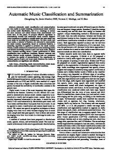
more than 70% of biopsies of suspected breast cancer lesion turn out to be benign. The number of efforts has been taken for the design and development of CAD system. These systems assist radiologist for interpreting mammograms for detection and classification of masses and so improve the breast cancer diagnosis and reduce mortality rate. The objective of the study is to investigate efficient methods for automatic detection and classification of masses in digital mammograms. The process adopted for detection and classification of masses in our work is described in Figure 1. At first step mammograms obtain from DDSM (Database of Digitized Screen-film Mammograms) acts as an input. Then Preprocessing is applied to remove labels and non-mass regions. After Preprocessing a combined approach is adopted for automatic detection of masses which consists of Otsu’s global thresholding technique, morphological operations and watershed transformation. Otsu’s
International Journal of Intelligent Engineering and Systems, Vol.10, No.1, 2017
DOI: 10.22266/ijies2017.0228.08
Received: May 30, 2016
global thresholding method and morphological operations are used to find location of suspicious mass. Then watershed transformation is applied to extract mass of exact size and shape. Once the masses are detected features based on Intensity, Texture and Shape are extracted from detected masses. The larger set of extracted features may hamper the performance of the classifiers so an optimal features set is selected using step-wise linear discriminant analysis. These features are used to classify masses using Fisher’s Linear Discriminate Analysis, Support Vector Machine and Multilayer Perceptron with two training algorithms Levenberg-Marquardt (MLP-LM) and Bayesian Regularization (MLP-RBF). The proposed method is far better than methods studied by other researchers in terms of rate of automatic detection, classification accuracy and execution time required for automatic detection and classification of masses. In proposed method the performance of the classifiers was evaluated using sensitivity, specificity, accuracy and AUC while other researchers used either AUC or accuracy with sensitivity and specificity. The remainder of the paper is organized as: Section 2 reviews of related work. Section 3 describes Preprocessing of mammograms. Automatic detection and Extraction of Masses is presented in section 4. Section 5 describes feature extraction from detected masses and selection of optimal features. Classification of masses into benign and malignant is described in section 6. In Section 7 results of the methods & discussion and conclusion of the paper in Section 8.
Figure. 1 Proposed Methodology
66
2. Related Work Automatic detection and classification of breast lesion is a challenging research area. Moayedi et al. [6] investigate the use of SEL weighted Support Vector machine for the classification of masses. The proposed method determines contourlet coefficients as features using contourlet transformation. The optimal features are selected by Genetic Algorithm. The accuracy of the classifiers reported was 91.5% and 81% for SVFNN and Kernel SVM respectively. The experiment was performed on set of images obtained from Mini-MIAS database. Arbach et al. [7] proposed backpropagation neural network (BNN) and KNN algorithm for the classification of masses. The experiment was conducted on 160 cases with ten texture and shape features. Author compares the results of classifiers with radiologist results. The KNN has 85.7% specificity and 84.6% sensitivity. The accuracy of BNN was determined by area under ROC curve 0.923.Christoyianni et al. [9] investigate the use of RBF and MLP Net for the classification masses using 12 texture features. The total classification accuracy achieved for MLP was 84.03%, 4% higher than RBF. Petrosian et al. [14] used modified decision-tree classifier to classify masses into benign or malign using texture features from GLCM. The optimal features are selected by leave-one-out (LOO) method [8]. The accuracy of the classifier obtained in terms of sensitivity and specificity was 76% and 64% respectively. Chan et al. [12] studied the importance of Texture features derived from GLCM matrix for classification of masses. The five optimal features out of eight features are select by stepwise linear discriminant analysis. The experiment was conducted on 168 malign and 504 normal cases. The accuracy of the classifier was evaluated using area under ROC curve and the average value of Az is 0.84 during training and 0.82 during testing. Kegelmeyer et al. [10] proposed method for detection of speculated masses using laws of texture measures. The experiment was conducted on 85 cases and the cases were screened by four radiologists to verify accuracy of proposed system. The accuracy of the method was 100% sensitivity and 82% specificity. Rangayyan et al. [11] proposed a technique that makes use of two shape factors, speculation index and fractional concavity. The method provides an accuracy of 81.5%. de Oliveira Martins et al. [13] proposed Ripley’s K function and support vector machine for the classification of masses. The best result obtained with proposed method was 94.94% of accuracy. Wong et al. [40] used ANN based technique for the classification of Masses. The four
International Journal of Intelligent Engineering and Systems, Vol.10, No.1, 2017
DOI: 10.22266/ijies2017.0228.08
Received: May 30, 2016
optimal features are selected using sequential forward selection technique. The classification accuracy of ANN using leave-one-out method is 86%. The experiment was conducted on fifty mammograms obtained from Mini-MIAS database. Zheng et al. [41] proposed hybrid support vector machine (K-SVM) for the classification of masses into begin or malign. The features are obtained by K-means algorithm for benign and malignant tumors separately. Then, generalized SVM is used for the classification with 10-fold cross validation and achieve accuracy 97.38% when tested on WDBC data set of 32 mammograms. Mohanty et al. [42] proposed a hybrid method for feature selection. The Experiment was conducted using decision tree classifier on reduce set of 26 features for 300 mammograms obtain from MIAS database and obtain an accuracy 97.7%. The motivations behind the proposed method are Required to improve rate of classification for automatic mass detection system. Extraction & Selection of most relevant features that will improve classification accuracy. Study of classifiers that will minimize the false positive rate Required to use large and balance data set (benign and malignant) because unbalanced data set may hamper the performance of classifiers [44][45]. The overall system should take minimum execution time. Design and development of CAD system that will assist radiologist.
67
4.1 Otsu’s global thresholding method Otsu’s global thresholding method is used to find out location of suspicious mass [17]. It basically convert gray level image into binary image. The thresholds that minimize inter class variance between black and white pixel is select automatically from image histogram. The resulting image is shown in Figure 3a. Then morphological operations are applied to find the location of the suspicious mass and extract the region of suspicious mass (cropping) from original image as shown in Figure 3b. 4.2 Morphological operations The Mathematical morphological operations are used to analyze the shapes and textures in images [15, 16]. Suppose I(s, t) be a gray scale image and S be a structuring element then Erosion (⊖) and Dilation (⊕) operations are defined as: Erosion: [I⊖S] (s,t)=min(u,v)∈SI(s+u,t+v) (1) Dilation: [I⊕S] (s,t)=max(u,v)∈SI(s−u,t−v) (2) Using above, the Opening morphological operation (o) is I o S= (I⊖S) ⊕ S. Similarly the closing operation (●) is I●S= (I⊕S)⊖ S. The TopHat and BotHat operations mentioned below are applied to enhance or suppress details of gray scale mammogram image smaller than structuring element: TopHat(G) = G - (GoS) BotHat(G) = G - (G●S)
(3) (4)
3. Preprocessing The basic objective of image preprocessing is to reduce noise and improve quality of images. Another goal is to remove labels and non-mass regions from breast area. A 3x3 median filter improve the quality of images by reducing both unipolar and bipolar impulse noise. Morphological operations are preformed on improved quality image to remove labels and borders. Figure 2 describes the preprocessing step.
Figure. 2 Preprocessing. (a) Original image; (b) A3x3 Median filtered Image; (c) Image after removing labels and border.
4. Mass Detection Segmentation is performed for extracting the Region of Interest (ROI) from the background of digital mammogram. The segmentation process is divided into following three steps-
Figure. 3 Suspicious mass detection. (a)Otsu’s thresholding method; (b) suspicious mass
International Journal of Intelligent Engineering and Systems, Vol.10, No.1, 2017
DOI: 10.22266/ijies2017.0228.08
Received: May 30, 2016
The TopHat image is added with original image and then subtracts BotHat to minimize the contrast and gaps between objects. Next step is to highlight the intensity valleys in image to detect the mass by performing watershed transformation to do this we enhanced the image by performing complement operation. The result of morphological operation is shown in Figure 4. 4.3 Watershed transformation & Extraction of Masses Watershed transformation is used for the detection of masses. It is based on mathematical morphology and it has many advantages compared to other image segmentation methods. Watershed transformation can find closed shape and exact position of objects. Vincent and Soille [18] proposed the algorithm for finding the watershed lines using the immersion simulation algorithm. Image segmentation process may affect due to presence of noise or other sort of non-uniformity that’s why some preprocessing steps are applied. A 3x3 median filter with contrast stretching transformation is applied to enhance image. The amount of contrast stretching is controlled by gamma parameter. It specifies the shape of mapping curve between input and output. In this work gamma value is set to 5. The foreground and background objects are marked by performing opening-byreconstruction and threshold opening-closing-byreconstruction. In this way the masses are detected. The mass of exact size and shape will be determined by performing morphological operations on result of watershed transformation. Figure 5 shows the result of watershed transformation.
68
5. Feature Extraction and Selection The performance of CAD system depends on features selection than classification methods. Radiologist diagnose a mass in mammograms to discriminate them into begin and malign with visually observed features such as shape, size and margins. But different radiologist may have different interpretation. The Computer Aided Diagnosis system will remove this problem by providing multiple methods to extract more discriminative and accurate features.
5.1 Feature Extraction The features extracted are classified into three types: Intensity features, Textural features and shape features. 5.1.1
Intensity features are the simplest features [19]. We have extracted six features F1-F6 from segmented masses using Histogram analysis. These features are Average gray level (F1), Average Contrast (F2), Smoothness (F3), Skewness or Third moment (F4), Uniformity (F5) and Entropy1 (F6) [3][20]. 5.1.2
Figure. 5 Watershed transformations. (a) Opening closing by reconstruction; (b) Threshold Opening closing by reconstruction; (c) Cropped Image; (d) Actual Extracted Mass
Textural features
Haralick introduced the Gray Level Co-occurrence Matrix (GLCM) and his texture features. It considers the association between two pixels at a time. The component of GLCM matrix P(x, y, d, Ө) is a joint probability between two pixels x and y with distance d and direction Ө [21,22]. The Textural based 11 features F7-F17 are extracted from extracted masses using GLCM for direction Ө=0o with distance d=1. These features are Energy (F7), Entropy2 (F8), Contrast (F9), Mean (F10), Standard deviation (F11), Variance (F12), Correlation (F13), Homogeneity (F14), Sum average (F15), Sum Variance (F16) and Sum entropy (F16) 5.1.3
Figure. 4 Morphological operations. (a) TopHat image; (b) BotHat image; (c) addition and subtraction of BotHat and TopHat image; (d) Complement image
Intensity features
Shape features
These features are based on shape of detected mass. We have extracted eight features (F18-F25) [23, 24, 25]. These features are Area (F18), Perimeter (F19), Compactness (F20), Normalized standard deviation– Dnrl (F21), Area ratio-RA (F22), Contour roughness-R (F23), Normalized Residual ValueNRV (F24) and Overlapping ratio-Mshape (F25). 5.1.4
Optimal Feature Selection
The optimal subset of features is selected before classification process because larger feature set will hamper the performance of classifiers [26]. The optimal features are selected based on four factors
International Journal of Intelligent Engineering and Systems, Vol.10, No.1, 2017
DOI: 10.22266/ijies2017.0228.08
Received: May 30, 2016
69
they are Discrimination, Reliability, Independence and Optimality [27]. The step-wise Linear Discriminate Analysis is used to select the most discriminative features from twenty five features derived in section 5.1 [28, 29]. The selection of optimal features is determined by minimization of Wilk’s lamda [30]. At each step of step-wise feature selection method feature is selected or removed one at a time. The entry of a feature in feature pool at entry step or removal of a feature from feature pool at removal step is determined by F-statistics. When a new feature is entered in feature pool, its significance is compared with Fenter. It is entered in feature pool only if its significance is higher than Fenter. Similarly a feature is removed from feature pool if its significance is lower than Fremove. The rank column in Table 1 of Box test indicates the number of independent variable and log determinants indicate how group covariance matrix differs. Large the value more it differs. The canonical correlation shown in Table 2 is a measure of association between the groups in dependent variables and discriminate function. A high value indicates high association. The result of Eigen values and Wilk’s lambda is shown in Table 3. Eigen values describe ratio between explained and unexplained variation and it must be greater than 1. Wilk’s lambda is used to test significance of the discriminate function. Smaller the value of Wilk’s lambda grater is the ability of discriminating. A Set of 25 features are reduced to 8 features using LDA. The selected features are shown in Table 4. The shape based features contribute 50% of the optimal set.
6. Classification A Linear discriminate analysis, Support Vector Machine and Artificial Neural Network (ANN) is used to classify masses. 6.1 Linear Discriminant Analysis Linear Discriminant Analysis is a fundamental technique of data classification. In this method objects are classified by constructing the decision boundaries. Decision boundaries are constructed by optimizing error criterion [31, 32]. Table 1. Box Test Log Determinants Type
Rank
Log Determinant
0 (Benign) 1 (Malignant) Pooled within-groups
8 8 8
-28.472 -26.766 -26.795
Table 2. Canonical Discriminant Functions Test Results Box's M 514.923 14.118 Approx. 36 df1 F 1402843.815 df2 Sig.
.000
Table 3. Eigen values and Wilk’s lambda
Eigen value 1.186 Wilk’s' Lambda 0.457
Eigen values % of Cumulative Canonical Variance % Correlation 100 100 0.737 Wilk’s' Lambda Chidf square 504.574 8
Sig. 0
Table 4. Selected features Skewness Uniformity Entropy2 Sum Entropy Perimeter Compactness Dnrl R
The discriminant equation is E =α0+ α1X1+ α2X2+ α3X3……+ αnXn + ε
(5)
Where ε is an error term and α0, α1, …….. αn are discriminant coefficients. Fisher’s linear discriminant analysis is used for the classification of masses. It makes use of ratio of between-class scatter to within-class scatter. The linear discriminant coefficients calculated for the classification of masses into two groups are shown in Table 5. 6.2 Artificial Neural Network ANN is a simplified model of biological neural Network [33, 34, 35]. It is the massively parallel distributed system which consists of large number of processing elements called nodes or neurons. ANN usually uses non-linear thresholding functions to generate desired output [36, 37]. The major feature of ANN is the ability to lean and adopt. Multilayer perceptron (MLP) and Radial bias function (RBF) network are the most commonly used methods for classification of masses [39].
International Journal of Intelligent Engineering and Systems, Vol.10, No.1, 2017
DOI: 10.22266/ijies2017.0228.08
Received: May 30, 2016
70
Table 5. Discriminant coefficients Classification Function Coefficients Features Type 0 (Benign) 1 (Malignant) Skewness -6.333 -6.246 Uniformity 1206.123 1187.442 Entropy2 123.360 114.730 Sum Entropy 14.597 22.626 Perimeter .502 .523 Compactness 40.560 54.613 Dnrl 2154.704 2007.636 R -1670.756 -1827.873 (Constant) -438.254 -426.287 Fisher's linear discriminant functions
An ANN consists of three layers: input, output and hidden. The proposed method used Multilayer perceptron with backpropagation (MLP) for the classification of masses. Multilayer perceptron Net is trained using two training algorithms LevenbergMarquardt (MLP-LM) and Bayesian Regularization (MLP-RBF). It consists of eight input neurons, ten hidden neurons and one output neuron. The feature set is divided as 70% for training 30% for validation & testing. The performance of the classifiers is determined by mean squared error (MSE). 6.3 Support Vector Machine(SVM) The foundations of Support Vector Machines (SVM) have been developed by Vapnik for solving classification task [38]. The basic goal of SVM is to find an optimal hyperplane. The optimal hyperplane means separate the data with maximal margin. The data points which are near the optimal hyperplane are called support vectors. The distance between the separating hyperplane and data points is called margin of the SVM classifier. An n-dimensional pattern x has m coordinates, x=(x1, x2, .., xm), where each xi is a real number, xiϵR for i = 1, 2, …,m and a class labels yjϵ{±1}. Consider a training set K of n sets with class labels, K={(x1, y1), (x2, y2), …, (xn, yn)}. Let S’ be a dot product space in which the patterns x are embedded. Then a hyperplane in the space S’ can be written as {𝑥 ∈ 𝑆 ′ |𝑤. 𝑥 + 𝑏 = 0}, 𝑤 ∈ 𝑆 ′ , 𝑏 ∈ 𝑅
(6)
& the dot product w● x is defined as𝑤. 𝑥 = ∑𝑚 𝑖=1 𝑤𝑖 𝑥𝑖
(7)
Where w is a weight normal to the line and b is a bias. In proposed method Kernel based SVM with K-fold (K=10) validation is used for the
classification of masses into begin or malign. The linear classifier is the hyperplane P(w• x+ b=0) with the maximum margin between two hyper planes P1 and P2. The Hyper plane P is defined as: xi●w+b ≥ +1 when yi = +1
(8)
xi●w+b≤ +1 when yi = -1
(9)
The SVM with linear Kernel classify the data as𝑐𝑙𝑎𝑠𝑠 (𝑥𝑖 ) = {
+1 −1
𝑖𝑓 𝑥𝑖 .𝑤+𝑏>0 𝑖𝑓 𝑥𝑖 .𝑤+𝑏