Benchmarks tion-specific gap penalties and weight matrix choice. Nucleic Acids Res. 22:4673-4680.
The authors would like to thank Sonja Scheffer for help with DNA sequencing and Roberta Henegar, Sharon Ochs, Nicole Guimond, and Patrice Armstrong for excellent technical assistance. Mention of a trade name or commercial product in this publication is solely for the purpose of providing specific information and does not imply recommendation or endorsement by the U.S. Department of Agriculture. Address correspondence to Dr. Andrea M. Skantar, USDA-ARS Nematology Lab, Bldg. 011-A BARC West, Rm. 130, Beltsville, MD 20705, USA. e-mail:
[email protected] Received 12 May 2000; accepted 1 September 2000.
Andrea M. Skantar and Lynn K. Carta US Department of AgricultureARS Nematology Lab Beltsville, MD, USA
Fluorescent Multiplex PCR and Capillary Electrophoresis for Analysis of PKD1 and PKD2 Associated Microsatellite Markers BioTechniques 29:1186-1190 (December 2000)
Autosomal dominant polycystic kidney disease (ADPKD) is one of the most common genetic disorders in man, with an incidence of 1 in 1000. It is characterized by progressive renal cystic disease typically leading to end stage renal disease (ESRD) in the sixth decade (4). There are at least two ADPKD loci responsible for the disease: PKD1 on 16p13.3 and PKD2 on 4q21q23 (7,10,16). However, there are also evidences of a third PKD locus (2). To perform mutation screening, the gene involved in the progression of the disease in a particular family must first be assigned by genetic linkage analysis. Analysis of PKD1 and PKD2 gene associated microsatellite markers is therefore crucial before performing mutation search in these families. In ADPKD linkage studies described to date, only classical polyacrylamide gel electrophoresis approaches were used to assess the lengths of the amplified al-
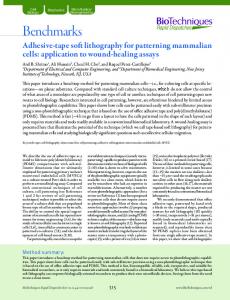
leles (References 1, 9, 12 and others). We developed three fluorescent multiplex PCRs for optimized simultaneous amplification of nine highly polymorphic PKD1 and PKD2 associated microsatellites: D16S291 (13); D16S664, D16S665, and D16S663 (9); D16S3252 (12); D4S423, D4S1534, D4S1563, and D4S2929 (3) (Figure 1). To get a better resolution and to reduce manual work, we used capillary electrophoresis instead of polyacrylamide electrophoresis to assess the lengths of amplified products. Due to different electrophoretic mobility of complementary strands of the amplified product, CA strands move faster on the denaturating polyacrylamide gels than GT strands (11). This can result in two bands instead of one for the same allele. In the case of capillary electrophoresis, only one of the strands is fluorescently labeled; therefore, only one can be visualized (6). Genomic DNA was extracted from white blood cells by a standard salting out procedure (8) or from blood spots by a Chelex 100 (Bio-Rad Laboratories, Hercules, CA, USA) based protocol (15). We used previously published primer pairs (http://www.gdb.org). One of the primers from each pair was endlabeled with a distinct fluorescent dye (Table 1). Multiplex PCRs were performed on a GeneAmp PCR System 9600 thermal cycler (Applied Biosys-
Figure 1. Map of the microsatellite markers around PKD1 (16p13.3) and PKD2 (4q21-q23) genes. Data are summarized from various sources (1,5,9,12). Approximate genetic distances are given in centiMorgans (cM). Vol. 29, No. 6 (2000)
Benchmarks Table 1. Primer Sequences and PCR Conditions
Amount of MgCl 2 Tann.c No. of Primera [mM] (°C) Cycles
Multiplex Microsatellites
Primer Sequences and Labeling
A
D16S664 (CW3)/PKD1
TET-5′-AATATCTCATCAGACTTTGC-3′
6
D16S291 (AC2.5)/PKD1
6FAM-5′-GCAGCCTCAGTTGTGTTTCCTAATC-3′
5
D4S423/PKD2 D4S1534/PKD2
5′-AGTGCTGGGATTACAGGCATGAACC-3′b
10
6FAM-5′-TTGAGTAGTTCCTGAAGCAGC-3′
5.5
5′-CAAAGTCCTCCATCTTGAGTG-3′
5.5
HEX-5′-ATTCAGTTTCAGCCCCAT-3′
3
5′-ACCAGCCCAAGGTAGAGG-3′ B
D16S665 (SM6)/PKD1 D4S1563/PKD2 D4S2929/PKD2
C
D16S663 (CW2)/PKD1 D16S3252 (KG8)/PKD1
1.8
59
30
1.25
55
35
1
57
35
3
HEX-5′-AGCTGGGGTCTCAGGGGAGCT-3′
2.5
5′-GCGCACACAGCACTAACACG-3′
2.5
HEX-5′-GCTGCCTGACACACTGG-3′
4
5′-ACTATTGCTGTTGCTGACCC-3′
4
TET-5′-GGCCAGGAGTTCAAAA-3′
2.5
5′-TGCAGCAAGTCCAACA-3′
2.5
TET-5′-GTCTTTCTAGGAATGAAATCAT-3′
5
5′-ATTGCAGCAAGACTCCATCT-3′
5
6FAM-5′-CTCCCAGGGTGGAGGAAGGTG-3′
4
5′-GTCCTGACCCCAGTGCACAGAC-3′
4
a Amount of primer in pmol/reaction. b Reverse primer for D16S291 (AC2.5) and D16S664 (CW3). cAnnealing temperature.
tems, Foster City, CA, USA) with 2.5–5 ng genomic DNA, 50 mM KCl, 10 mM Tris-HCl (pH 8.3), 0.4 mM dNTPs, and 0.5 U AmpliTaq Gold in a total volume of 12.5 µL. The amounts of MgCl2 are given in Table 1. After initial denaturation of 12 min at 95°C, the fragments were amplified by 30–35 cycles of denaturation at 95°C (98°C for multiplex A) for 30 s, annealing at temperatures as designated in Table 1 for 30 s, and extension at 72°C for 30 s. To avoid length differences resulting from the addition of single deoxyadenosine to the 3′ ends of the products, which could compromise interpretation of microsatellite electropherograms, we prolonged the final extension step to 15 min at 72°C. In addition, the products were left for several hours in darkness at room temperature. In case of the Chelex-isolated DNA, 20 µL isolate and double amounts of primers were used in a total volume of 50 µL. The lengths of the PCR products were as1188 BioTechniques
sessed by capillary electrophoresis, using an ABI PR I S M 310 genetic analyzer (Applied Biosystems). One microliter of each sample was mixed with 12.8 µL formamide (Amresco, Solon, OH, USA) and 0.2 µL GeneScan 500 [TAMRA] size standard (Applied Biosystems), denaturated at 90ºC for 2 min and then chilled on ice. The described approach was used for screening 51 patients and 66 healthy family members from 19 Slovene ADPKD families. An example of a result is summarized in Figure 2, which shows electropherograms of the three multiplex reactions revealing the genotype of an ADPKD patient. Most of the microsatellites typed are dinucleotide (CA) n repeats known to be troublesome because of the presence of stutter products (14). In some of the cases, when persons were heterozygous with alleles differing in a single repeat unit (2 bp), the interpretation was made difficult because of these stutter products. The
most troublesome, in that respect, were microsatellites D16S663 (CW2) and D16S664 (CW3). To get a good resolution of peaks, the quantity of template DNA was minimized, and primer concentration was optimized. Under the described amplifying conditions, the commonly used KG8 primer pair (12) resulted in a highly competitive product compared to other fragments in the mixture. To get a longer and less competitive product, we used an alternative reverse primer designed according to the PKD1 gene sequence (L39891, database Entrez). This primer lays 178 bp downstream from the intragenic microsatellite marker (Table 1). The same reverse primer was used for the amplification of microsatellites CW3 and AC2.5 because part of the downstream sequences is almost identical. It becomes evident in Figure 2 that the investigated patient is heterozygous for the majority of microsatellite markers we used. This indicates a high degree Vol. 29, No. 6 (2000)
Benchmarks
Figure 2. Electropherograms showing amplified PKD1 and PKD2 associated microsatellite markers. The X-axis shows the size of the fragments (bp), and the Y-axis shows units of fluorescence. Amplification products are shown in cross section as peaks. Names of microsatellites are indicated in the figure, and shaded peaks correspond to the amplified alleles (shown by arrows). Red peaks correspond to the size standard. Remaining unshaded peaks represent artifactual stutter or nonspecific products.
of informativity for the patient’s family. The described approach simplifies the analysis of the PKD1 and PKD2 gene associated microsatellite markers that are important for quick presymptomatic and prenatal diagnosis in ADPKD families. REFERENCES 1.Coto, E., S. Sanz de Castro, S. Aguado, J. Alvarez, M. Arias, M.J. Menendez, and C. Lopez-Larrea. 1995. DNA microsatellite analysis of families with autosomal dominant polycystic kidney disease types 1 and 2: evaluation of clinical heterogeneity between both forms of the disease. J. Med. Genet. 32:442445. 2.Daoust, M.C., D.M. Reynolds, D.G. Bichet, and S. Somlo. 1995. Evidence for a third genetic locus for autosomal dominant polycystic kidney disease. Genomics 25:733-736. 3.Dib, C., S. Faure, C. Fizames, D. Samson, N. 1190 BioTechniques
Drouot, A. Vignal, P. Millaseau, S. Marc et al. 1996. Acomprehensive genetic map of the human genome based on 5,264 microsatellites. Nature 380:152-154. 4.Harris, P.C. 1995. Molecular genetics of ADPKD. Contrib. Nephrol. 115:8-15. 5.Iglesias, D.M., R.S. Martin, A. Fraga, M. Virginillo, A.R. Kornblihtt, E. Arrzurieta, M. Viribay, J.L. San Milian, M. Herrera, and V. Bernath. 1997. Genetic heterogeneity of autosomal dominant polycystic kidney disease in Argentina. J. Med. Genet. 34:827-830. 6.Inman, K. and N. Rudin. 1997. An introduction to forensic DNA analysis, p. 256. CRC Press, New York. 7.Kimberling, W.J., S. Kumar, P.A. Gabow, J.B. Kenyon, C.J. Connolly and S. Somlo. 1993. Autosomal dominant polycystic kidney disease: localization of a second gene to chromosome 4q13-q23. Genomics 18:467-472. 8.Miller, S.A., D.D. Dykes, and H.F. Polesky. 1988. A simple salting out procedure for extracting DNA from human nucleated cells. Nucleic Acids Res. 16:1215. 9.Peral, B., C.J. Ward, J.L. San Millan, S. Thomas, R.L. Stallings, F. Moreno, and
P.C. Harris. 1994. Evidence of linkage disequilibrium in the Spanish polycystc kidney disease 1 population. Am. J. Hum. Genet. 54:899-908. 10.Peters, D.J., L. Spruit, J.J. Saris, D. Ravine, L.A. Sandkuijl, R. Fossdal, J. Boersma, R. Van Eijk et al. 1993. Chromosome 4 localization of a second gene for autosomal dominant polycystic kidney disease. Nat. Genet. 5:359362. 11.Saitoh, H., S. Ueda, K. Kurusaki, and M. Kiuchi. 1998. The different mobility of complementary strands depends on the proportion AC/GT. Forensic Sci. Int. 2:81-90. 12.Snarey, A., S. Thomas, M.C. Schneider, S.E. Pound, N. Barton, A.F. Wright, S. Somlo, G.G. Germino, P.C.Harris, S.T. Reeders and A.-M. Frischauf. 1994. Linkage disequilibrium in the region of the autosomal dominant polycystic kidney disease gene (PKD1). Am. J. Hum. Genet. 55:365-371. 13.Thompson, A.D., Y. Shen, K. Holman, G.R. Sutherland, D.F. Callen, and R.I. Richards. 1992. Isolation and characterization of (AC)n microsatellite genetic markers from human chromosome 16. Genomics 13:402-408. 14.Walsh, P.S., N.J. Fildes, and R. Reynolds. 1996. Sequence analysis and characterization of stutter products at the tetranucleotide repeat locus vWA. Nucleic Acids Res. 24:28072812. 15.Walsh, P.S., D.A. Metzger, and R. Higuchi. 1991. Chelex 100 as a medium for simple extraction of DNA for PCR-based typing from forensic material. BioTechniques 4:506-513. 16.The European Polycystic Kidney Disease Consortium. 1994. The polycystic kidney disease 1 gene encodes a 14 kb transcript and lies within a duplicated region on chromosome 16. Cell 77:881-894.
The work was supported from the Ministry of Science of Slovenia by research program P1-0527-0381 and by a “young researcher grant” to K.V. Address correspondence to Prof. Radovan Komel, Medical Centre for Molecular Biology, Institute of Biochemistry, Faculty of Medicine, Vrazov trg 2, SI-1000 Ljubljana, Slovenia. e-mail:
[email protected] Received 15 May 2000; accepted 25 August 2000.
Katja Vouk, Barbara Gazvoda and Radovan Komel Medical Centre for Molecular Biology Ljubljana, Slovenia
Vol. 29, No. 6 (2000)