Hindawi Publishing Corporation International Journal of Breast Cancer Volume 2016, Article ID 2469523, 10 pages http://dx.doi.org/10.1155/2016/2469523
Review Article Beyond DNA: An Integrated and Functional Approach for Classifying Germline Variants in Breast Cancer Genes T. Pesaran, R. Karam, R. Huether, S. Li, S. Farber-Katz, A. Chamberlin, H. Chong, H. LaDuca, and A. Elliott Ambry Genetics Corp., 15 Argonaut, Aliso Viejo, CA 92656, USA Correspondence should be addressed to T. Pesaran;
[email protected] Received 4 June 2016; Revised 4 September 2016; Accepted 19 September 2016 Academic Editor: Zsuzsanna Kah´an Copyright © 2016 T. Pesaran et al. This is an open access article distributed under the Creative Commons Attribution License, which permits unrestricted use, distribution, and reproduction in any medium, provided the original work is properly cited. Genetic testing for hereditary breast cancer is an integral part of individualized care in the new era of precision medicine. The accuracy of an assay is reliant on not only the technology and bioinformatics analysis utilized but also the experience and infrastructure required to correctly classify genetic variants as disease-causing. Interpreting the clinical significance of germline variants identified by hereditary cancer testing is complex and has a significant impact on the management of patients who are at increased cancer risk. In this review we give an overview of our clinical laboratory’s integrated approach to variant assessment. We discuss some of the nuances that should be considered in the assessment of genomic variants. In addition, we highlight lines of evidence such as functional assays and structural analysis that can be useful in the assessment of rare and complex variants.
1. Introduction The landscape of genetic testing for hereditary breast cancer susceptibility has changed drastically with the application of massively parallel sequencing based tests in clinical diagnostics. Clinical genomic laboratories are performing an increasing number of massively parallel sequencing assays for cancer predisposition genes [1], which has led to an intensified application of these assays in clinical and research settings [2]. Breast cancer gene panels and exome sequencing generate vast amounts of genetic alteration data, thereby presenting a significant challenge to determine which variants are responsible for the disease or phenotype. Multigene breast cancer panels in particular have gained in popularity over the past few years and are now routinely ordered by genetics, oncology, and breast surgical clinics. These tests allow for simultaneous analysis of numerous cancer genes that, when mutated, can have a significant impact on cancer risk stratification and management [3]. A major component of clinical molecular diagnostic testing is accurate assessment and interpretation of genetic variants. Ambry Genetics’ BreastNext Cancer panel analyzes 17 genes (ATM, BARD1, BRCA1, BRCA2, BRIP1, CDH1, CHEK2,
MRE11A, MUTYH, NBN, NF1, PALB2, PTEN, RAD50, RAD51C, RAD51D, and TP53) by massively parallel sequencing of all coding exons and a minimum of 5 base pairs into the flanking 5 and 3 ends of all introns and untranslated regions. In addition, clinically significant intronic mutations beyond 5 base pairs and the promoter region of PTEN (c.-1300 to c.745) are always sequenced and reported. Sequencing is conducted on the Illumina HiSeq2500 or NextSeq using 150 bp paired-end conditions as described in the manufacturer’s standard workflow (Illumina). After initial data processing, all clinical samples had to pass minimum thresholds to be included in the analysis. The three parameters were as follows: mean base calling quality score is greater than 30, the percentage of passes that reached over 30 had to be 75% overall, and the percentage of perfectly matched indexes needed to be greater than 85%. For each gene, a minimum coverage of 20x is required for candidate variants to be called. In an effort to help standardize the interpretation and reporting of genetic testing results, organizations such as the American College of Medical Genetics and Genomics (ACMG), Association for Molecular Pathology (AMP), and the International Agency for Research and Cancer (IARC) have proposed criteria for the interpretation and reporting of
2 sequence variants [7–9]. These criteria weigh multiple lines of evidence to categorize variants under a five-tier classification algorithm using terms such as pathogenic (P), variant, likely pathogenic (VLP), variant of unknown significance (VUS), variant, likely benign (VLB), and benign (B) to indicate the likelihood of association with disease. Per ACMG guidelines, the term “likely” refers to a classification tier that equates to a >90% likelihood of a variant being disease-causing or benign [7, 8]. Recently the clinical utility of the ACMG guidelines was demonstrated in a cohort of individuals undergoing sequencing for inherited cancer risk [10]. While the ACMG guidelines provide a basic framework for variant assessment, gene and syndrome-specific factors such as penetrance, prevalence, inheritance pattern, disease mechanism, and protein structure and function need to be considered. In addition, when considering the phenotype of the patients in which a variant is identified, one must take into account the prevalence of the disease and how the patients are ascertained to account for potential phenocopies. For example, many genes on hereditary breast cancer panels are considered to be moderate penetrance and are associated with a 2- to 5-fold increased breast cancer risk. Given the relatively high prevalence of breast cancer (1/8 women in the US), traditional segregation methods are confounded by phenocopies and are even more difficult to employ with genes that have reduced penetrance. These confounders indicate that these genes require large numbers of segregation events to provide meaningful results. Consideration should also be given to gene-specific factors such as frequency of germline and somatic de novo alterations, additional tests in tumors such as loss of heterozygosity studies, variation in nonsensemediated decay, and alternate splicing. For example, in genes such as TP53 and PTEN, germline de novo variants are known to be a relatively common cause of disease [11, 12]. However, with breast cancer genes such as ATM, CHEK2, and PALB2, the de novo rate is unknown. This is confounded by the fact that breast cancer is a common disease and one cannot infer if the de novo event in these genes directly correlates with disease or occurred by chance. In addition, although somatic de novo data is available for some genes [13] its incorporation into germline variant analysis has yet to be standardized and will need to be performed on a gene-by-gene basis. Consortia such as the Evidence Based Network for the Interpretation of Germline Mutant Alleles (ENIGMA) have demonstrated the power of a collaborative approach to variant assessment and have made great strides in the reclassification of VUS in breast cancer genes as pathogenic or benign. However even these groups are limited by the rate at which data is accumulated. Open-access databases such as ClinVar and the Leiden Open (source) Variant Database (LOVD) can be useful in identifying additional cases or publications related to a variant. These databases have also helped standardize the interpretation of variants between laboratories by identifying discrepancies in classifications. Collaborative efforts by clinical laboratories including Ambry Genetics, GeneDx, University of Chicago, and Laboratory for Molecular Medicine have resulted in the sharing of internal data consisting of segregation and cooccurrences with mutations in the same gene or other genes and de novo
International Journal of Breast Cancer observations have led to the resolution of 78% of clinically actionable differences (VUS versus VLP/mutation) and 92% of VUS versus likely benign/benign differences (internal data). Despite these efforts, one of the challenges faced by molecular laboratories and clinicians is that many genetic variants are very rare and do not have enough published data to be classified beyond VUS. We present here our laboratory’s integrated approach to variant assessment and review tools used to assess the impact of variants on protein function.
2. Integrated Approach to Variant Assessment Ambry Genetics has developed and implemented an integrated approach to variant assessment (Table 1) that encompasses a five-tier variant classification algorithm similar to those presented by ACMG and IARC. Although the foundation of Ambry Genetics’ classification algorithm is based on the ACMG guidelines, we have adopted stringent thresholds similar to those proposed by the IARC, where “likely” refers to a >95% confidence of a variant being disease-causing or benign [9]. In this algorithm, both pathogenic and likely pathogenic variants are interpreted as clinically actionable with recommendations for medical management and family member testing. Ambry Genetics’ algorithm incorporates multiple lines of evidence aimed at assessing both the impact of the variant on the protein and the pathogenicity of the variant in relation to a disease phenotype (Figure 1 and Table 2). These lines of evidence are weighted as stand-alone (categories A and F), strong (categories B and D), or supportive (categories C and E) and when combined as described in Table 1, they can lead to a classification of likely benign, benign, likely pathogenic, or pathogenic. When the evidence is limited or conflicting, the variants remain classified as VUS. Lines of evidence such as its location, structure-function, and functional and RNA studies reflect the functional impact on the mRNA or protein. Evolutionary conservation, in silico models such as Polyphen and SIFT, and general population frequency reflect fitness, that is, reproductive success and survival as measured by a lack of allelic diversity. The observed phenotype in variant carriers and the cosegregation of the variant with disease and the cooccurrence with other pathogenic variants reflect the pathogenicity of the variant (Figure 1). Some of this evidence is readily available via databases such as allele frequency data in the Exome Aggregation Consortium (ExAC) or the data in published literature [14]. However published literature generally contains data for common variants and the data supporting pathogenicity for rare variants is scarce and frequently only available internally. For most genes on breast cancer panels, computational data from in silico models, evolutionary conservation, and protein structural analysis are readily available. Population frequency data has been accumulating at a fast pace due to major contributions from 1000 Genomes, NHLBI Exome Sequencing Project (ESP), and ExAC. These data have had a significant impact on the identification of benign variants at high frequencies that are too frequent to be pathogenic based on disease incidence alone, particularly for historically understudied ethnic groups. For breast cancer genes, this
Pathogenic
Pathogenic
Likely pathogenic
Likely pathogenic
VUS
Likely benign
Likely benign
Benign
5
5
4
4
3
3
3
1
Criteria (i) Alterations resulting in premature truncation (e.g., reading frame shift, nonsense) (ii) Other ACMG-defined mutations (i.e., initiation codon or gross deletion) (iii) Strong segregation with disease (LOD >3 = >10 meioses) (i) Confirmed de novo alteration in the setting of a new disease (appropriate phenotype) in the family (ii) Significant disease association in appropriately sized case-control study(ies) (iii) Being detected in individuals satisfying established diagnostic criteria for classic disease without a clear mutation (iv) Last nucleotide of exon (v) Good segregation with disease (LOD 1.5–3 = 5–9 meioses) (vi) Deficient protein function in appropriate functional assay(s) (vii) Functionally validated splicing mutation (viii) Well-characterized mutation at the same position (ix) Other strong data supporting pathogenic classification (e.g., structural) (i) Alterations at the canonical donor/acceptor sites (± 1, 2) without another strong (B-level) evidence supporting pathogenicity (i) Rarity in general population databases (dbSNP, ESP, 1000 Genomes, ExAC) (ii) In silico models in agreement (deleterious) and/or completely conserved position in appropriate species (iii) Moderate segregation with disease (at least 3 informative meioses) for rare diseases (iv) Other data supporting pathogenic classification (e.g., structural)
3 of B 2 of B and at least 1 of C 1 of B and at least 3 of C Insufficient or conflicting evidence Gross duplications without strong evidence for pathogenic or benign D (i) Intronic alteration with no splicing impact by RT-PCR analysis or another splicing assay 1 needed (ii) Other strong data supporting benign classification (strong) (i) Cooccurrences with mutations in the same gene (phase unknown) (ii) Cooccurrences with mutations in other high penetrant genes that clearly explain a proband’s phenotype (iii) Subpopulation frequency in support of benign classification E (iv) Intact protein function observed in appropriate functional assay(s) 2 needed (v) In silico models in agreement (benign) (supportive) (vi) Not segregating with disease in family study (genes with incomplete penetrance) (vii) No disease association in small case-control study (viii) Other data supporting benign classification (i) General population or subpopulation frequency is too high to be a pathogenic mutation based on disease/syndrome prevalence and penetrance F (ii) Not segregating with disease in family study (genes with complete penetrance) 1 needed (iii) Internal frequency is too high to be a pathogenic mutation based on disease/syndrome prevalence and penetrance (stand-alone) (iv) Being seen in trans with a mutation or in homozygous state in individuals without severe disease for that gene (v) No disease association in appropriately sized case-control study(ies) 1 of D and at least 2 of E 2 or more of D >3 of E w/o conflicting data >4 of E w/conflicting data
C 4 needed (supportive)
1 needed
B 4 needed (strong)
Category A 1 needed (stand-alone)
The variant classification scheme is not intended for the interpretation of alterations complicated by epigenetic factors including genetic modifiers, multifactorial disease, or low-risk disease association alleles and may be limited in the interpretation of alterations confounded by incomplete penetrance, variable expressivity, phenocopies, and triallelic or oligogenic inheritance.
Classification
Class
Table 1: Classification scheme for high penetrance autosomal dominant breast cancer genes.
International Journal of Breast Cancer 3
4
International Journal of Breast Cancer Function Structural function RNA studies
Uniprot, wwPDB, internal data
Published literature, internal data
Functional assays Published literature, internal data
In silico assessment
Cooccurrence & segregation
Evolutionary conservation UCSC genome browser, NCBI
ess Fitn
Published literature, internal data
Variant classification
Population frequency data
Phenotype Published literature, internal data Case-control studies
1000 Genomes, ESP, Published literature, ExAC internal data
Pa th og en ic ity
Polyphen, SIFT, mutationAssessor, Provean, ESE, fruitfly
Figure 1: An integrated approach for variant classification. Lines of evidence such as structural function, RNA studies, and functional studies assess the functional impact on the mRNA and protein. Cooccurrence, segregation, case-control studies, and the observed phenotype in variant carriers reflect the pathogenicity of a variant. Population frequency, in silico models, and evolutionary conservation assess fitness of the amino acid or nucleotide position.
threshold has been conservatively set at an allele frequency of 1% in large population cohorts if used as a stand-alone line of evidence supporting benign classification (Table 1, category F). Careful consideration of population cohort size is needed to attain a high confidence (lower 95% CI is above 1% with 𝑝 value 99%) and provide unbiased molecular evidence to elucidate the functional impact of a VUS (Figure 2). As an example of a convincingly validated assay, Guidugli and colleagues determined the
sensitivity of their homology-directed DNA break repair (HDR) functional assay to be 100% (95% confidence interval (CI): 75.3%–100%) and the specificity to be 100% (95% CI: 81.5%–100%) [17]. 3.1. RNA Studies for Splicing VUS. While some splicing variants, such as canonical ±1 or 2 splice sites, are often assumed to disrupt gene function by leading to the reduced expression of the abnormal allele due to nonsense-mediated decay (NMD) [18] or abnormal protein truncations [19], comprehensive evaluation of splicing alterations is essential for accurate clinical interpretation. For canonical splice site ±1 and 2 variants, one must also consider the possibility of an in-frame deletion/insertion, which could retain the critical regions of the protein and hence lead to a mild, neutral, or gain-offunction effect. In addition, variants that are predicted to impact splicing but that are not located at the canonical sites (±1 and 2) require additional strong evidence (see details in Section 2) to be classified as pathogenic or benign [7]. Bioinformatics software has been developed to predict putative splice sites [20]. In general, these in silico tools are more sensitive (∼90–100%) than being specific (∼60–80%) when predicting the impact of a variant on splicing [21, 22]. However, by nature in silico tools can only provide supporting evidence which restricts their use [7]. Consequently, data from RNA splicing assays, designed to provide quantitative and qualitative characterization of transcripts, are usually necessary to evaluate the pathogenicity of these variants. Since published RNA data is not available for every variant, clinical genomic laboratories can more accurately classify splicing alterations by implementing their own RNA protocols and assays to provide accurate classification of splicing alterations. Reliability, in which an assay yields the same results in repeated trials, is a key issue when implementing mRNA assays in a clinical functional lab for evaluation of VUS.
6 To improve reliability, the ENIGMA consortium conducted a multicenter investigation to compare mRNA splicing assay protocols used by its members [23]. The consortium provided several recommendations for best practices in clinical testing of splicing alterations, including the standardization of protocols and the use of analytically sensitive detection methods [23]. Of the detection methods evaluated, capillary electrophoresis (CE) was shown to yield the highest analytic sensitivity. However, a major limitation of CE is its inability to harvest and subsequently perform sequence analysis of the RT-PCR product. In order to perform sequence analysis and full characterization of alternatively spliced transcripts, the consortium concluded that cloning single PCR products into a vector system is a useful alternative for isolating single transcripts for sequencing, which improves sensitivity over band excision and sequencing alone. Even in cases that appear straightforward, the consortium recommends using in vivo, in vitro, and clinical analysis to predict with 99% likelihood that a variant is benign or pathogenic [23]. For example, although most canonical splice site variants are considered a priori to be at least likely pathogenic, the presence of naturally occurring alternative splicing that mimics a pathogenic alteration and results in a similar impact on splicing (e.g., exon skipping) needs to be carefully evaluated, as it may result in diminished pathogenicity. Care must be taken to determine whether a transcript is present in normal controls. As the functional lab obtains more data on each gene, a more accurate picture of splicing patterns will emerge, thereby leading to improved classification of splice site variations. 3.2. Functional Assays for Missense VUS. Missense alterations with no impact on splicing can be evaluated by utilizing wet lab assays or experimental structure data. While functional studies can be a powerful tool in support of pathogenicity, not all functional studies accurately predict impacts on gene or protein function. For this reason ACMG/AMP provides recommendations for assessing the validity of functional assays, in order to confirm that the functional assay accurately measures a function that leads to disease [7]. One must consider how closely the functional assay reflects the biological environment. This is important when deciding whether to test patient samples or to perform in vitro assays. It is important to consider the known biological functions of the protein, while also examining whether those functions actually contribute to tumorigenicity. For example, many functional assays have been developed to interrogate BRCA1 VUS [24]. Some assays focus on the known DNA repair functions of BRCA1, such as the HDR assay [25, 26] and the radiation resistance assay [27]. Others examine BRCA1 localization [28, 29] and the ability of cells with BRCA1 variants to generate Rad51 foci [30, 31] in the presence of DNA damage as surrogates for BRCA1 function. Additional assays focus on one functional component of BRCA1 instead of the full protein, including the transcription activation assay, which employs the C-terminal BRCT domains, and the ubiquitin ligase assay, which utilizes the N-terminal region [32–35]. These two assays are limited by their inability to account for effects of the entire protein,
International Journal of Breast Cancer and others have noted that certain variants that lost ubiquitin ligase activity were not classified as pathogenic by genetic studies [36, 37]. Similarly, protein or peptide binding assays may resolve the ability of a variant to bind to a protein target in vitro, but these data should be incorporated into a multifactorial model that takes into account other functional in vivo data [38, 39]. In addition, validation data that assess the analytical performance of the assay and account for specimen integrity are important factors to consider when implementing functional assays in a functional clinical genomic laboratory and in using these results in classification of variants [7]. To investigate the effect of missense variants on BRCA1 function, Lu et al. tested 68 missense variants using an in vitro HDR assay [26]. The analysis showed that the HDR defective or partial defective missense variants from the BRCT domain are positioned either in the center of the structure or on the surface responsible for protein-protein interactions, while the HDR-WT variants from the BRCT domain were surface exposed or partially surface exposed variants [26]. This highlights the complexity of interpreting missense germline variants, indicating that an integrated approach, by compiling the results of functional assays, structure evaluation, and analysis of clinical parameters, should identify the most functionally and clinically relevant alterations. 3.3. Analysis of Insertion Breakpoints for Gross Duplications. Most gross deletions in high-risk cancer genes, larger than 3∼ 5 megabases, fall within microarray reporting guidelines and are reported as deleterious [38, 39]; however, without breakpoint information gross duplications are mostly reported as VUS. While array comparative genomic hybridization (aCGH) is a method used in cancer research for the detection of gross chromosomal aberrations in cancer genes, it cannot accurately determine the exact genomic breakpoints of the amplification [40–43]. To map the exact insertion breakpoints, paired-end high throughput sequencing can be used. Gross genomic amplifications may occur as a tandem duplication within the cancer gene itself, resulting in a novel function, or as a nontandem duplication inserted in a novel location of the genome. Therefore, identifying the exact breakpoints of tandem duplications in high-risk cancer genes can lead to VUS being reclassified as likely pathogenic or likely benign. To identify the exact breakpoints of tandem duplications, Ambry Genetics is currently utilizing the paired-end sequencing method to further characterize gross duplications. Probe sets are designed to capture the target regions with the suggested breakpoints identified by aCGH. Captured DNA is then sequenced by paired-end massively parallel sequencing and mapped to the human genome. The Ambry Genetics pipeline identifies read pairs that are in the wrong orientation, indicating a tandem duplication (Figure 3(a)). Clusters of read pairs with soft clipping that span breakpoints can indicate rearrangement breakpoints down to the exact coordinates (Figure 3(b)). As an example, an exon 11 duplication in BRCA1 previously classified as VUS can be reclassified as likely pathogenic if the breakpoint is identified to cause a frameshift in the gene (Figure 3).
International Journal of Breast Cancer
7 Exon 11
Reference
Paired sequence read aligned to target gene BRCA1 gene Tandem duplication (a)
BRCA1
Exon 11
(b)
Figure 3: Identification of tandem duplication insertion breakpoints spanning BRCA1 exon 11, using paired-end sequencing. (a) Mapped read pairs in the wrong orientation indicate a tandem duplication. (b) Ambry’s breakpoint detection tools can identify clusters of read pairs with soft clipping which indicate rearrangement breakpoints.
4. Computational Structural Analysis Computational structural algorithms offer a unique solution for assessing a variant’s impact on protein function in that they are faster than experimental studies and often use data from many scientific disciplines [44]. However, the quality of the information provided by computational analyses varies depending on the information source. For instance, primary sequence analyses using evolutionary tools can identify the likely impact of a variant. By comparing an altered human sequence to proteins with a similar primary sequence or related structural shape, the fitness of the variant can be predicted based on the variability of that position and other aspects such as the chemical similarity of the wild type and variant amino acids. Ambry Genetics relies on multiple tools, including the “Sorting Intolerant from Tolerant” (SIFT) and Polyphen2 programs [40]. We use the consensus of two programs, usually SIFT and Polyphen2 where applicable, and consider only concordant results as a line of evidence. If only one program is applicable such as Provean [45] with indels we incorporate predictions from the single program with conservative thresholds determined by analysis of our internal data. Alternatively, analyses of the secondary and tertiary structures of the protein increase the reliability of interpretation. The most reliable computational algorithms focus on biophysics methods which are more oriented towards direct simulation of the physical processes occurring in a protein [46–48]. In many regards, computational methods are the most diverse in the range of properties that they can quantify; however, they come at the expense of computational requirements and speed with which accurate properties can be derived. One of the most common and easily identified
sources of disruption induced by a variant is the influence on protein stability [46, 47, 49–51]. Protein stability can be affected in multiple ways, such as misfolding or unfolding of the protein structure, which commonly results in either loss of function or premature degradation and haploinsufficiency. As an example, protein stability has been used by Karchin et al. to generate a predictive tool for the likelihood of the effect of an alteration in the breast cancer gene BRCA2 [52]. There are other significant ways that variants exert their pathogenic effect which can be described through structure. For instance, a variant may not significantly affect the resting state structure of the protein but rather affect the movement of the protein in the course of its function. It may impact its binding with other target proteins or substrates or it may induce aggregation [47, 48]. Detailed understanding of biophysical principles illuminated through structure is crucial to evaluate and interpret the impact of alterations.
5. Tertiary and Quaternary Sequence of Breast Cancer Genes The use of biophysical methods to predict the impact of a variant on a protein often requires the availability of structures for the target gene or benefits significantly from it. Among the 17 genes represented in the BreastNext Cancer panel, there are a total of 247 experimentally derived structures, tabulated per gene in the PDBs (Protein DataBank files) column of Table 2, using either Nuclear Magnetic Resonance Imaging (NMR) or X-ray crystallographic methods [5, 53]. The coverage described above corresponds to the total range of residues covered by all experimental measurements divided by the total length of the protein. While there are notable exceptions
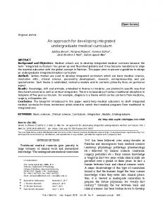
8 where no experimental structures have been determined, the majority of the genes have been partially and in the case of TP53 completely elucidated experimentally. The structure of TP53 is highly ordered throughout the protein, allowing for complete measurement of one low-energy form; however some proteins in this set such as BRCA1 and BRCA2 are composed of regions which have no characteristic fixed structure. The ordered regions within the structured protein, such as in the N (Really Interesting New Gene, i.e., RING, domain) and C terminus (BRCA1 C Terminus, i.e., BRCT, repeats) of BRCA1, offer higher quality means to define domain boundaries. These can be analyzed as a folded functional unit rather than through conservation techniques that are used in the Protein Families (Pfam) database [54] or using meta predictors such as InterPro [55]. The structural coverage for the genes in Table 2 does not take into account that long stretches of some proteins have little intrinsic globular structure, so the numbers can be seen as a very conservative estimate of the range of available residues covered. In addition, there remain some proteins, such as ATM or NBN, where no or lowresolution structures have been experimentally measured [5, 6]. For these systems, structural analysis incorporates the use of homology models built on the structures of known related proteins. This significantly increases the effective range of structural coverage and the insights available. Disruption in the folding of a domain in a protein by a missense pathogenic variant is well known to result in a loss of function. The clinically observed alteration c.5509T>G (p.Trp1837Gly) (ClinVar: SCV000077040) represents a case where structural features explain the disruption of the BRCT repeat region in BRCA1. The C-terminal portion of BRCA1 contains a pair of BRCT repeat domains, BRCT1 and BRCT2, which are described in atomic detail, including the arrangement of amino acids that make up these domains, by 26 different crystal structures [56–59]. The side chain of amino acid Trp 1837 (W1837; magenta stick) is buried in the core of the BRCT2 domain surrounded by hydrophobic amino acids (green sticks), while the backbone participates in a helix involved in binding BACH1 (Figure 4) [56]. The alteration W1837 to G1837 (W1837G) would result in the loss of the large stabilizing hydrophobic side chain and is anticipated to be very destabilizing. The instability introduced by this alteration has been quantitatively calculated by computational folding algorithms which indicate it to be very destabilizing [5]. Indeed, E. coli expressed with in vitro mutants of p.W1837G produce an unfolded protein that was present only in inclusion bodies which could not be refolded [60]. In another set of biochemical and cell-based transcriptional experiments, this alteration resulted in compromised proteolysis and phosphopeptide-binding [39, 58, 59, 61]. Together, these functional data support the qualitative and quantitative structural observation that the variant would create a very unfavorable cavity within this domain, thereby disrupting folding and protein function. This example demonstrates how detailed structural analysis on publically available data can facilitate the understanding and interpretation of alterations on the function of a protein and can be supported by both computational and experimental observations.
International Journal of Breast Cancer BACH1
p.W1837G
BRCA1-BRCT2
Figure 4: The structure of BRCA1 p.Trp1837 (shown in magenta with sticks) in the BRCA-BRCT domain (PDB: 1T15 [6]). Nearby hydrophobic amino acids sidechains from residue 1837 are shown as sticks. Bound BACH1 peptide is shown as teal stick.
6. Conclusion Although cancer genetic testing has traditionally been limited to highly penetrant and well-characterized susceptibility genes, the application of multigene panels using massively parallel sequencing is steadily becoming more common in genetic cancer risk assessment due to reduced costs and increased efficiency. Multigene panels, in turn, tend to result in the identification of more variants per individual, the clinical significance of which needs to be assessed using multiple lines of weighted evidence. We present an integrated approach for assessing variants observed on hereditary breast cancer panels and believe that this improves the clinical management of patients with personal and family histories of breast cancer due to more accurate variant classification. Comprehensive variant assessment programs that integrate multiple lines of evidence aimed at assessing a variant’s impact on protein function, fitness, and pathogenicity facilitate high-quality and efficient variant classification, providing increased benefit and reliability for patients.
Competing Interests Ambry Genetics Corp. is a CLIA approved clinical genetics testing laboratory. The authors of this review are all full time paid employees of Ambry Genetics.
References [1] H. LaDuca, A. J. Stuenkel, J. S. Dolinsky et al., “Utilization of multigene panels in hereditary cancer predisposition testing: analysis of more than 2,000 patients,” Genetics in Medicine, vol. 16, no. 11, pp. 830–837, 2014.
International Journal of Breast Cancer [2] M. B. Daly, J. E. Axilbund, S. Buys et al., “Genetic/familial highrisk assessment: breast and ovarian,” The National Comprehensive Cancer Network, vol. 8, pp. 562–594, 2010. [3] “National Comprehensive Cancer Network Genetic/Familial High-Risk Assessment: Breast and Ovarian (Version 2.2016),” 2016, https://www.nccn.org/professionals/physician gls/pdf/ genetics screening.pdf. [4] M. Magrane and UniProt Consortium, “UniProt knowledgebase: a hub of integrated protein data,” Database, vol. 2011, Article ID bar009, 2011. [5] H. M. Berman, J. Westbrook, Z. Feng et al., “The protein data bank,” Nucleic Acids Research, vol. 28, no. 1, pp. 235–242, 2000. [6] W. C. Lau, Y. Li, Z. Liu, Y. Gao, Q. Zhang, and M. S. Huen, “Structure of the human dimeric ATM kinase,” Cell Cycle, vol. 15, no. 8, pp. 1117–1124, 2016. [7] S. Richards, N. Aziz, S. Bale et al., “Standards and guidelines for the interpretation of sequence variants: a joint consensus recommendation of the American College of Medical Genetics and Genomics and the Association for Molecular Pathology,” Genetics in Medicine, vol. 17, no. 5, pp. 405–424, 2015. [8] C. S. Richards, S. Bale, D. B. Bellissimo et al., “ACMG recommendations for standards for interpretation and reporting of sequence variations: revisions 2007,” Genetics in Medicine, vol. 10, no. 4, pp. 294–300, 2008. [9] S. V. Tavtigian, M. S. Greenblatt, D. E. Goldgar, and P. Boffetta, “Assessing pathogenicity: overview of results from the IARC unclassified genetic variants working group,” Human Mutation, vol. 29, no. 11, pp. 1261–1264, 2008. [10] K. N. Maxwell, S. N. Hart, J. Vijai et al., “Evaluation of ACMGguideline-based variant classification of cancer susceptibility and non-cancer-associated genes in families affected by breast cancer,” The American Journal of Human Genetics, vol. 98, no. 5, pp. 801–817, 2016. [11] K. D. Gonzalez, C. H. Buzin, K. A. Noltner et al., “High frequency of de novo mutations in Li-Fraumeni syndrome,” Journal of Medical Genetics, vol. 46, no. 10, pp. 689–693, 2009. [12] J. Mester and C. Eng, “Estimate of de novo mutation frequency in probands with PTEN hamartoma tumor syndrome,” Genetics in Medicine, vol. 14, no. 9, pp. 819–822, 2012. [13] D. R. Walker, J. P. Bond, R. E. Tarone et al., “Evolutionary conservation and somatic mutation hotspot maps of p53: correlation with p53 protein structural and functional features,” Oncogene, vol. 18, no. 1, pp. 211–218, 1999. [14] M. Lek, K. J. Karczewski, E. V. Minikel et al., “Analysis of protein-coding genetic variation in 60,706 humans,” Nature, vol. 536, no. 7616, pp. 285–291, 2016. [15] F. P. Li, J. F. Fraumeni Jr., J. J. Mulvihill et al., “A cancer family syndrome in twenty-four kindreds,” Cancer Research, vol. 48, no. 18, pp. 5358–5362, 1988. [16] D. Pruss, B. Morris, E. Hughes et al., “Development and validation of a new algorithm for the reclassification of genetic variants identified in the BRCA1 and BRCA2 genes,” Breast Cancer Research and Treatment, vol. 147, no. 1, pp. 119–132, 2014. [17] L. Guidugli, V. S. Pankratz, N. Singh et al., “A classification model for BRCA2 DNA binding domain missense variants based on homology-directed repair activity,” Cancer Research, vol. 73, no. 1, pp. 265–275, 2013. [18] R. Karam, J. Wengrod, L. B. Gardner, and M. F. Wilkinson, “Regulation of nonsense-mediated mRNA decay: implications for physiology and disease,” Biochimica et Biophysica Acta— Gene Regulatory Mechanisms, vol. 1829, no. 6-7, pp. 624–633, 2013.
9 [19] O. Anczuk´ow, M. D. Ware, M. Buisson et al., “Does the nonsense-mediated mRNA decay mechanism prevent the synthesis of truncated BRCA1, CHK2, and p53 proteins?” Human Mutation, vol. 29, no. 1, pp. 65–73, 2008. [20] X. Jian, E. Boerwinkle, and X. Liu, “In silico tools for splicing defect prediction: a survey from the viewpoint of end users,” Genetics in Medicine, vol. 16, no. 7, pp. 497–503, 2014. [21] C. Houdayer, V. Caux-Moncoutier, S. Krieger et al., “Guidelines for splicing analysis in molecular diagnosis derived from a set of 327 combined in silico/in vitro studies on BRCA1 and BRCA2 variants,” Human Mutation, vol. 33, no. 8, pp. 1228–1238, 2012. [22] M. P. G. Vreeswijk, J. N. Kraan, H. M. van der Klift et al., “Intronic variants in BRCA1 and BRCA2 that affect RNA splicing can be reliably selected by splice-site prediction programs,” Human Mutation, vol. 30, no. 1, pp. 107–114, 2009. [23] P. J. Whiley, M. de la Hoya, M. Thomassen et al., “Comparison of mRNA splicing assay protocols across multiple laboratories: recommendations for best practice in standardized clinical testing,” Clinical Chemistry, vol. 60, no. 2, pp. 341–352, 2014. [24] G. A. Millot, M. A. Carvalho, S. M. Caputo et al., “A guide for functional analysis of BRCA1 variants of uncertain significance,” Human Mutation, vol. 33, no. 11, pp. 1526–1537, 2012. [25] D. J. R. Ransburgh, N. Chiba, C. Ishioka, A. E. Toland, and J. D. Parvin, “Identification of breast tumor mutations in BRCA1 that abolish its function in homologous DNA recombination,” Cancer Research, vol. 70, no. 3, pp. 988–995, 2010. [26] C. Lu, M. Xie, M. C. Wendl et al., “Patterns and functional implications of rare germline variants across 12 cancer types,” Nature Communications, vol. 6, Article ID 10086, 2015. [27] R. Scully, S. Ganesan, K. Vlasakova, J. Chen, M. Socolovsky, and D. M. Livingston, “Genetic analysis of BRCA1 function in a defined tumor cell line,” Molecular Cell, vol. 4, no. 6, pp. 1093– 1099, 1999. [28] W. W. Y. Au and B. R. Henderson, “The BRCA1 RING and BRCT domains cooperate in targeting BRCA1 to ionizing radiationinduced nuclear foci,” The Journal of Biological Chemistry, vol. 280, no. 8, pp. 6993–7001, 2005. [29] J. A. Rodriguez, W. W. Y. Au, and B. R. Henderson, “Cytoplasmic mislocalization of BRCA1 caused by cancer-associated mutations in the BRCT domain,” Experimental Cell Research, vol. 293, no. 1, pp. 14–21, 2004. [30] R. Drost, P. Bouwman, S. Rottenberg et al., “BRCA1 RING function is essential for tumor suppression but dispensable for therapy resistance,” Cancer Cell, vol. 20, no. 6, pp. 797–809, 2011. [31] K. A. T. Naipal, N. S. Verkaik, N. Ameziane et al., “Functional Ex vivo assay to select homologous recombination-deficient breast tumors for PARP inhibitor treatment,” Clinical Cancer Research, vol. 20, no. 18, pp. 4816–4826, 2014. [32] A. N. A. Monteiro, A. August, and H. Hanafusa, “Evidence for a transcriptional activation function of BRCA1 C-terminal region,” Proceedings of the National Academy of Sciences of the United States of America, vol. 93, no. 24, pp. 13595–13599, 1996. [33] J. Vallon-Christersson, C. Cayanan, K. Haraldsson et al., “Functional analysis of BRCA1 C-terminal missense mutations identified in breast and ovarian cancer families,” Human Molecular Genetics, vol. 10, no. 4, pp. 353–360, 2001. [34] C. M. Phelan, V. Dapic, B. Tice et al., “Classification of BRCA1 missense variants of unknown clinical significance,” Journal of Medical Genetics, vol. 42, no. 2, pp. 138–146, 2005. [35] M. A. Carvalho, S. M. Marsillac, R. Karchin et al., “Determination of cancer risk associated with germ line BRCA1 missense
10
[36]
[37]
[38]
[39]
[40]
[41]
[42]
[43]
[44]
[45]
[46]
[47]
[48]
[49]
[50]
International Journal of Breast Cancer variants by functional analysis,” Cancer Research, vol. 67, no. 4, pp. 1494–1501, 2007. P. S. Brzovic, J. R. Keeffe, H. Nishikawa et al., “Binding and recognition in the assembly of an active BRCA1/BARD1 ubiquitin-ligase complex,” Proceedings of the National Academy of Sciences of the United States of America, vol. 100, no. 10, pp. 5646–5651, 2003. J. R. Morris, L. Pangon, C. Boutell, T. Katagiri, N. H. Keep, and E. Solomon, “Genetic analysis of BRCA1 ubiquitin ligase activity and its relationship to breast cancer susceptibility,” Human Molecular Genetics, vol. 15, no. 4, pp. 599–606, 2006. S. T. South, C. Lee, A. N. Lamb et al., “ACMG standards and guidelines for constitutional cytogenomic microarray analysis, including postnatal and prenatal applications: revision 2013,” Genetics in Medicine, vol. 15, no. 11, pp. 901–909, 2013. M. S. Lee, R. Green, S. M. Marsillac et al., “Comprehensive analysis of missense variations in the BRCT domain of BRCA1 by structural and functional assays,” Cancer Research, vol. 70, no. 12, pp. 4880–4890, 2010. A. Kallioniemi, O.-P. Kallioniemi, D. Sudar et al., “Comparative genomic hybridization for molecular cytogenetic analysis of solid tumors,” Science, vol. 258, no. 5083, pp. 818–821, 1992. D. Pinkel and D. G. Albertson, “Array comparative genomic hybridization and its applications in cancer,” Nature Genetics, vol. 37, no. 6, supplement, pp. S11–S17, 2005. Y. Saillour, M. Coss´ee, F. Leturcq et al., “Detection of exonic copy-number changes using a highly efficient oligonucleotidebased comparative genomic hybridization-array method,” Human Mutation, vol. 29, no. 9, pp. 1083–1090, 2008. J. Staaf, T. T¨orngren, E. Rambech et al., “Detection and precise mapping of germline rearrangements in BRCA1, BRCA2, MSH2, and MLH1 using zoom-in array comparative genomic hybridization (aCGH),” Human Mutation, vol. 29, no. 4, pp. 555–564, 2008. T. A. Peterson, E. Doughty, and M. G. Kann, “Towards precision medicine: advances in computational approaches for the analysis of human variants,” Journal of Molecular Biology, vol. 425, no. 21, pp. 4047–4063, 2013. Y. Choi, G. E. Sims, S. Murphy, J. R. Miller, and A. P. Chan, “Predicting the functional effect of amino acid substitutions and indels,” PLoS ONE, vol. 7, no. 10, Article ID e46688, 2012. S. Stefl, H. Nishi, M. Petukh, A. R. Panchenko, and E. Alexov, “Molecular mechanisms of disease-causing missense mutations,” Journal of Molecular Biology, vol. 425, no. 21, pp. 3919– 3936, 2013. T. G. Kucukkal, Y. Yang, S. C. Chapman, W. Cao, and E. Alexov, “Computational and experimental approaches to reveal the effects of single nucleotide polymorphisms with respect to disease diagnostics,” International Journal of Molecular Sciences, vol. 15, no. 6, pp. 9670–9717, 2014. B. M. Kroncke, C. G. Vanoye, J. Meiler, A. L. George Jr., and C. R. Sanders, “Personalized biochemistry and biophysics,” Biochemistry, vol. 54, no. 16, pp. 2551–2559, 2015. J. Reumers, J. Schymkowitz, and F. Rousseau, “Using structural bioinformatics to investigate the impact of non synonymous SNPs and disease mutations: scope and limitations,” BMC Bioinformatics, vol. 10, supplement 8, article S9, 2009. R. Casadio, M. Vassura, S. Tiwari, P. Fariselli, and P. Luigi Martelli, “Correlating disease-related mutations to their effect on protein stability: a large-scale analysis of the human proteome,” Human Mutation, vol. 32, no. 10, pp. 1161–1170, 2011.
[51] D. E. V. Pires, J. Chen, T. L. Blundell, and D. B. Ascher, “In silico functional dissection of saturation mutagenesis: interpreting the relationship between phenotypes and changes in protein stability, interactions and activity,” Scientific Reports, vol. 6, Article ID 19848, 2016. [52] R. Karchin, M. Agarwal, A. Sali, F. Couch, and M. S. Beattie, “Classifying variants of undetermined significance in BRCA2 with protein likelihood ratios,” Cancer Informatics, vol. 6, pp. 203–216, 2008. [53] A. A. Yee, A. Savchenko, A. Ignachenko et al., “NMR and X-ray crystallography, complementary tools in structural proteomics of small proteins,” Journal of the American Chemical Society, vol. 127, no. 47, pp. 16512–16517, 2005. [54] R. D. Finn, P. Coggill, R. Y. Eberhardt et al., “The Pfam protein families database: towards a more sustainable future,” Nucleic Acids Research, vol. 44, no. 1, pp. D279–D285, 2016. [55] S. Hunter, R. Apweiler, T. K. Attwood et al., “InterPro: the integrative protein signature database,” Nucleic Acids Research, vol. 37, no. 1, pp. D211–D215, 2009. [56] J. A. Clapperton, I. A. Manke, D. M. Lowery et al., “Structure and mechanism of BRCA1 BRCT domain recognition of phosphorylated BACH1 with implications for cancer,” Nature Structural & Molecular Biology, vol. 11, no. 6, pp. 512–518, 2004. [57] S. L. Clark, A. M. Rodriguez, R. R. Snyder, G. D. V. Hankins, and D. Boehning, “Structure-function of the tumor suppressor BRCA1,” Computational and Structural Biotechnology Journal, vol. 1, no. 1, pp. 1–8, 2012. [58] R. S. Williams, R. Green, and J. N. M. Glover, “Crystal structure of the BRCT repeat region from the breast cancer-associated protein BRCA1,” Nature Structural Biology, vol. 8, no. 10, pp. 838–842, 2001. [59] R. S. Williams, M. S. Lee, D. D. Hau, and J. N. M. Glover, “Structural basis of phosphopeptide recognition by the BRCT domain of BRCA1,” Nature Structural & Molecular Biology, vol. 11, no. 6, pp. 519–525, 2004. [60] P. J. E. Rowling, R. Cook, and L. S. Itzhaki, “Toward classification of BRCA1 missense variants using a biophysical approach,” The Journal of Biological Chemistry, vol. 285, no. 26, pp. 20080– 20087, 2010. [61] J. N. M. Glover, “Insights into the molecular basis of human hereditary breast cancer from studies of the BRCA1 BRCT domain,” Familial Cancer, vol. 5, no. 1, pp. 89–93, 2006.
MEDIATORS of
INFLAMMATION
The Scientific World Journal Hindawi Publishing Corporation http://www.hindawi.com
Volume 2014
Gastroenterology Research and Practice Hindawi Publishing Corporation http://www.hindawi.com
Volume 2014
Journal of
Hindawi Publishing Corporation http://www.hindawi.com
Diabetes Research Volume 2014
Hindawi Publishing Corporation http://www.hindawi.com
Volume 2014
Hindawi Publishing Corporation http://www.hindawi.com
Volume 2014
International Journal of
Journal of
Endocrinology
Immunology Research Hindawi Publishing Corporation http://www.hindawi.com
Disease Markers
Hindawi Publishing Corporation http://www.hindawi.com
Volume 2014
Volume 2014
Submit your manuscripts at http://www.hindawi.com BioMed Research International
PPAR Research Hindawi Publishing Corporation http://www.hindawi.com
Hindawi Publishing Corporation http://www.hindawi.com
Volume 2014
Volume 2014
Journal of
Obesity
Journal of
Ophthalmology Hindawi Publishing Corporation http://www.hindawi.com
Volume 2014
Evidence-Based Complementary and Alternative Medicine
Stem Cells International Hindawi Publishing Corporation http://www.hindawi.com
Volume 2014
Hindawi Publishing Corporation http://www.hindawi.com
Volume 2014
Journal of
Oncology Hindawi Publishing Corporation http://www.hindawi.com
Volume 2014
Hindawi Publishing Corporation http://www.hindawi.com
Volume 2014
Parkinson’s Disease
Computational and Mathematical Methods in Medicine Hindawi Publishing Corporation http://www.hindawi.com
Volume 2014
AIDS
Behavioural Neurology Hindawi Publishing Corporation http://www.hindawi.com
Research and Treatment Volume 2014
Hindawi Publishing Corporation http://www.hindawi.com
Volume 2014
Hindawi Publishing Corporation http://www.hindawi.com
Volume 2014
Oxidative Medicine and Cellular Longevity Hindawi Publishing Corporation http://www.hindawi.com
Volume 2014