differential equations using ModelMaker 3.0.4 software. (Cherwell Scientific ..... application des articles R. 1321-26 a` R. 1321-36 du code de la santé publique.
Subscriber access provided by IRSN
Article
Biodynamics, Subcellular Partitioning, and Ultrastructural Effects of Organic Selenium in a Freshwater Bivalve Christelle Adam-Guillermin, Elodie Fournier, Magali Floriani, Virginie Camilleri, Jean-Charles Massabuau, and Jacqueline Garnier-Laplace Environ. Sci. Technol., 2009, 43 (6), 2112-2117• DOI: 10.1021/es802891j • Publication Date (Web): 06 February 2009 Downloaded from http://pubs.acs.org on April 15, 2009
More About This Article Additional resources and features associated with this article are available within the HTML version: • • • •
Supporting Information Access to high resolution figures Links to articles and content related to this article Copyright permission to reproduce figures and/or text from this article
Environmental Science & Technology is published by the American Chemical Society. 1155 Sixteenth Street N.W., Washington, DC 20036
Environ. Sci. Technol. 2009, 43, 2112–2117
Biodynamics, Subcellular Partitioning, and Ultrastructural Effects of Organic Selenium in a Freshwater Bivalve C H R I S T E L L E A D A M - G U I L L E R M I N , * ,† ELODIE FOURNIER,† MAGALI FLORIANI,† VIRGINIE CAMILLERI,† JEAN-CHARLES MASSABUAU,‡ AND JACQUELINE GARNIER-LAPLACE† Laboratory of Radioecology and Ecotoxicology, IRSN/DEI/SECRE, Cadarache, Baˆt. 186, BP3, 13115 St Paul lez Durance, France, and Universite´ Bordeaux 1, CNRS, UMR 5805 EPOC, Place du Dr Peyneau, 33120, Arcachon, France
Received April 22, 2008. Revised manuscript received December 12, 2008. Accepted January 8, 2009.
Selenium is a trace element characterized by concentrations that narrowly range between being essential and being toxic. Even though inorganic selenite and selenate are the predominant chemical forms of Se in surface waters, the toxicity of Se to aquatic organisms is mostly governed by the bioavailability of organic selenium within food webs. The present study was designed to evaluate organic selenium bioaccumulation and toxicity patterns in the freshwater sentinel species Corbicula fluminea. Waterborne selenomethionine (SeMet) exposure was used to mimic dietary organo-Se uptake. Our results demonstrate that SeMet is accumulated to a relatively high extent with a concentration factor of 770 (wet weight basis). Higher uptake than depuration rates suggest that bivalves deal with high Se amounts using a strategy of detoxification based on Se sequestration that could involve granules, as shown by a strong increase of Se in the particulate subcellular fraction. Selenium is persistent in the cytosol of bivalves exposed to SeMet where it is found in proteins of a wide range of molecular mass, indicating a possible replacement of methionine by selenomethionine. A subsequent alteration of protein function might be one of the mechanisms of Se toxicity that could explain the histopathological effects we observed in gills by using transmission electronic microscopy. Those analyses showed changes in gill filament ultrastructure and suggested mitochondria as the first target for SeMet cytotoxicity, with alterations of the outer membrane and of cristae morphology. Organo-Se would thus not only be toxic via indirect mechanisms of maternal transfer as it was suggested for fish but also directly. Our results on Se distribution agree with studies that used dietary Se transfer, and highlight the relevance (and less expensive way) of using SeMet water-only exposure protocols to mimic the real environment.
Introduction Selenium is a naturally occurring trace element found in association with mineral formations such as coal seams or phosphate deposits. Typical concentrations in unpolluted freshwaters range from 0.01 to 0.20 µg L-1 (1, 2), but may be highly variable as a function of the geochemical background. As such, an excess of the French drinking water quality guideline set at 10 µg L-1 is tolerated for Se concentrations up to 70 µg L-1 (3). Natural background concentrations of Se may also be enhanced due to anthropogenic activities, including mining, irrigation, petroleum refining, and coal combustion. Selenium occurs in surface waters mainly as the oxidized inorganic forms selenite Se(+IV) (SeO32-) and selenate Se(+VI) (SeO42-). The reduced forms include volatile methylated selenides, Se(-II), seleno-amino-acids (e.g., selenomethionine [SeMet]) and their associated proteins, and inorganic selenides. Organic selenides usually occur at lower concentrations in water than inorganic Se species, but exhibit a greater bioavailability (4). Selenium is an essential micronutrient for a wide variety of plants and animals, ranging from algae (5) to fish (6). It plays an important role in antioxidative defense in the form of glutathione peroxidase and as such can have a protective effect on metal toxicity (7). However, its essential role may shift to one of toxicity within a very narrow range of concentrations. For example, selenium concentrations lower than 0.1 µg Se g-1 (dry weight) in food of rainbow trout may lead to severe signs of deficiency; whereas, above 10 µg Se g-1, toxic effects begin to appear (6). Most of the studies conducted on selenium ecotoxicity have focused on fish and birds while little attention has been given to invertebrates, considered most often only as food sources of Se to higher trophic levels (8). Within this general context, a research program was conducted to characterize the links between Se speciation and toxic effects in bivalves, in terms of ventilatory behavior, histopathologies, enzyme activity, and gene expression. The biological model chosen, Corbicula fluminea, is a benthic, freshwater clam species that daily filters large amounts of water, and thus is able to accumulate high concentrations of contaminants. Here, we explore selenium bioaccumulation and toxicity patterns in bivalves exposed to organic selenium. Selenomethionine, the main form of Se compounds in algae and higher forms of plants (9), was used to mimic a trophic transfer from algae. Selenium uptake and depuration dynamics were studied at the tissular levels. To depict toxicity mechanisms, selenium partitioning between soluble and insoluble fractions of gills and visceral mass was also measured, together with its distribution among proteins pools. Histopathological effects were studied using transmission electronic microscopy (TEM) in gills. This research is important because it shows that there may be a direct toxicity from organic Se; whereas data in the literature have concentrated on indirect mechanisms of toxicity via biotransformation of Se within food webs. Additionally, this is the first documentation of alterations in mitochondria morphology and cristae organization following organo-Se exposure.
Experimental Section * Corresponding author e-mail: christelle.adam-guillermin@ irsn.fr; phone: (+33) 4 42 19 94 01; fax: (+33) 4 42 19 91 51. † Laboratory of Radioecology and Ecotoxicology, IRSN/DEI/ SECRE. ‡ Universite´ Bordeaux 1, CNRS. 2112
9
ENVIRONMENTAL SCIENCE & TECHNOLOGY / VOL. 43, NO. 6, 2009
Experimental Conditions. C. fluminea specimens (soft body weight: 0.55 ( 0.11 g wet weight) were collected from a noncontaminated site on a channel close to the River Garonne (Fontet, France) and acclimated to laboratory conditions for at least one month before the experiment. They were fed 10.1021/es802891j CCC: $40.75
2009 American Chemical Society
Published on Web 02/06/2009
daily with a suspension of unicellular algae Chlamydomonas reinhardtii (total density of 1-2 × 10⁵ cells mL-1 within the tank just after algae addition) and maintained under a 14-h light/10-h dark photoperiod (4.0 ( 0.5 µmol m-2 s-1) at 20.0 ( 0.5 °C. Air was bubbled continuously into the tanks to ensure algal mixing, gas-water equilibration, and to control the respiratory status of the bivalves. During the experiments, the animals were isolated from external vibrations in the laboratory using an antivibrating bench. Experiments were performed in glass tanks filled with 10 L of synthetic water (Ca2+ 290 µM; Mg2+ 195 µM; Na+ 500 µM; K+ 165 µM; Cl- 882 µM; SO42- 100 µM; NO3- 500 µM; CO324 µM; PO43- 5 µM, pH 7.0 at equilibrium) over washed quartz sand (SILAQ, Mios, France). The artificial water was made according to the method of Smith et al. (10). The pH was regulated at 7.0 ( 0.1 by addition of NaOH using a pH-stat (CONSORT R301, Illkirch, Belgium). Conditions of temperature and algae supply were similar to those used during the acclimation phase. Variations in the ionic composition of the water and the algal cells supply were limited by daily renewal of half the water volume. Bivalves were exposed to a waterborne concentration of 50 µg Se L-1. Seleno-L-methionine (C5H11NO2Se) was purchased from Sigma-Aldrich (Steinheim, Germany). Selenium dynamics were explored in the mantle, gills, visceral mass, and muscle (including the foot) throughout an uptake phase of 20 days and a depuration phase of 50 days. Selenium subcellular and cytosolic distributions were studied in gills and visceral mass after a short accumulation time (1 day), at the end of the uptake phase (20 days), and after 30 days of depuration. Subcellular Selenium Partitioning. Whole organisms were frozen in liquid nitrogen and stored at -80 °C until the homogenization step (maximal storage time: 3 months). Partially thawed gills and visceral mass from three individuals (n ) 3) were then dissected, and homogenized on ice with a motor-driven glass tissue grinder (Ika Labortechnik, Germany). To limit organelle disruption, homogenization was performed for 3 min at 2000 rev s-1. Homogenization was done within an ice-cold homogenization buffer (TrisBase 25 mM, Pefabloc 1 mM, dithiothreitol (DTT) 2 mM, sodium azide 0.2%) adjusted to pH 7.2. The tissue-to-buffer ratio was 1:3 (w w-1). To separate the cytosolic and pellet fractions, two centrifugation steps were used: a first centrifugation was done at 45,000g for 40 min at 4 °C and a second one at 100,000g for 1 h. The supernatant obtained at the end of the second centrifugation step was then divided into a 1-mL sample that was analyzed the same day for cytosolic Se concentration and into a 2-mL sample used to determine Se cytosolic protein partitioning. The two pellets fractions were combined into the insoluble, particulate Se fraction. Cytosolic Partitioning. Determinations of the subcellular partitioning of selenium were performed on the gill and visceral mass cytosol fraction. The cytosol was fractionated by low pressure chromatography (GradiFrac System) on a steric exclusion column (200 Hiload 16/60, Amersham Pharmacia Biotech) with an effective size separation range of 10-600 kDa and a bed volume of 120 mL (Vt). The column was calibrated from 16 to 438 kDa using a molecular marker kit (Amersham 17-0441-01) and the void volume (V0) was determined using blue dextran (2000 kDa). Absorbance of proteins was quantified at 280 nm using an online ultraviolet (UV) absorbance detector. The column was eluted with a mobile phase of 25 mM TRIS-HCl and 150 mM KCl, adjusted to pH 7.2, at a flow rate of 1 mL min-1. Eluting fractions of 5 mL were then collected to analyze for Se. Quality control tests were performed to determine the recovery of metals initially present in the tissue cytosols. The mass balance recoveries were in the 90-130% range.
Selenium Analysis. Selenium concentrations were measured using the hydride generation-quartz furnace atomic absorption spectrometry (HG-QFAAS) method (FIAS-100/ AAS 4110ZL Perkin-Elmer, Shelton, CT; detection limit 0.04 µg L-1). Because HG-QFAAS only detects selenium in the selenite form, a procedure was required to convert reduced selenium into the suitable valence. First, samples were digested by boiling a 1:1 mixture of nitric acid and hydrogen peroxide at 100 °C for 90 min in a sealed system. Samples were then reduced to selenite with boiling hydrochloric acid (final concentration of 4 M HCl) at 90 °C for 45 min. Digestion blanks and a reference sample (Dolt, U.S. National Institute of Standards and Technology) were processed during each analysis run. Blanks indicated negligible concentrations (below detection limits), while the reference concentration recovery was 95%. Preparation of Tissues for Microscopy. Three clams were removed from each tank after 20 days of exposure. After dissection, the gill tissues were fixed with 4% glutaraldehyde in 0.1 M sodium cacodylate buffer (pH 7.4) for two days at 4 °C. The samples were washed for 5 min three times with the same buffer. Samples were postosmicated with 1% osmium tetroxyde in cacodylate buffer for 1 h, dehydrated in ethanol, and embedded in Epon 812. Ultrathin sections for TEM analysis (80 nm) were stained with uranyl acetate and examined with an optical microscope or a Tecnai G2 Biotwin (FEI) using an accelerating voltage of 120 kV. At least 70 photographs of filaments and 200 photographs of mitochondria were taken, analyzed, and compared, from three different organisms (n ) 3). Selenium Biodynamics Modeling. The accumulation of selenium in clams was described using a compartmental model, where the organism and its environment are represented by a group of compartments between which exchanges occur. Such compartmental models usually have little physiological or anatomical reality, but simply characterize the dynamics of the chemicals. The biodynamics were modeled using first-order kinetics (11). Assuming no growth during the experiment, the change in selenium concentration C(t) over time, for m independent compartments, is a function of uptake minus loss: dC(t) ) dt
m
∑ i)1
dCi(t) ) dt
m
∑A ·C i
w - λi · Ci(t)
(1)
i)1
The uptake rate is defined for each compartment i by the uptake rate constant Ai (mL g-1 d-1, or µg g-1 tissue d-1 per µg mL-1 of water) multiplied by the selenium concentration in water Cw (µg mL-1). The loss rate is defined by a depuration rate constant λi (d-1) multiplied by the selenium concentration (µg g-1 w.w.) in compartment i. The steady-state concentration factor CF can be calculated from eq 1 as: dC(t) C(t) ) ) 0 and hence CF ) dt Cw
m
Ai
∑λ i)1
(2)
i
The kinetic parameters Ai and λi were estimated from the differential equations using ModelMaker 3.0.4 software (Cherwell Scientific Ltd.) and a fourth-order Runge-Kutta integration method. The values are given with their associated uncertainties. The time needed by the bivalves to depurate was calculated using this kinetic model, which was run until the background Se value was reached (taking into account the typical standard deviation associated with the mean). Statistical Analysis. Results are given as arithmetic means ( 1 standard error. The statistical significance of treatment differences (p e 0.05) was evaluated using one-way analysis of variance (ANOVA) after checking assumptions of normality VOL. 43, NO. 6, 2009 / ENVIRONMENTAL SCIENCE & TECHNOLOGY
9
2113
FIGURE 2. Selenium subcellular partitioning in gills of C. fluminea exposed to 50 µg Se L-1 of waterborne SeMet. Se concentrations in the fractions are presented as a function of time (left panel) or of the total Se concentrations in the gills (right panel). Black symbols represent the particulate fractions and white symbols represent the cytosolic fractions. On the right panel, the solid line represents the linear function depicting Se concentration in the particulate fraction. Values are mean ( SD (n ) 3). Similar patterns were observed for the visceral mass.
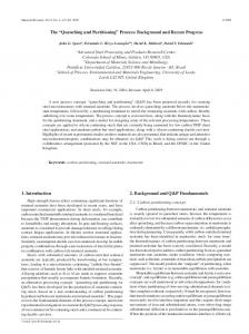
FIGURE 1. Selenium uptake and depuration in four organs and in the whole body of C. fluminea exposed to 50 µg Se L-1 of waterborne SeMet for 20 days, followed by a 50-day depuration period. For the four organs, values are mean ( SD (n ) 5). For the whole body, the closed circles represent the observed concentrations and the curve represents the modeled concentrations. The first point is the Se background concentration. and homoscedasticity of error terms (SigmaStat 2.0, SPSS Science, Chicago, IL).
Results Kinetics of Selenium Accumulation. Concentration of Se in control animals was 0.37 ( 0.02 µg Se g-1 (n ) 10), and did not change significantly during the course of the experiment. Bioaccumulation of SeMet was high since from the onset of the exposure phase (day 1), Se concentrations in the tissues of exposed animals increased 10- to 19-fold above those exposed to background concentrations (Figure 1). Se was persistent in the tissues, since Se measured at the end of the depuration phase (day 70) still represented 30% of Se accumulated at day 20 for mantle and gills, and 40 and 45% for muscle and visceral mass. A two-compartment model was used to depict Se kinetics at the whole body level, with a rapid compartment defined by uptake and depuration kinetic rate constants of 200,000 ( 106,000 mL g-1 w.w. d-1 and 1940 ( 1030 d-1 respectively, and a slower compartment described by uptake and depuration kinetic rate constants of 11.5 ( 1.8 mL g-1 w.w. d-1 and 0.0173 ( 0.00655 d-1 (R2 ) 0.81). The corresponding steady-state concentration factor was 769 mL g-1 (w.w.). Based on the kinetic model, it would take approximately 220 days for Se concentrations to reach values not significantly different from those of controls (0.37 µg Se g-1 w.w.). Selenium Partitioning in the Cytosolic and Particulate Fractions. The cytosolic fraction of Se was stable at a value of ca. 1.2 µg Se g-1 w.w. from the first exposure day (Figure 2). To the contrary, Se concentrations in the particulate fraction continually increased and reached more than 20 times the initial concentration at the end of the exposure 2114
9
ENVIRONMENTAL SCIENCE & TECHNOLOGY / VOL. 43, NO. 6, 2009
FIGURE 3. Size exclusion chromatography-FIAS chromatograms of Se-containing proteins in SeMet (left panel: gills and right panel: visceral mass) contaminated C. fluminea. Chromatograms presented are the mean of two to three chromatograms. phase. During the depuration phase, particulate Se concentrations decreased and Se was homogeneously distributed between both fractions after 1 month of depuration. These concentrations were of the order of 1.2 µg Se g-1 w.w, still 0.8 µg Se g-1 over controls. These trends were particularly apparent when Se concentrations in each fraction were plotted against total tissue Se, with a linear increase of particulate Se and a saturation for cytosolic Se. Selenium Partitioning in the Cytosolic Fraction. Figure 3 displays the size exclusion chromatograms for SeMet contaminated bivalves. The Se-containing proteins in the SeMet contaminated bivalves were largely distributed across the molecular separation range of the chromatographic column. At day one, gill tissues contained three major bands of Se compounds. A large peak eluted in the low molecular weight (LMW) range (600 kDa). At the end of the accumulation phase, this later peak increased to reach a proportion equal to the peak near the Vt of the column (LMW range compounds), while a third small peak was observed around 30 kDa. Finally, after 30 days of depuration, the protein profile was similar to the profile observed at day one, with a major peak in the LMW range. In the visceral mass, the same tendency was observed, i.e., Se-containing peaks eluted across the entire column volume, with a higher peak in the LMW range and a wide
FIGURE 4. Thin cross section through filaments observed with a TEM, showing two filaments. (A) Control group; (B) SeMet exposed group. band that ranged on the whole column. No change of this profile was observed with time. Histopathological Effects. Thin cross sections through filaments were observed with an optical microscope and showed that in SeMet exposed organisms a general disorganization occurred of lamellae and broken interfilament junctions (not shown). 92% of junctions were preserved in the control group vs 33% in the SeMet exposed group. Effects on the filament ultrastructure were analyzed by TEM (Figure 4). The control group (Figure 4A) displayed filaments with two areas, frontal and abfrontal, edged by a polymorphous epithelium. Frontal, laterofrontal, and lateral cilia were observed on the frontal face whereas rare cilia were seen on the abfrontal face. In the SeMet exposed group (Figure 4B), the frontal zone became very dense, vacuoles disappeared, and the central zone of the filament was swollen illustrating the existence of an edema. This observation demonstrates the occurrence of large disturbances of either the microcirculatory blood flow in the area and/or the impairment of the local cellular ionic exchange system. Alteration of gill filament cell mitochondria were also seen (Figure 5). In the SeMet exposed group (Figure 5B), ultrastructural studies revealed disorganized mitochondrial crests and an irregularly indented outer membrane.
Discussion Relatively High Uptake and Persistence of SeMet at the Tissular Level. SeMet concentration factor of 770 is higher than the value of 150 found for Cd in C. fluminea but lower than the concentration factor of 4,000 found for methylmercury for the same species (12). Its accumulation seems to be linked to a high uptake rate constant (11.5 mL g-1 d-1) and a low depuration rate constant (0.0173 d-1). The estimated depuration rate constant is close to the value (0.035
FIGURE 5. Thin cross section through filaments observed with a TEM, showing the aspect of the mitochondria of gill filaments. (A) Control group; (B) SeMet exposed group. d-1) found by Roditi and Fisher in zebra mussels exposed to contaminated algae (13). This high uptake rate constant may be explained by an inadvertent bioaccumulation of SeMet in place of methionine, which has been shown to be accumulated in another bivalve species via an active transport system (14). Such an inadvertent uptake of SeMet in place of methionine was also demonstrated for mammals (15). Bioaccumulation reflects different strategies of detoxification (16). Whereas some organisms are regulators that prevent metals from entering or expel them efficiently once taken up, others are accumulators that for example synthetize ligands that bind the metals. Accumulators tend to show elevated body burdens, which is generally the case for bivalves. The kinetic parameters estimated for C. fluminea tend to confirm this hypothesis, with uptake rate constants significantly higher than depuration rate constants. Se Detoxification in the Particulate Fraction and Persistence in the Cytosol. An average of 71% of the total gill Se was found in the particulate fraction at the end of the exposure phase and it was even more important in the visceral mass (77%). These values are close to the 72% of watersoluble Se found in macroinvertebrates sampled in situ in agricultural drain waters by Fan et al. (17). In the cytosol, a plateau of selenium concentration was observed around 1.2 µg g-1, which might correspond to a saturation of the cytosol Se binding sites. Above that saturation threshold, Se would be detoxified into the particulate phase, since the particulate fraction increases linearly with increasing Se total concentrations in the organ. Several authors have shown that the subcellular distribution of metals and metalloids in the prey directly alters trophic transfer to predators. In particular, metal in soluble fractions of prey has been shown to be more bioavailable to predators than metal bound to nonsoluble fractions (i.e., exoskeleton and metal concretions) (16, 18). Our VOL. 43, NO. 6, 2009 / ENVIRONMENTAL SCIENCE & TECHNOLOGY
9
2115
results show that selenium in the soluble fraction can increase rapidly and to a high extent in SeMet contaminated bivalves and additionally, it is more persistent than selenite (data not shown), which might explain the higher bioavailability and toxicity of SeMet observed in aquatic ecosystems (17). SeMet Might Replace Methionine in the General Protein Pool. Se-containing peaks eluted across the entire column volume, with the highest peak in the LMW range and a wide band that ranged on the whole column, containing some major peaks around 30 and 60 kDa and in the void volume. A similar distribution pattern was observed by Unrine et al. (19) in western fence lizards fed with selenomethionine exposed crickets. They found, using the same column as in the present study, a major peak in the LMW range (600 kDa). A wide range of studies have been done on mammal models to investigate selenoproteins. About 30 seleniumcontaining proteins have been identified, with known functions for about 12 of them (20). The molecular weights of the compounds that occurred around 30 and 60 kDa in our study may correspond to those of glutathione peroxidase and selenoprotein P identified for aquatic organisms. For example, in the freshwater bivalve Unio tumidus, Doyen et al. (21) identified a selenium-dependent peroxidase transcript with a theoretical molecular weight of 26.4 kDa. A similar glutathione peroxidase and a selenoprotein P (MW of =50 kDa) were also identified in zebrafish (22). However, the wide elution range observed for SeMet indicates that it may not only be integrated into selenoproteins but also in the general protein pool in place of methionine, in contrast to selenite that is mainly found in small molecular weight compounds (Supporting Information, Figure S1). This may alter the activity of enzymes or the substrate access if the replacement occurs in the vicinity of the active site (23). From an environmental perspective, this substitution of SeMet into proteins in place of methionine may have consequences on bivalves and their predator reproduction by enhancing teratogenic effects (24). Direct Effects of SeMet on Gill Filament and Mitochondria Morphology. Important histopathological effects were observed after 20 days of exposure to 50 µg L-1 of SeMet, with swollen gill filaments. Gill filament alteration may affect this organ’s function and as such, may explain the drastic decrease in ventilatory flow rates observed by Fournier et al. (25) in C. fluminea exposed to SeMet. Pathological effects were also observed in gills of the teleostean fish Lepomis cyanellus exposed to selenium contamination in Belews Lake, with dilated sinusoids and swollen lamellae. This Se-induced dilatation of gill lamellae caused impaired blood flow, inactive gas exchange, and a metabolic stress response (increased respiratory demand and oxygen consumption) that can lead to death (24). Another striking result is the alteration of mitochondria morphology and cristae organization, which has to our knowledge never been observed in organo-Se exposed organisms. These changes may result in primary metabolism impairment. As such, selenium might be toxic not only indirectly through maternal transfer of proteins where SeMet may replace methionine as it was suggested for fish, but also directly on the exposed organisms. From a more general perspective it must be underlined that our results are very similar to data obtained after Se trophic transfer in the field (24) or in controlled conditions (13, 19). This similarity clearly highlights, for the first time, that SeMet water-only exposure produces results that 2116
9
ENVIRONMENTAL SCIENCE & TECHNOLOGY / VOL. 43, NO. 6, 2009
mimic data obtained for dietary organo-Se exposures. Thus, this shortened (and less expensive) experimental protocol of using water exposures to test Se kinetics and effects can be used to produce environmentally relevant insights.
Acknowledgments We thank Tom Hinton for his useful help and advice in the revision process. This work was supported by IRSN as part of the ENVIRHOM program.
Supporting Information Available Additional figure. This information is available free of charge via the Internet at http://pubs.acs.org.
Literature Cited (1) Conde, J. E.; Sanz Alaejos, M. Selenium concentrations in natural and environmental waters. Chem. Rev. 1997, 97, 1979– 2002. (2) Lemly, A. D. Aquatic selenium pollution is a global environmental safety issue. Ecotox. Environ. Saf. 2004, 59, 44–56. (3) Ministe`re des solidarite´s, de la sant e´ et de la famille. Circulaire NDGS/SD7A n° 2004-602 du 15 de´cembre 2004 relative a` la gestion du risque sanitaire en cas de de´passement des limites de qualite´ des eaux destine´es a` la consommation humaine pour les parame`tres antimoine, arsenic, fluor, plomb et se´le´nium en application des articles R. 1321-26 a` R. 1321-36 du code de la sante´ publique. In Bulletin officiel N°2005-1, Annonce N°30, 2005. (4) Wang, C.; Lovell, R. T. Organic selenium sources, selenomethionine and selenoyeast, have higher bioavailability than an inorganic selenium source, sodium selenite, in diets for channel catfish (Ictalurus punctatus). Aquaculture 1997, 152, 223. (5) Novoselov, S.; Rao, M.; Onoshko, N.; Zhi, H.; Kryukov, G.; Xiang, Y.; Weeks, D.; Hatfield, D.; Gladyshev, V. Selenoproteins and selenocysteine insertion system in the model plant cell system Chlamydomonas reinhardtii. EMBO 2002, 21, 3681– 3693. (6) Hodson, P. V.; Hilton, J. W. The nutritional requirements and toxicity to fish of dietary and waterborne selenium. Ecol. Bull. 1983, 35, 335–339. (7) Tran, D.; Moody, A. J.; Fisher, A. S.; Foulkes, M. E.; Jha, A. N. Protective effects of selenium on mercury-induced DNA damage in mussel haemocytes. Aquat. Toxicol. 2007, 84, 11–18. (8) deBruyn, A. M. H.; Chapman, P. M. Selenium toxicity to invertebrates: Will proposed thresholds for toxicity to fish and birds also protect their prey? Environ. Sci. Technol. 2007, 41, 1766–1770. (9) Tinggi, B. Essentiality and toxicity of selenium and its status in Australia: a review. Toxicol. Lett. 2003, 137, 103–110. (10) Smith, E. J.; Davison, W.; Hamilton-Taylor, J. Methods for preparing synthetic freshwaters. Water Res. 2002, 36, 1286– 1296. (11) Garnier-Laplace, J.; Vray, F.; Baudin, J. P. Dynamic model for radionuclide transfer from water to freshwater fish. Estimation of accumulation and depuration kinetic parameters for 137Cs and 106Ru in carp (Cyprinus carpio L.). Water, Air Soil Pollut. 1997, 98, 141–166. (12) Inza, B.; Ribeyre, F.; Maury-Brachet, R.; Boudou, A. Tissue distribution of inorganic mercury, methylmercury and cadmium in the Asiatic clam (Corbicula fluminea) in relation to the contamination levels of the water column and sediment. Chemosphere 1997, 35, 2817–2836. (13) Roditi, H. A.; Fisher, N. S. Rates and routes of trace element uptake in zebra mussels. Limnol. Oceanogr. 1999, 44, 1730– 1749. (14) Stewart, M. G. Kinetics of neutral amino-acid transport by isolated gill tissue of the bivalve Mya arenaria (L.). J. Exp. Mar. Biol. Ecol. 1978, 32, 39–52. (15) Ducros, V.; Favier, A. Selenium metabolism. EMC-endocrinologie 2004, 1, 19–28. (16) Wallace, W. G.; Luoma, S. N. Subcellular compartmentalization of Cd and Zn in two bivalves. I. Significance of metalsensitive fractions (MSF) and biologically detoxified metal (BDM). Mar. Ecol.: Prog. Ser. 2003, 249, 183–197. (17) Fan, T. W.; Teh, S. J.; Hinton, D. E.; Higashi, R. M. Selenium biotransformations into proteinaceous forms by foodweb
(18)
(19)
(20) (21)
organisms of selenium-laden drainage waters in California. Aquat. Toxicol. 2002, 57, 65–84. Wallace, W. G.; Luoma, S. N. Subcellular compartmentalization of Cd and Zn in two bivalves. II. Significance of trophically available metal (TAM). Mar. Ecol.: Prog. Ser. 2003, 257, 125– 137. Unrine, J. M.; Jackson, B. P.; Hopkins, W. A. Selenomethionine biotransformation and incorporation into proteins along a simulated terrestrial food chain. Environ. Sci. Technol. 2007, 41, 3601–3606. Arteel, G. E.; Sies, H. The biochemistry of selenium and the glutathione system. Environ. Toxicol. Pharmacol. 2001, 10, 153– 158. Doyen, P.; Vasseur, P.; Rodius, F. Identification, sequencing and expression of selenium-dependent glutathione peroxidase transcript in the freshwater bivalve Unio tumidus exposed to Aroclor 1254. Comp. Biochem. Physiol. 2006, 144, 122–129.
(22) Kryukov, G. V.; Gladyshev, V. N. Selenium metabolism in zebrafish: multiplicity of selenoprotein genes and expression of a protein containing 17 selenocysteine residues. Genes Cells 2000, 5, 1049–1060. (23) Schrauzer, G. N. Selenomethionine: A review of its nutritional significance, metabolism and toxicity. J. Nutr. 2000, 130, 1653– 1656. (24) Lemly, A. D. Teratogenic effects of selenium in natural populations of freshwater fish. Ecotoxicol. Environ. Saf. 1993, 26, 181– 204. (25) Fournier, E.; Adam, C.; Massabuau, J.-C.; Garnier-Laplace, J. Bioaccumulation of waterborne selenium in the Asiatic clam Corbicula fluminea: influence of feeding-induced ventila tory activity and selenium species. Aquat. Toxicol. 2005, 72, 251–260.
ES802891J
VOL. 43, NO. 6, 2009 / ENVIRONMENTAL SCIENCE & TECHNOLOGY
9
2117