pituitary apoplexy, pituitary tumours and mucocele or pyocele of the paranasal sinuses, may closely resemble retrobulbar neuritis.7 Further diagnostic confusion ...
I Case Reports
Carotid - ophthalmic aneurysm: an uncommon cause of acute monocular blindness Ruben Kuzniecky, MD Calvin Melmed, MD, FRCPC Hyman Schipper, MD PhD
arotid-ophthalmic aneurysms may present with a variety of visual deficits, which are usually progressive. Sudden visual disturbances may indicate aneurysmal rupture, sudden expansion of the dome of the aneurysm or an embolus originating from the aneurysmal sac. We describe a patient with a carotid-ophthalmic aneurysm who presented with acute loss of vision. C
Case report A 32-year-old right-handed woman was admitted to hospital complaining of sudden loss of vision in her right eye. She had suffered from migraines for 10 years, experiencing vague blurring of vision followed by pounding temporal headaches and nausea. On the day of admission the patient sensed a sudden bright flash of light in her right eye, which was followed by light-headedness and syncope that lasted 1 to 2 minutes. Immediately thereafter she had blurred vision in her right eye, right retrobulbar pain associated with eye movements in all directions and a mild headache. There were no symptoms of a seizure or complaints of focal numbness or weakness. The findings at physical examination were unremarkable. Neurologic examination revealed an afferent pupillary defect, a central scotoma, the ability only to count fingers, and loss of colour vision in her right eye. FundusFrom the Department of Neurology and Neurosurgery, McGill University and Sir Mortimer B. Davis Jewish General Hospital, Montreal
Reprint requests to: Dr. Calvin Melmed, Department of Neurology and Neurosurgery, Sir Mortimer B. Davis Jewish General Hospital, 3755 C6te Ste-Catherine Rd., Montreal, PQ H3T 1E2
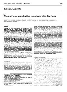
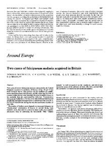
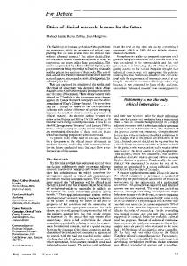
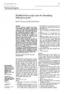
copy revealed a normal optic disk with sharp margins and normal venous pulsations. Acute retrobulbar neuropathy and syncope of unknown origin were diagnosed. The results of laboratory investigations were unremarkable. A non-contrast-enhanced computed tomogram of the head appeared normal, but a subsequent contrast study revealed a round, hyperdense lesion to the right of the optic chiasm (Fig. 1). A four-vessel angiogram showed a single aneurysm, 17 mm in diameter, at the junction of the right ophthalmic and internal carotid arteries (Fig. 2). During a right frontal craniotomy the right optic nerve appeared flattened and stretched over the dome of the aneurysm, and there was a small hemorrhage or hemorrhagic infarction at the nerve's superior aspect. The aneurysm was clipped, but the patient's vision had not returned 2 months after surgery.
Comments
Carotid-ophthalmic aneurysms are rare.'-3 They originate from the anteromedial wall of the internal carotid artery between the point of the artery's bifurcation and the origin of the ophthalmic artery. At presentation, 50% of the aneurysms are greater than 25 mm and 75% greater than 10 mm in diameter. They occur more frequently in women and are more likely to be multiple compared with other intracranial aneurysms. Most reports have emphasized the slow progression of visual symptoms. Kothandaram and associates4 found that most of their patients presented with a monocular central visual field defect, which could progress to complete blindness, along with a contralateral defect in the upper temporal CMAJ, VOL. 136, APRIL 1, 1987
727
[v~ .
aspect of the field (junctional scotoma). These symptoms are thought to arise from the progressive displacement of and secondary damage to all the fibres in the optic nerve, including the decussating lower nasal fibres, which loop forward into
I 2~.1
1.t'~~~~~~~~
#4
Fig. 1 -Contrast-enhanced computed tomogram showing well-defined hyperdense lesion to right of optic chiasm (arrow).
;'1
~~~~~~~~~4
-~~~~~~~~~~~~~~~~~~~~~~-
.,
the contralateral optic nerve. Sudden loss of central vision has been reported in only one other patient, after a carotid-ophthalmic aneurysm ruptured;5 pathological examination of the optic nerve revealed compression and necrosis of the fibres. In our patient, sudden expansion of the aneurysm could have stretched the optic nerve and interrupted its blood supply, resulting in fibre infarction and a sudden central scotoma. The hemorrhage observed at surgical exploration is consistent with this mechanism since there was no evidence of rupture of the aneurysm's dome. A relatively sudden loss of central vision in one eye, afferent pupillary defect, normal findings at funduscopy and retrobulbar pain associated with eye movements suggest retrobulbar neuritis, which is usually encountered in people with multiple sclerosis or other demyelinating diseases.6 In rare instances compressive lesions may produce an acute monocular loss of vision, presumably because of interruption of the blood supply to the optic nerve. This phenomenon, reported in cases of pituitary apoplexy, pituitary tumours and mucocele or pyocele of the paranasal sinuses, may closely resemble retrobulbar neuritis.7 Further diagnostic confusion may arise when patients who present with acute loss of vision secondary to compressive lesions such as craniopharyngiomas and medulloblastomas respond initially to steroid therapy.8 Carotid-ophthalmic aneurysm should be considered in patients who present with acute loss of vision, especially if they have atypical features such as apoplexy and photopsia and no other signs or symptoms of demyelinating disease. References 1. Locksley HB: Natural history of subarachnoid hemorrhage, intracranial aneurysms and arteriovenous malformations. Based on 6368 cases in the cooperative study. J Neurosurg 1966; 25: 219-239
AW
2. Krayenbuhl H, Yasargil MG: Das Hiraneurysms, Geigy, Basel, 1958: 15-16
17pue tmmgrin Angogtramstehownanceurs of ophhlifnditra right dhoiameterefne atedes juncion aoptid arteiesm (arrow).
Fig. 21
3. Pool JI, Potts DC: Aneurysms and Arteriovenous Anomalies of the Brain: Diagnosis and Treatment, Har-Row, New York, 1965: 52-77 4. Kothandaram P, Dawson BH, Kruyt RC: Carotid-ophthalmic aneurysm. A study of 19 patients. J Neurosurg 1971; 34:
544-548 5. Stern W, Ernest G: Intracranial ophthalmic artery aneurysm. Am J Ophthalmol 1975; 80: 203-206 6. Wray S: Neuro-ophthalmologic diseases. In Rosenberg R (ed): The Clinical Neurosciences, vol 1, Churchill, New York, 1983: 797-840 7. Robinson J: Sudden blindness with pituitary tumors. J Neurosurg 1972; 36: 83-85
8. Hirst L, Miller N, Kumar A et al: Medulloblastoma causing a corticosteroid responsive optic neuropathy. Am J Ophthalmol 1980; 89: 437-442 728
CMAJ, VOL. 136, APRIL 1, 1987