Basidiobolomycosis
BASIDIOBOLOMYCOSIS. PRESENTATIONS
REPORT
OF
A
108
CASE
WITH
UNIQUE
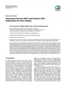
Saeed RR1*, Mustafa H1, Majid R2 1. General University Teaching Hospital of Sulaimania, Kurdistan, Iraq 2. Shorsh Hospital of Sulaimania- Department of Histopathology, Sulaimania, Kurdistan, Iraq Correspondence Dr. Rebeen Rahim Saeed. General University Teaching Hospital of Sulaimania, Kurdistan, Iraq Email:
[email protected] Saeed RR, Mustafa H, Majid R. Basidiobolomycosis. Report of a case with unique presentations. Case Study and Case Report 2013; 3(2): 108 - 115. ABSTRACT The simultaneous involvement of oral cavity, pharynx, larynx, and vocal cords in particular; all together by Basidiobolomycosis is very rare; to our knowledge such presentation is not reported in the literature. Basidiobolomycosis is a rare invasive fungal infection that affects gastrointestinal tract, mucosa, subcutaneous tissue and skin commonly in children, less often in adolescence and very rarely in adult. We report a 39 years old male who presented with gradual swelling of the right side of the face and neck. Subsequently it was complicated by respiratory distress and difficulty in swallowing in the late course of the illness. IV Itraconazole was given for two weeks and then switched to oral Itraconazole for 10 months. After 10 months of follow up both clinically and by CT scan, the patient showed complete recovery from the infection. Key words: Basidiobolomycosis, fungal infection INTRODUCTION Most cases of Basidiobolomycosis are usually under-diagnosed as they present like tonsillitis, malignancy or TB. It's very rare; only about 172 cases are reported all over the world. It can cause invasive disease of lungs, gastrointestinal system, and skin, and can even be disseminated throughout the body. CASE REPORT Case presentation A 39- year- old man was brought to A&E by his wife. He was complaining of sore throat for one month duration associated with swelling of the right side of face extending from neck and mandible to the right cheek. The swelling was painless and started to increase in
Case Study and Case Report 2013; 3(2): 108 - 115.
108
Basidiobolomycosis
109
size gradually. He did not respond to the usual antibiotics as he was diagnosed as a case of acute tonsillitis initially. There was no constitutional symptoms and furthermore as the mass increased in size gradually, he subsequently developed voice changes, dysphagea and weight loss at the late course of the disease. He never travelled abroad and vaccination history was up-to-date. He was not diabetic and he has never taken immunosuppressive or any other drugs. On examination, there was right side facial swelling, which was extended from right upper cervical region toward the cheek, and there were palpable few lymph nodes in both cervical submandibular region, they were firm in consistency. His vital signs were within reference range, the rest of physical exam were normal apart from tracheostomy and jejunostomy tube which he had it for the nutrition considering having stridor and dysphagea in the late course of the disease. Investigations Investigations revealed polymorphonuclear leucocytosis (total leukocyte count 14 700/mm3) with mild eosinophilia 7% and increased platelet count of 537000/ mm3. Liver function tests and renal function tests were within normal limits. Blood glucose and electrolytes were within reference range. Blood film was normal apart from leucocystosis and thrombocytosis. HCV, HIV, HBV markers were negative. ESR was 71mm/hr. when specimen(FNA) taken from the oro-pharyngeal lesion; it only showed pyogenic inflammatory collection and then the FNA of the cervical lymph nodes showed abundant necrotic eosinophilic material, scattered giant cells, occasional clusters of epitheloid histiocytes and reactive lymphoid cells. No definite malignant cells were seen. The picture was suggestive for TB, however PCR for TB was negative and the patient did not respond to trial of Anti-TB medications. The follicular tonsils were removed and sent for histopathology analysis. Moreover, we biopsied the peritonsillar area the oro-pharyngeal area again of the evolving mass around the tonsillar area after the tonsillectomy which was extending to pharynx and larynx. The histopathologist excluded Malignancy and also PCR was negative for TB. The histopathologist—In her first report suggested chronic granulomatous infection then after a week of diagnosis Basidiobolomycosis was confirmed (Figure 1, 2). Before the result of biopsy getting back, he was admitted one day to the emergency room for having haematamesis. OGD was done and it showed gastric outlet obstruction due to chronic active bulbar ulcer with severe GERD and lower esophageal ulceration. When gastrojujenostomy had been decided by the surgeon and in the operation they noticed a small yellow colored mass on the outer side transverse colon which was sent for histopathological exam and result was basidiobolomycosis (Figure 3). He was arranged for CT scan of head and neck—the description is explained in the image in detail (Figure 4). Meanwhile, MRI was arranged and the description is mentioned in (Figure 4).
Case Study and Case Report 2013; 3(2): 108 - 115.
109
Basidiobolomycosis
110
Figure 1. Neck mass: The microscopy of a neck mass biopsy reveals an intense eosinophilic inflammatory reaction containing fungal hyphae surrounded by the Splendore-Hoeppli phenomenon (Black arrow). (Hematoxylin and eosin stain ×100).
Figure 2. Tonsil: The fungi consist of broad pauci septate hyphae consistent with basidiobolomycosis (Black arrow). (Periodic acid-Schiff hematoxylin stain ×400).
Case Study and Case Report 2013; 3(2): 108 - 115.
110
Basidiobolomycosis
111
Figure 3. Transeverse colon: The wall of the transverse colon demonstrates an abscess with a necrotic center containing eosinophils surrounded by granulomatous inflammation with Langerhans giant cells (Black arrow). (Fungal elements can be seen in the surrounding). Treatment The patient was managed with proton pump inhibitors, Intravenous fluids. When the patient developed stridor and dysphagea, tracheostomy and jejunostomy was done for providing a good airway and nutrition respectively as the patient had gastric outlet obstruction due to active bulber ulcer with severe GERD and lower esophageal ulceration, and he also had vocal cord involvement by the fungus. The patient was put on a treatment of Itraconazole; first IV 200 mg twice daily for two weeks and then orally 200mg twice daily for 10 months. Outcome and follow-up Patient had started responding within two weeks of treatment dramatically, and the treatment was continued for 10 months with regular monitoring of clinical features, biochemical parameters, and imaging. The patient is now almost cured and is free of any recurrence on a follow-up.
Case Study and Case Report 2013; 3(2): 108 - 115.
111
Basidiobolomycosis
112
Figure 4. Neck MRIs in Figure A and B and neck CT scans in the Figure C, D and E. In Figure A and B, neck MRIs with Gd show ill - defined soft tissue mass depicted arising from the right tonsilar bed, measuring (6.73 × 15.39 cm), heterogeneously enhancing after Gd IV extended downward to both supraglottic and glottic region reaching). In Figure C, D and E, neck CT scans show evidence of large heterogenous irregular outlined soft tissue density mass lesion involving the right side of the nasophayrnx mostly involving the fossa of rosenmuller, opening of Eustachian tube and the pre-vertebral muscle causing marked narrowing of the nasopharynx. The bellow mass lesion shows marked downward extensions in to the supraglottic and glottis spaces, mostly at the right side greatly involving the hard and soft palate, floor of the mouth, tongue base, phayrngoepiglottic folds and even involving the right vocal cord resulting in nearly total obstruction of the laryngo-pharynx measuring (20×9×8cm) in size associated with adjacent matted LNs, the largest one is measured 20mm in diameter. Few small enlarged left posterior triangle LNs are seen and the figure D and E shows CT follow up for the patient after taking Itraconazole which was taken in, June, July and September of 2012 respectively and the last CT (Figure E) shows significant shrinkage of the mass and this was on after 4 months of the Diagnosis of the condition.
Case Study and Case Report 2013; 3(2): 108 - 115.
112
Basidiobolomycosis
113
DISCUSSION Basidiobolomycosis is a rare disease caused by the fungus Basidiobolus ranarum, an environmental saprophyte found worldwide 1 - 2. B. ranarum was first isolated in1955 from decaying plants in the United States 3 - 4. The first recognized case of infection in human being was one of the subcutaneous mycosis reported in1956 in Indonesia 4 - 5. The first culture-proved case of invasive basidiobolomycosis of the the palate and maxillary sinus was reported in the United States in 1978 4, 6. Basidiobolomycosis is unusual fungal infection caused by B. ranarum that occurs exclusively in healthy individuals 4, 7. It commonly involves the trunk and extremities, however its role in causing gastrointestinal and intra abdominal infections is also increasing 8 - 9. The portal of entry is believed to be through skin after insect bites, scratches or minor cuts. Infection is characterized by a hardened firm nodule that expands and spreads locally 10. The case we present here is unique because the infection was in the oro-phyarngeo-laryngeal region and involvement of such an area with Basidiobolomycosis has not been reported in the literature. What we learn from this case, as it was initially diagnosed as acute follicular tonsillitis and then the course of the disease prolonged and there was no response to antibiotics—TB and malignancy like lymphoma had come to our mind as the second and third differential diagnosis as it was explained above, we have excluded TB and malignancy; for instance lymphoma, rhabdomyosarcoma through the biopsy. Then finally the histopathologist had reached to a conclusion that the finding is consistent with basiobolomycosis as it is explained in Figure 1,2 and 3 although the clinical appearance of the lesions of zygomycosis due to B. ranarum is usually very suggestive, histopathology and culture can only make a definitive diagnosis. The demonstration of hyaline, wide, thin walled, non-septate or scarcely septate hyphae or hyphal fragments in KOH digests or in H & E stained sections of a biopsy from the actively growing part of a lesion is a very characteristic feature 11. Immunoflourescent techniques using fungus specific antibody conjugated to flourescein, can be employed in identifying hyphae in clinical specimens, especially when they are scarce and when no biopsied tissue is available for culture 12. Treatments of Basidiobolomycosis include medical and surgical interventions, surgery may not be necessary. Oral Potassium iodide (KI) can be used in a dose of 30 mg/kg, however most of the case reports show good response of the infection toward Itraconazole. Surgical excision may also be curative. The usual treatment of choice has been potassium iodide (KI) in a dose of 30 mg/kg daily given as a single daily dose or divided into three daily doses 13 - 17. Treatment is usually continued for 6-12 months. Beneficial effects have also been achieved with itraconazole in a dose of 100 200 mg daily 16 - 17. In conclusion we have to put fungal infection like basidiobolomycosis in mind especially if there is an unusual lesion in the oropharyngolarynx which does not respond to antibiotics. Itraconazole was used in our case which was very effective as it is clearly seen in CT follow up from the start of the disease. Furthermore, surgical debridement of the lesion in addition to the medical treatment is recommended by some experts to terminate the infection, however our case does not have any surgical intervention yet he improved very well with the medical treatment.
CONCLUSION
Case Study and Case Report 2013; 3(2): 108 - 115.
113
Basidiobolomycosis
114
There are many learning points from this case: Basidiobolomycosis is a rare infection should be put in the differential diagnosis of any swelling in oropharyngeolaryngeal area when not responding to usual treatment like TB and lymphoma. Oropharyngeolaryngeal involvement with Basidiobolomycosis is very rare Basidiobolomycosis infection could be found in more than one site in the body during presentation. It's true the infection should be treated with both surgical debridement and antifungal medications, but in our case the patient responded to medical treatment without surgical debridement. COMPETING INTERESTS The authors declare that they have no competing interests. REFERENCES 1. Kwon-Chung KJ, Bennett JE. Medical Mycology. Philadelphia: Lea and Febiger, 1992. 2. Lyon GM, Smilack JD, Komatsu KK, Pasha TM, Leighton JA, Guarner J, Colby TV, Lindsley MD, Phelan M, Warnock DW, Hajjeh RA. Gastrointestinal basidiobolomycosis in Arizona: clinical and epidemiological characteristics and review of the literature. Clin Infect Dis. 2001; 32:1448 - 55. 3. Hussein MR, Musalam AO, Assiry MH, Eid RA, El Motawa AM, Gamel AM. Histological and ultrastructural features of gastro-intestinal basidiobolomycosis. Mycol Res. 2007; 111:926 - 30. 4. Al Jarie A, Al Azraki T, Al Mohsen I, Al Jumaah S, Almutawa A, Mohd Fahim Y, Al Shehri M, Abu Dayah A, Ibrahim A, Maw Shabana M, Rezk Abd-Elwahed Hussein M. Basidiobolomycosis: Case series. J Med Mycol. 2011; 21: 37 - 45. 5. Kian Joe L ,Pohan A, Tjoei Eng NI, Van Der Meulen H. Basidio-bolus ranarum as a cause of subcutaneous mycosis in Indonesia. AMA Arch Derm. 1956; 74: 378 - 83. 6. Dworzack DL, Pollock AS, Hodges GR, Barnes WG, Ajello L, Padhye A. Zygomycosis of the maxillary sinus and palate caused by Basidiobolus haptosporus. Arch Intern Med. 1978; 138:1274 - 6. 7. Carr EJ, Scott P, Gradon JD. Fatal gastrointestinal mucor mycosis that invaded the post operative abdominal wall wound in an immunocompetent host. Clin Infect Dis. 1999; 29: 956 - 7. 8. Khan ZU, Khoursheed M, Makar R, Al Waheeb S,Al-Bader I, Al-muzaini A, et al. Basidiobolus ranarum as an etiological agent of gastrointestinal zygomycosis. J Clin Microbiol. 2001; 39: 2360 - 3. 9. Goyal A, Gupta N, Das S, Jain S. Basidiobolomycosis of the nose and face: a case report and a mini-review of unusual cases of basidiobolomycosis. Mycopathologia. 2010; 170: 165 - 8.
Case Study and Case Report 2013; 3(2): 108 - 115.
114
Basidiobolomycosis
115
10. Mathew R, Kumaravel S, Kuruvilla S, Varghese RG, Shashikala, Srinivasan S, et al. Successful treatment of extensive basidiobolomycosis with oral itraconazole in a child. Int J Dermatol. 2005; 44: 572 – 5. 11. Rippon JW. Medical Mycology: The pathogenic fungiand actinomycetes, 3rd edn, Philadelphia: WB Saunders, 1988; 681-713. 12. Kwon-Chung KJ, Bennet JE. Medical mycology. Philadelphia: Lea & Febiger, 1992; 447-463. 13. Vismer HF, DeBeer HA, Dreyer L. Subcutaneous phycomycosis caused by Basidiobolus haptosporus (Drechsler, 1947). S Afr Med J. 1980; 58: 64 - 647. 14. Pasha TM, Leighton JA, Smilack JD, Heppel J, Colby TV, Kaufman L. Basidiobolomycosis: An unusualfungal infection mimicking in¯ammatory bowel disease. Gastroenterology. 1997; 112: 250 - 4. 15. Kamalam A, Thambia AS. Entomophthtorae basidiobolae successfully treated with KI. Mykoen. 1979; 22: 82 - 4. 16. Van Cutsem J, Van Gerven F, Janssen PAJ. Activity of orally and parentally administered itraconazole in the treatment of superficial and deep mycoses: Animal models. Rev Infect Dis. 1987; 9 (Suppl 1): 515 - 32. 17. Gugnani HC. A review of zygomycosis due to Basidiobolus ranarum. Eur J Epidemiol. 1999; 15: 923 -9.
Case Study and Case Report 2013; 3(2): 108 - 115.
115